Find Duplicates
 Check Document
 Submission(new)
 Experts Office
 Editorial Office
2019 Vol. 42, No. 4
Display Method:
2019, 42(4): 423-429.
doi: 10.12122/j.issn.1674-4500.2019.04.01
Abstract:
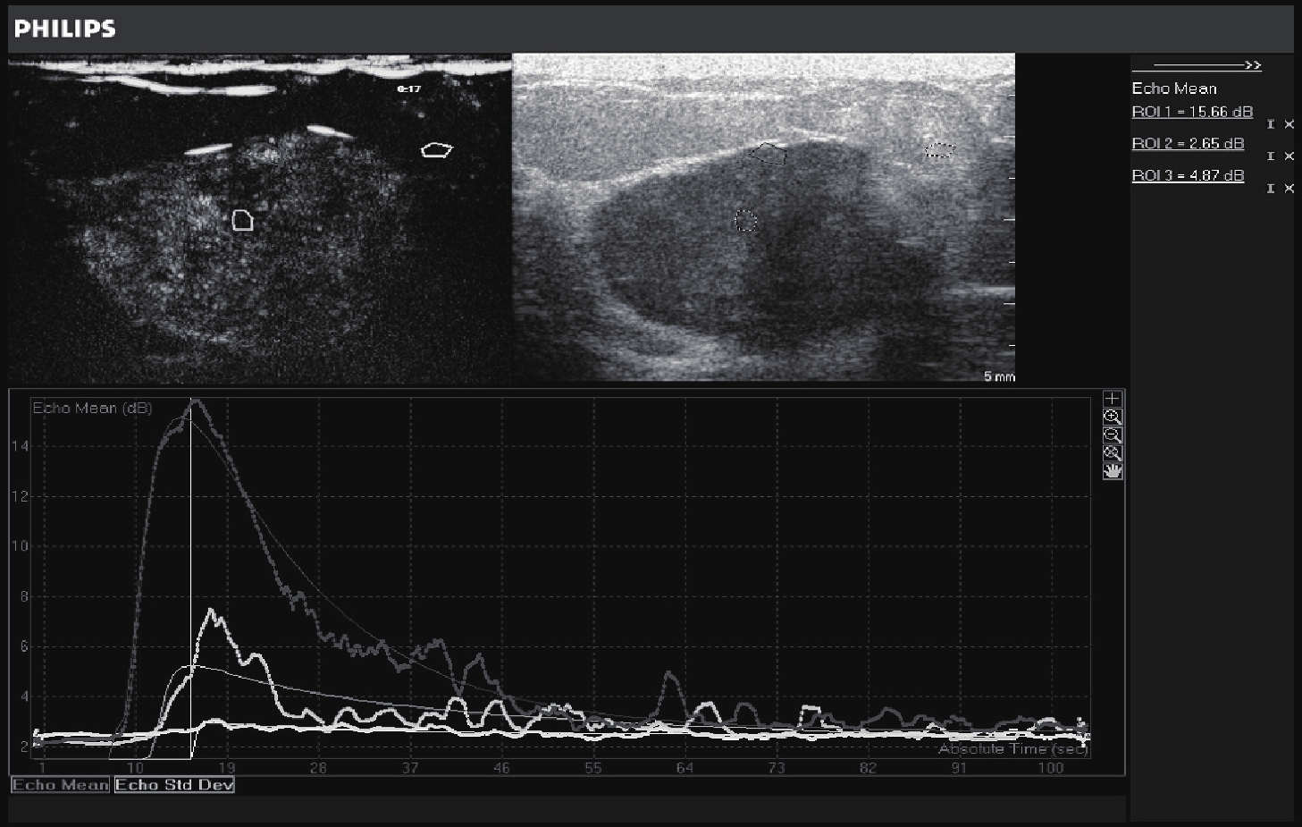
ObjectiveTo explore the differences between contrast-enhanced features and time-intensity curve quantitative parameters in different molecular types of breast cancer. MethodsWe retrospectivly analyzed 142 cases of breast cancer patients (142 tumors) by contrast-enhanced data. The estrogen receptor, progesterone receptor, human epidermal growth factor receptor-2 (HER-2), Ki-67 expression status were analyzed. The breast cancer were divided into four types: Luminal A, B (luminal epithelium), triple negative, and HER-2 overexpression. The contrast-enhanced features and time-intensity curve of breast cancer patients with different molecular classification were analyzed. ResultsLuminal type A showed a low or equal enhancement ratio of 80.8% (21/26), with a radial aggregation ratio of 69.2% (18/26). Luminal type B showed a low or equal enhancement ratio of 60.3% (35/58). The proportion of radial aggregation was 56.9% (33/58). The ratio of high enhancement of HER-2 overexpression was 90.0% (27/30), and the proportion of internal perfusion defect was 73.3% (22/30). The proportion of blood flow was 70.0% (21/30). The proportion of triple-yin showed high enhancement rate was 92.9% (26/28), and the clear ratio of enhanced boundary was 92.8% (26/28). The differences of the enhancement of borderline, enhancement intensity, perforation blood flow and perfusion defects between different molecular types of breast cancer were significant (P<0.05). But the differences of range of enhancement, contrast agent distribution, contrast agent enhancement sequence between angiographic pattern and molecular typing were not significant (P>0.05). ConclusionThere are differences in the features of contrast-enhanced ultrasound in different molecular types of breast cancer. It can provide valuable imaging information for preoperative prediction of breast cancer molecular classification.
2019, 42(4): 430-433.
doi: 10.12122/j.issn.1674-4500.2019.04.02
Abstract:
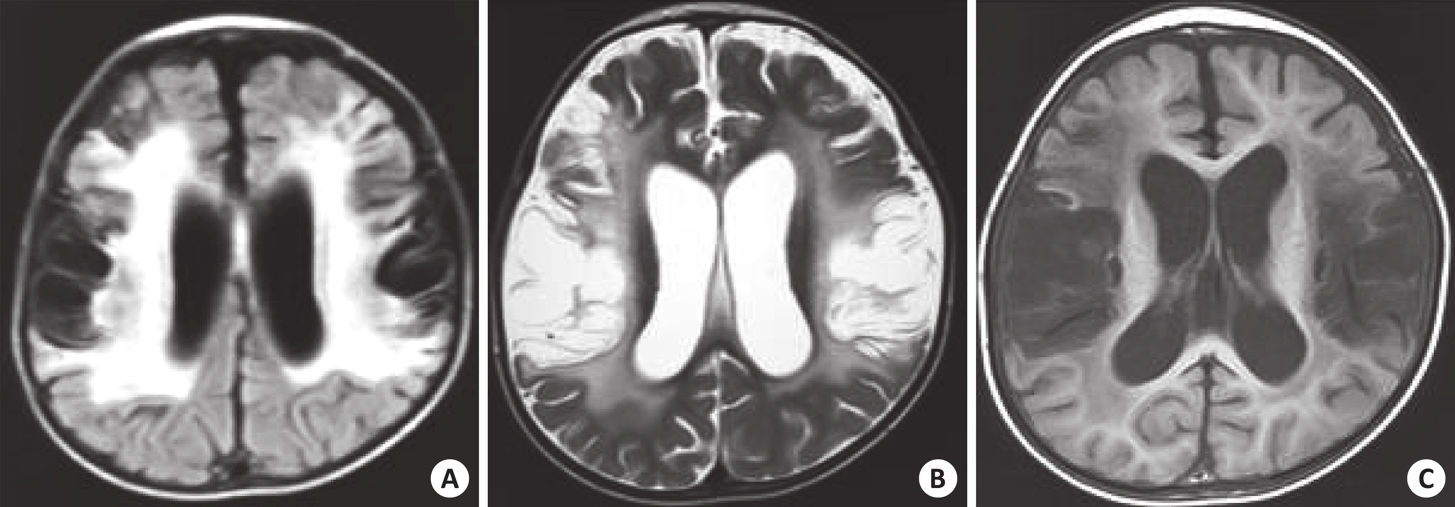
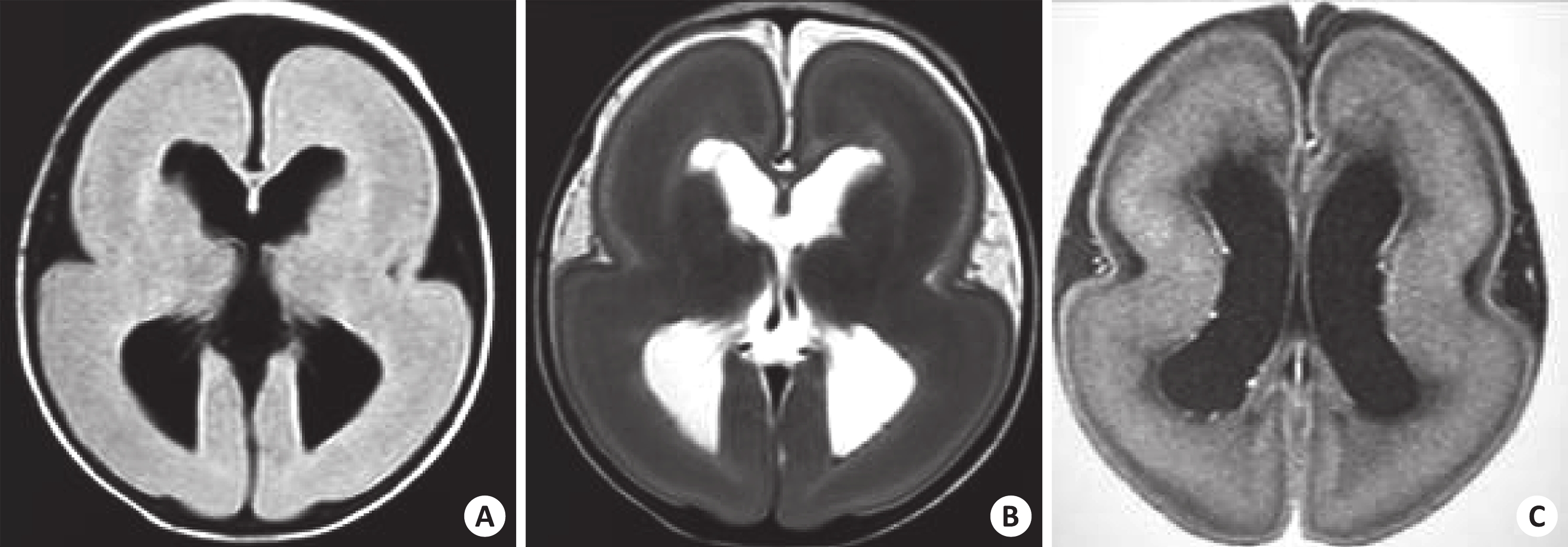
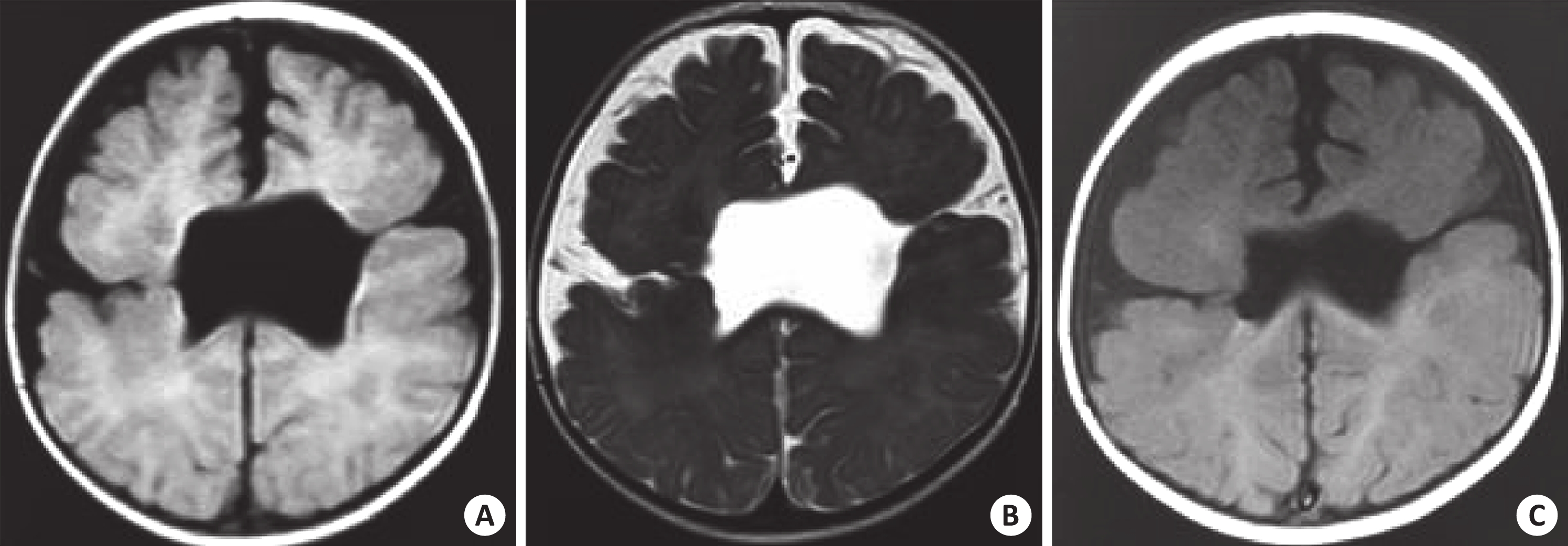
ObjectiveTo explore the value of MRI in the TORCH of children. MethodsThe imaging and clinical data of 36 children confirmed TORCH were retrospectively analyzed. Nineteen boys and 17 girls were included, ranging in age from 5 d to 3 years old. All the children underwent head scan of MRI. ResultsDiffuse cystic encephalomalacia accounted for 20 cases and penetrating cyst were 7 cases. There were 27 cases with varying degrees of calcification in the brain parenchyma, which were striated and spotted. A few cases with ileocentric fusion calcification, mainly located in the subependymal and periventricular areas. Microcephaly, smooth brain accounted for 11 cases, giant gyrus malformation 15 cases, cerebellar hypoplasia 3 cases, cleft brain malformation 6 cases,hydroanencephaly 3 cases, demyelination and gliosis of brain parenchyma 13 cases. There were 17 cases of hydrocephalus, 9 cases of subdural effusion and 21 cases of diffuse cerebral atrophy. ConclusionMRI could visually display various signs in TORCH syndrome. It is a preferred imaging examination for this disease.
2019, 42(4): 434-438.
doi: 10.12122/j.issn.1674-4500.2019.04.03
Abstract:
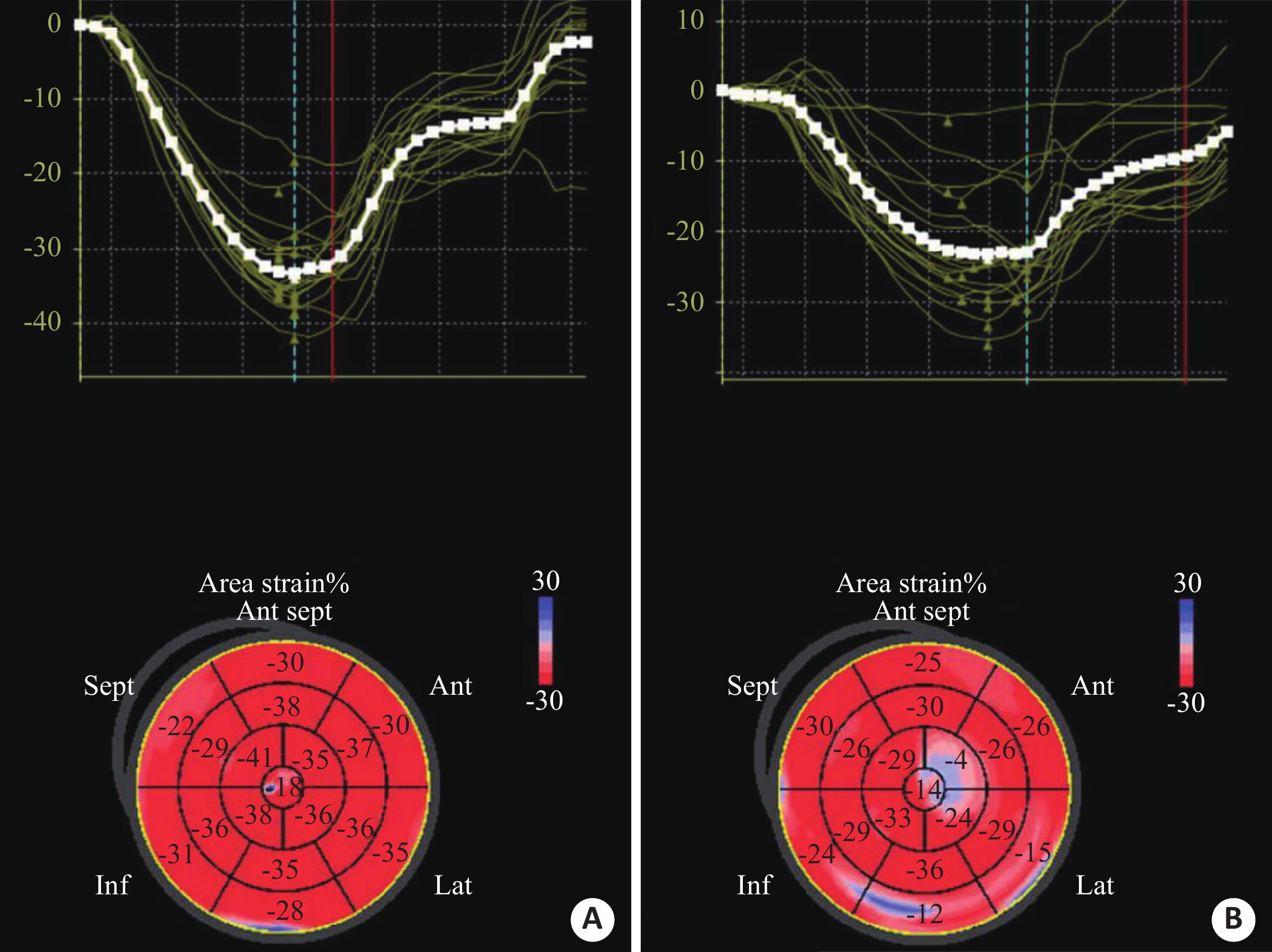
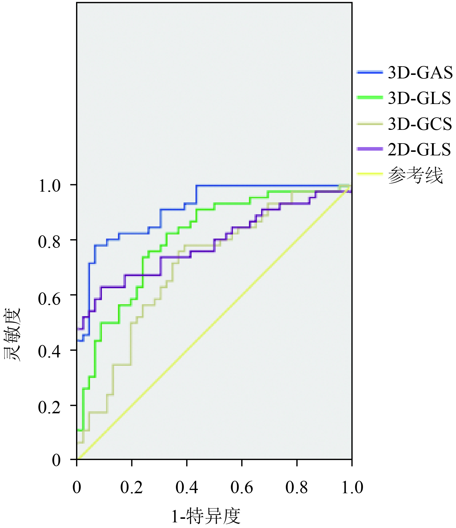
ObjectiveTo explore three-dimensional speckle tracking echocardiography (3D-STE) in evaluating the ventricular function changes in breast cancer patients before and after the administration of anthracyclines. MethodsRoutine echocardiography, two-dimensional speckle tracking echocardiography (2D-STE) and 3D-STE were separately applied to test the left ventricular function 24 h before, within 24 h of the end of the 2nd and the 4th cycle of anthracycline chemotherapy. The parameters obtained at these 3 time points were compared longitudinally. The correlation with the cumulative dose of anthracyclines was analyzed. Their sensitivity to the detection of early heart impairment caused by cardiotoxicity of anthracycline was analyzed. ResultsThe left ventricular global area strain (3D-GAS) and the global longitudinal strain (3D-GLS) within 24 h after the end of the 2nd and 4th cycle of chemotherapy were lower than those before chemotherapy (P<0.05). The left ventricular global circumferential strain (3D-GCS) within 24 h after the end of the 4th cycle of chemotherapy was lower than that before 24 h of chemotherapy (P<0.05). The change in global radial strain (3D-GRS) during the whole chemotherapy process was not significant (P>0.05). The sensitivity and specificity for 3D-GAS were 81.23% and 81.98%, respectively; 62.15% and 65.02% for 3D-GLS, respectively; 66.09% and 55.08% for 3D-GCS, respectively. 3D-GAS, 3D-GLS, 3D-GCS were negatively correlated with the cumulative dose of anthracyclines(P<0.05). Conclusion3D-STE for evaluating ventricular function changes in breast cancer patients before and after using anthracyclines is worth popularizing in clinical imaging diagnosis. It is a sensitive and reliable imaging method for monitoring the heart impairment caused by anthracyclines.
2019, 42(4): 439-443.
doi: 10.12122/j.issn.1674-4500.2019.04.04
Abstract:
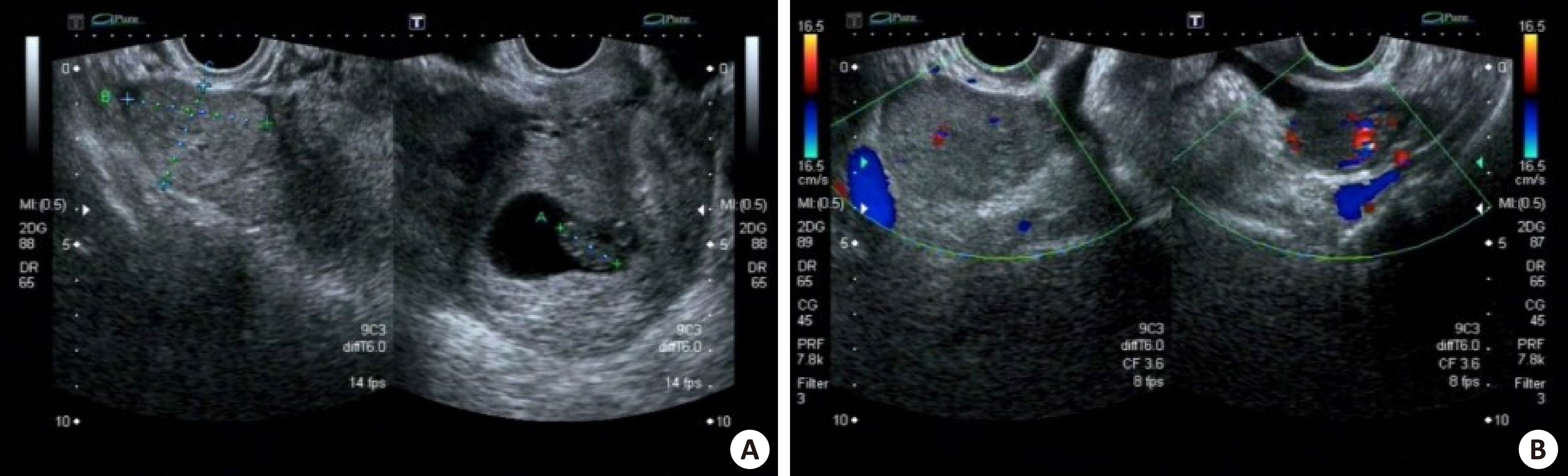
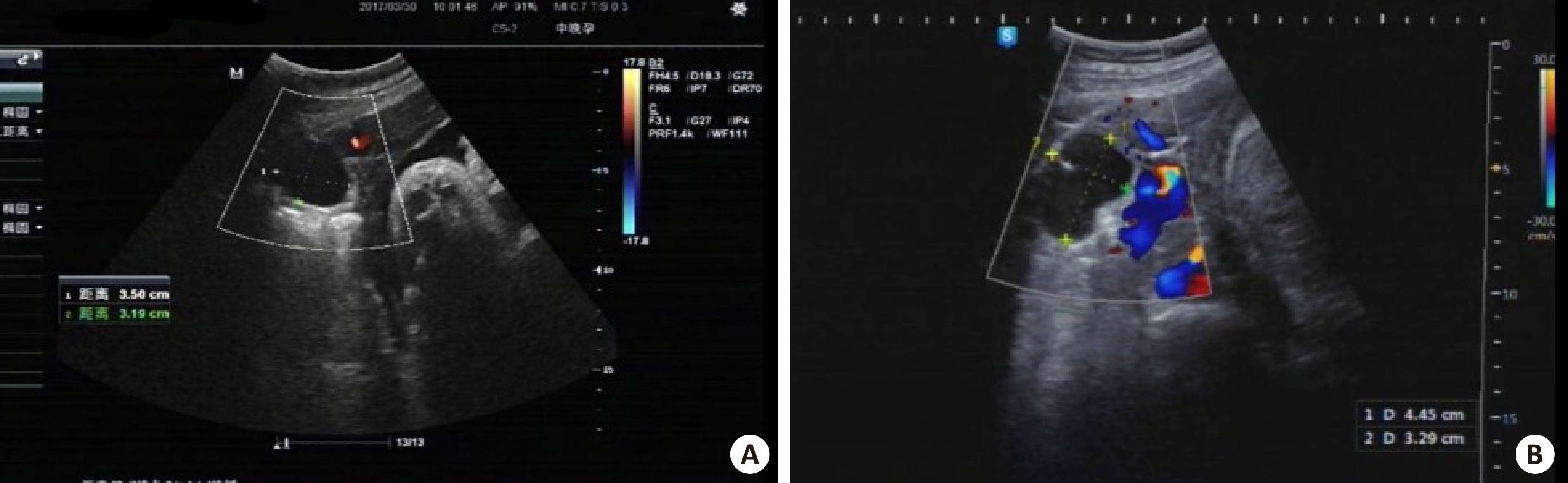
ObjectiveTo explore diagnostic and surveillant value of ultrasound in ovarian tumors during pregnancy. MethodsClinical and ultrasound data of 48 patients with pathologically proven ovarian tumors during pregnancy were retrospectively reviewed. The patients aged from 21 to 41 years old (30.5±4.7 years old) had undertaken ultrasound examinations during pregnancy between January 2016 to December 2018. The diagnostic and surveillant value of sonographic examinations were summarized. ResultsFifty-one tumors were detected by sonographic examinations in 48 patients. These lesions included 27 mature teratomas, 8 mucinous cystadenomas, 10 serous cystadenomas, 2 hemorrhagic corpus luteum cysts, 1 serous - mucinous tumor with endometrioid cyst, 1 simple cyst, and 1 struma ovarii. The ultrasound examinations would well document the imaging characteristics of these tumors. On sonography, the diametre of tumors ranged from 2.2 cm to 12.1 cm (5.5±2.1 cm). Based on sonographic features, 3 cases were probably malignant, and 7 cases were probably torsion of ovarian tumor or hemorrhagic corpus luteum cysts. Among 48 patients, 3 patients underwent pregnancy termination precession only by their personal willingness, and the resection of ovarian tumors were performed, and 12 patients underwent tumor resection through laparoscopic surgery before delivery due to abdominal pain, probably malignant, or large volumes of tumors. The other 33 patients underwent expectant treatment, and ovarian tumors were resected while cesarean delivery. The majority of patients achieved a satisfying pregnancy outcome. ConclusionUltrasound examinations could measure the lesions’ diametre, detect malignant lesions, differentiate the tumors, reveal the complications, and surveil the tumors during pregnancy, which provide pivotal evidence for clinical decision.
2019, 42(4): 444-448.
doi: 10.12122/j.issn.1674-4500.2019.04.05
Abstract:
ObjectiveTo explore the 2 years follow-up results of arthroscopic autologous chondrocyte transplantation in the treatment of degenerative knee articular cartilage injury. MethodsFifty-four patients with degenerative knee articular cartilage injury who underwent arthroscopic autologous chondrocyte transplantation in our hospital from October 2014 to October 2016 were enrolled in this study. Two years follow-up results were analyzed. All patients were recorded the dominant blood loss in the operation, hospitalization time, observation treatment efficiency. The knee pain visual analog score (VAS), Hss knee function score, Tegner motor function score and knee joint activity at the time before surgery, 3 months, 6 months, 12 months and 24 months after surgery were compared. MRI results before and after surgery were recorded. According to the treatment effect, the patients were divided into the effective group (effectiveness+improvement) and the ineffective group (ineffectiveness). The factors of the surgical effect were analyzed. ResultsThe dominant blood loss was 92.3±11.8 mL at 48 h postoperatively. The hospital stay were 8.3±2.5 d. The total effective rate was 79.63%. After the operation, the patients' VAS scores were significantly decreased, HSS score and Tegner score were significantly increased (P<0 01="" the="" knee="" joint="" activity="" was="" lower="" than="" that="" before="" surgery="" at="" the="" time="" of="" 3="" months="" after="" surgery="" and="" returned="" to="" normal="" at="" the="" time="" of="" 6="" months="" after="" surgery="" in="" 6="" months="" after="" surgery="" 19="" patients="" had="" a="" repaired="" tissue="" thickness="" which="" was="" higher="" than="" the="" normal="" cartilage="" thickness="" in="" 12="" months="" after="" surgery="" 6="" patients="" still="" had="" repaired="" cartilage="" hypertrophy="" in="" 24="" months="" after="" surgery="" 2="" patients="" had="" cartilage="" hypertrophy="" and="" were="" treated="" with="" arthroscopic="" surgery="" no="" other="" surgery-related="" complications="" were="" observed="" after="" the="" postoperative="" examination="" there="" were="" 43="" patients="" in="" the="" effective="" group="" and="" 11="" patients="" in="" the="" ineffective="" group="" univariate="" analysis="" showed="" that="" age="" duration="" of="" disease="" degree="" of="" cartilage="" damage="" postoperative="" joint="" weight="" and="" postoperative="" joint="" motion="" were="" the="" relevant="" factors="" that="" affected="" the="" surgical="" outcome="" logistic="" multivariate="" regression="" analysis="" showed="" that="" age="">70 years, disease course>1 year, cartilage injury III~IV, postoperative joint weight bearing, postoperative joint activity too early or too little were independent risk factors affecting the surgical outcome. ConclusionArthroscopic autologous chondrocyte transplantation is effective in the treatment of degenerative knee articular cartilage injury. Rehabilitation training should be followed in the postoperative rehabilitation training, which can effectively improve the treatment effects.
2019, 42(4): 449-452.
doi: 10.12122/j.issn.1674-4500.2019.04.06
Abstract:
ObjectiveTo explore the imaging characteristics of high frequency ultrasound in meniscus cyst of knee joint, guide minimally invasive treatment, and provide a method for diagnosis and treatment of this disease. MethodsThe ultrasonographic findings of 86 cases of meniscus cyst of knee joint from January 2017 to June 2019 were retrospectively analyzed. The ultrasonographic findings of meniscus cyst of knee joint were compared with those of MRI and arthroscopy. Eight cases were treated with ultrasound-guided minimally invasive therapy. ResultsAmong 86 cases of meniscus cysts diagnosed by high frequency ultrasound, compared with MRI and arthroscopy, ultrasound was highly sensitive to meniscal cysts. But 1 case was misdiagnosed, with a specificity of about 98.83%. In 82 cases of meniscal cysts, ultrasound showed 78 cases with meniscal tear, but 4 case was missed diagnossis, with a sensitivity of about 95.12%. The effect of ultrasound-guided minimally invasive treatment was improved in 8 cases. One case recurred, but no obvious clinical symptoms were found. ConclusionHigh-frequency ultrasound has a high clinical value in the diagnosis of meniscal cysts. It can be used for ultrasound-guided minimally invasive treatment.
2019, 42(4): 453-456.
doi: 10.12122/j.issn.1674-4500.2019.04.07
Abstract:
ObjectiveTo investigate the value of image texture analysis of T2 Fat-suppression sequence (FS T2WI) in differential diagnosis of benign and malignant breast nodules. MethodsA total of 61 images of breast nodules FS T2WI were retrospectively analyzed in 60 patients with surgical pathology. The ROC curves for differential diagnosis of benign and malignant breast nodules were drawn and compared with the pathological results. Results61 breast nodules in 60 patients, The FS T2WI texture parameter gray region matrix focuses on the operation of high gray level to determine the AUC value (0.701) of benign and malignant breast nodules with high diagnostic accuracy. The sensitivity of the diagnosis of malignant breast nodules was 65.52 % (19/29), and the specificity was 71.88 % (23/32), and the misjudgment rate was 31.15 % (19/61). The diagnostic sensitivity of FS T2WI images to malignant nodules in the breast was 71.43 %(20/28), and the specificity was 63.64 % (21/33), and the miscalculation rate was 32.79 %(20/61); The combined application sensitivity of the two is 64.29 %(18/28), the specificity is 78.79 % (26/33), and the miscalculation rate is 27.87 %(15/61). Compared with individual T2, the difference is statistically significant (χ2=72.255, P=0.000). ConclusionFS T2WI sequence combined with texture analysis can improve the specificity of diagnosis of benign and malignant breast nodules, which can reduce the rate of misjudgment, and improve the accuracy of diagnosis.
2019, 42(4): 457-460.
doi: 10.12122/j.issn.1674-4500.2019.04.08
Abstract:
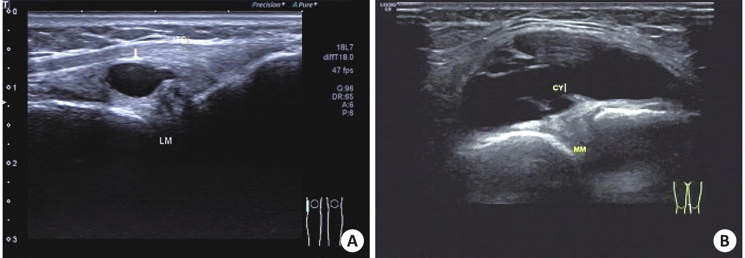
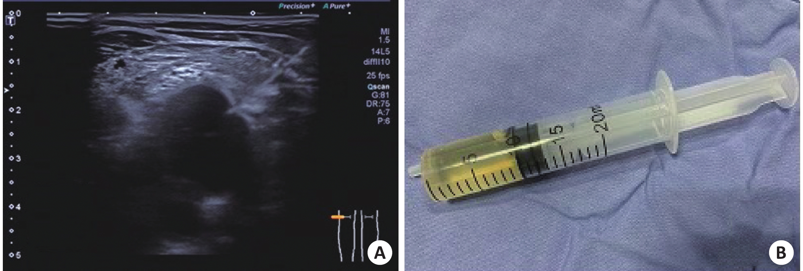
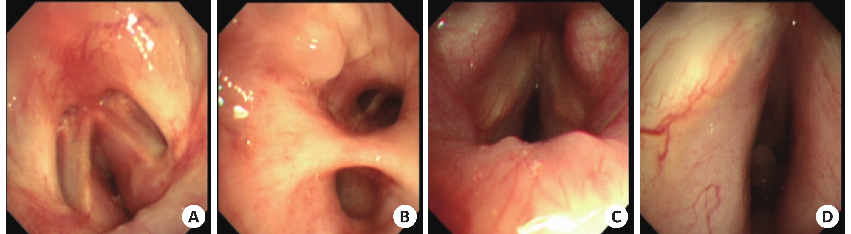
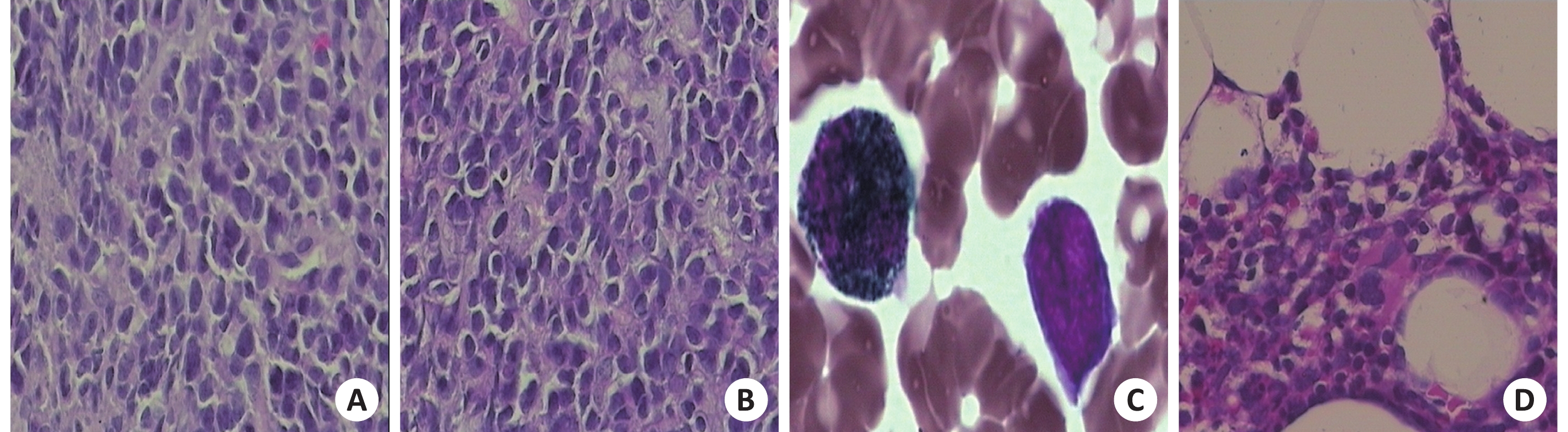
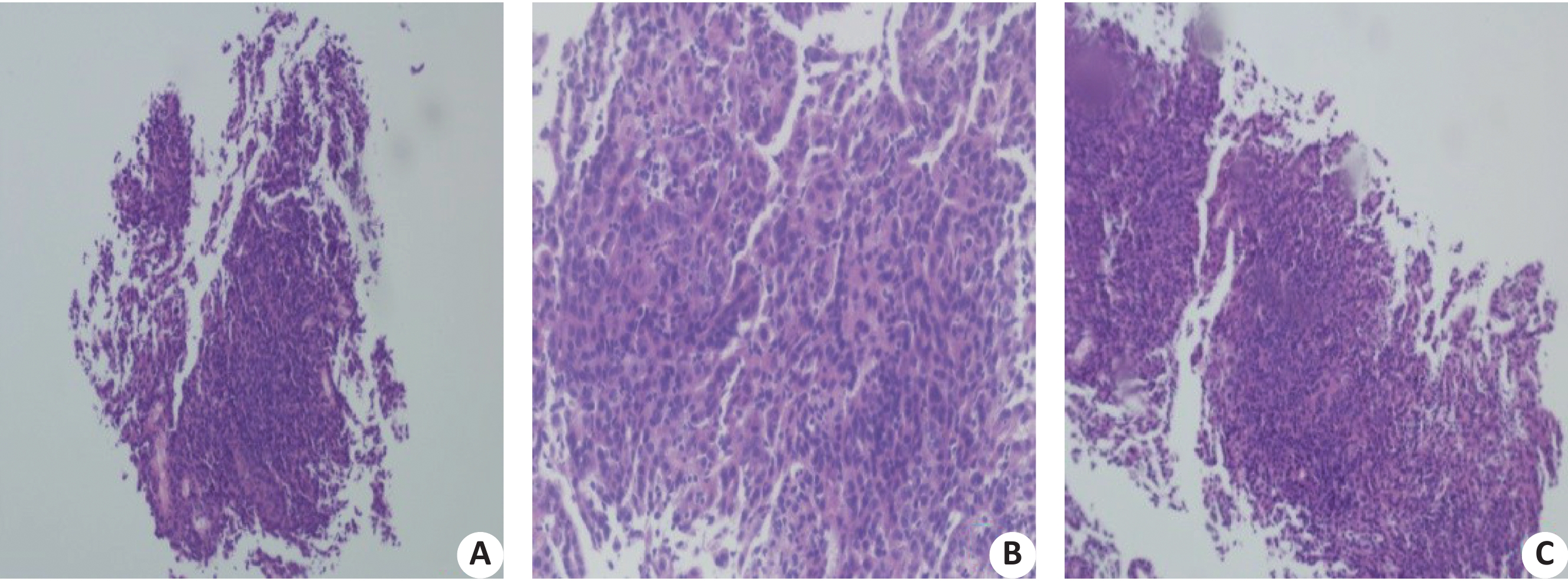
Diffuse large B cell lymphoma (DLBCL) is the main type of non Hodgkin lymphoma. The most common part of the organs and tissues outside the lymph nodes is gastrointestinal tract. In the case report, most of the DLBCL, which were first diagnosed with respiratory symptoms, were primary lymphoma of the lungs and mediastinum. We report 1 cases of diffuse large B lymphoma first diagnosed by respiratory symptoms. Combined with literature review, we aim to improve our understanding of DLBCL and avoid delayed treatment.
Diffuse large B cell lymphoma (DLBCL) is the main type of non Hodgkin lymphoma. The most common part of the organs and tissues outside the lymph nodes is gastrointestinal tract. In the case report, most of the DLBCL, which were first diagnosed with respiratory symptoms, were primary lymphoma of the lungs and mediastinum. We report 1 cases of diffuse large B lymphoma first diagnosed by respiratory symptoms. Combined with literature review, we aim to improve our understanding of DLBCL and avoid delayed treatment.
2019, 42(4): 461-464.
doi: 10.12122/j.issn.1674-4500.2019.04.09
Abstract:
ObjectiveTo explore the differential diagnostic value of macromolecular positive lymphophilic contrast agent diaminoglycol ether DTPA amide copolymer gadolinium complex (Gd-poly-DTPA-EOEA) enhanced MR lymphography (MRL) in lymph node imaging of tumor metastasis and inflammatory hyperplasia. MethodsA total of 14 New Zealand white rabbits. Seven rabbits were injected with complete immune adjuvant at the hind limb to establish an inflammatory hyperplasia group model of popliteal fossa lymph node. The Other 7 were inoculated with VX2 tumor in the posterior limb muscle, suggesting the popliteal fossa lymph node metastasis model and the opposite normal popliteal fossa lymph node as the control. Two magnetic resonance lymphography were performed before and after inoculation. After 3D reconstruction, the signal to noise ratio (SNR) of the largest rabbit popliteal fossa nodes was measured. The SNR differences between normal control side, inflammatory hyperplasia side and metastatic side lymph nodes at each observation time point were compared with the results of pathological examination. ResultOne rabbit was not successfully inoculated with tumor, and other inoculations were good. All the model rabbits completed the secondary MR lymphography. On the MR lymphangiography image, there was no significant difference between the inflammatory side and the control side popliteal fossa nodes SNR at all scanning time points in the inflammatory hyperplasia group (P>0.05). In the tumor metastasis group, there were significant differences between the tumor side and the control side popliteal fossa lymph node SNR at each scanning time point (P<0.01). Further comparison was made between the inflammatory side and the tumor side popliteal fossa lymph nodes. The difference between the inflammatory side and the tumor side popliteal fossa lymph nodes SNR was significant at each scanning time point (P<0.01). ConclusionMR lymphography (MRL) based on macromolecular positive lymphophilic contrast medium (Gd-poly-DTPA-EOEA) enhancement is a sensitive method for differentiating benign from malignant lymph nodes.

2019, 42(4): 465-468.
doi: 10.12122/j.issn.1674-4500.2019.04.10
Abstract:
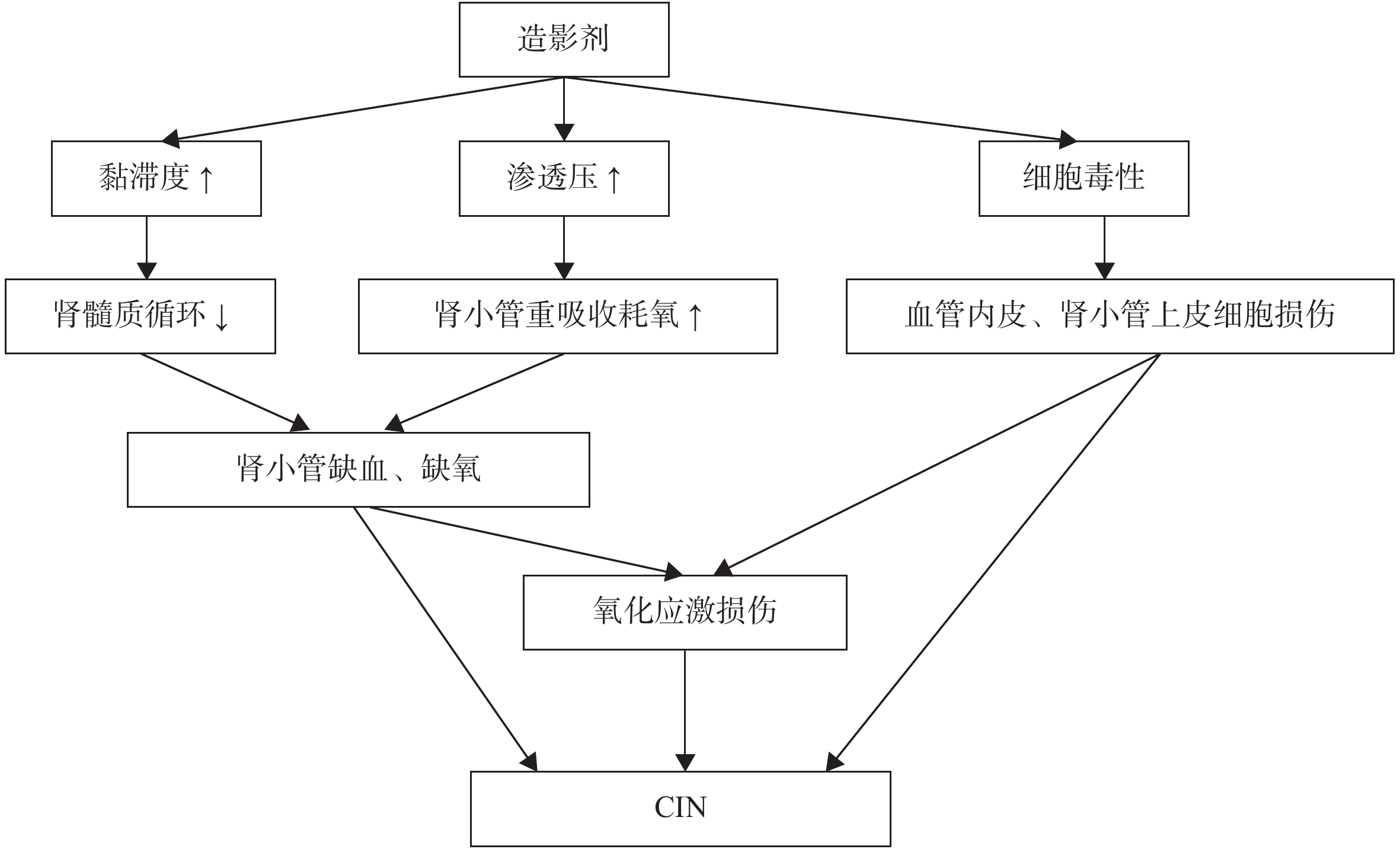
With the application of coronary angiography or intervention in the diagnosis, evaluation and treatment of coronary heart diseases, the incidence of contrast induced nephropathy is increasing. The contrast induced nephropathy are reversible in most patients, but it will increase the incidence of renal and cardiovascular adverse events. Therefore, how to effectively prevent and reduce the incidence of contrast induced acute kidney injury and reduce the incidence of adverse events in high-risk patients has become the focus of the current researches. According to the recommendations for the prevention and treatment of contrast nephropathy in the 2018 guidelines of the European Heart Association, this paper reviewed and summarized the current research progress in the prevention and treatment of the contrast induced nephropathy.
With the application of coronary angiography or intervention in the diagnosis, evaluation and treatment of coronary heart diseases, the incidence of contrast induced nephropathy is increasing. The contrast induced nephropathy are reversible in most patients, but it will increase the incidence of renal and cardiovascular adverse events. Therefore, how to effectively prevent and reduce the incidence of contrast induced acute kidney injury and reduce the incidence of adverse events in high-risk patients has become the focus of the current researches. According to the recommendations for the prevention and treatment of contrast nephropathy in the 2018 guidelines of the European Heart Association, this paper reviewed and summarized the current research progress in the prevention and treatment of the contrast induced nephropathy.
2019, 42(4): 469-472.
doi: 10.12122/j.issn.1674-4500.2019.04.11
Abstract:
ObjectivesTo evaluate the current situation of MRI equipment in China, provide a basis for the government to formulate the management policy of MRI equipment, and provide a reference for the use and management of MRI equipment in medical and health institutions. MethodsThe weights of evaluation indexes were determined by analytic hierarchy process and fuzzy comprehensive evaluation.The weighted rank sum ratio was used to evaluate 275 MRI equipment in 124 hospitals in China. ResultsThe comprehensive evaluation effect of MRI equipment was divided into three grades:good,medium and poor.There were 46 178 and 51 MRI equipment in each grade.WRSR results showed that the configuration structure of MRI equipment in China was unreasonable.Most of the 0.5 T field intensity MRI equipment came from domestic manufacturers and basic hospitals, while the 3.0 T field intensity was the opposite.The regional difference of MRI equipment was not obvious.The import dependence of MRI equipment was higher,and the imported equipment was generally better than the domestic equipment. The higher the field intensity,the better the comprehensive evaluation was. ConclusionAccording to regional health planning, health departments at all levels should allocate health resources rationally,allocate MRI equipment scientifically,control the field strength of MRI configuration,and avoid blindly introducing high-end models.In addition,we should vigorously promote the localization of MRI configuration and give full play to its advantages.
2019, 42(4): 473-475.
doi: 10.12122/j.issn.1674-4500.2019.04.12
Abstract:
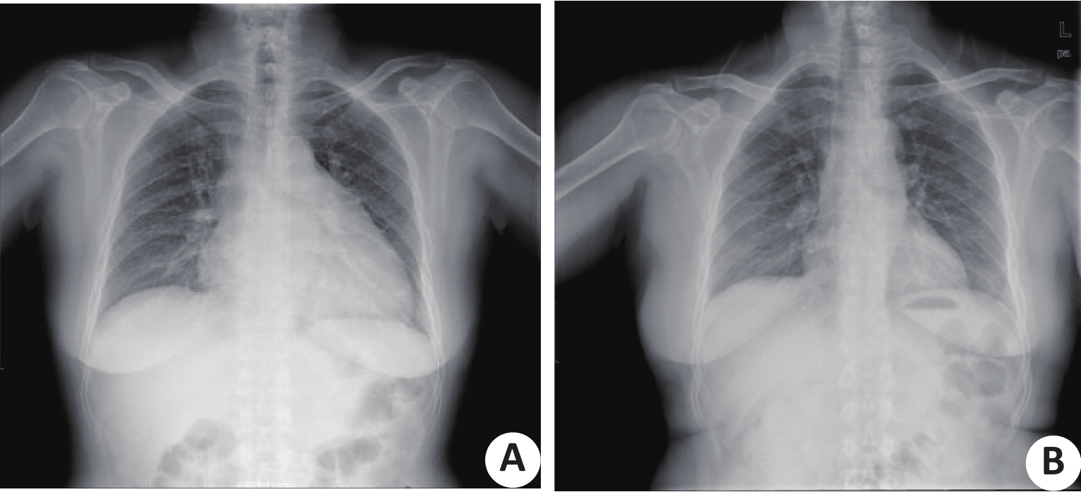
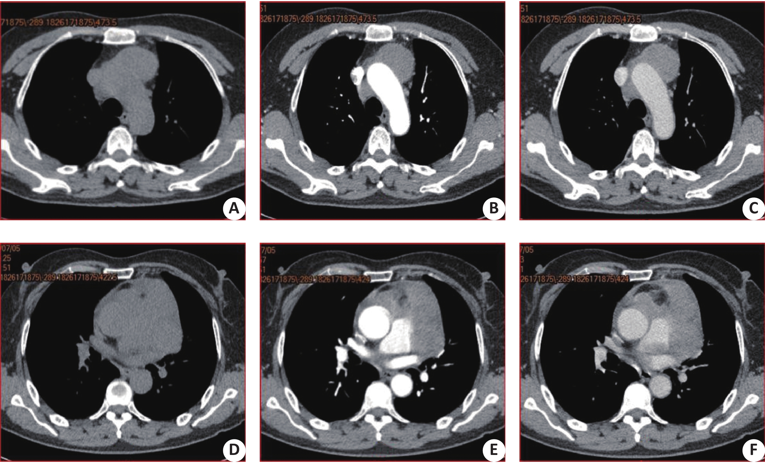
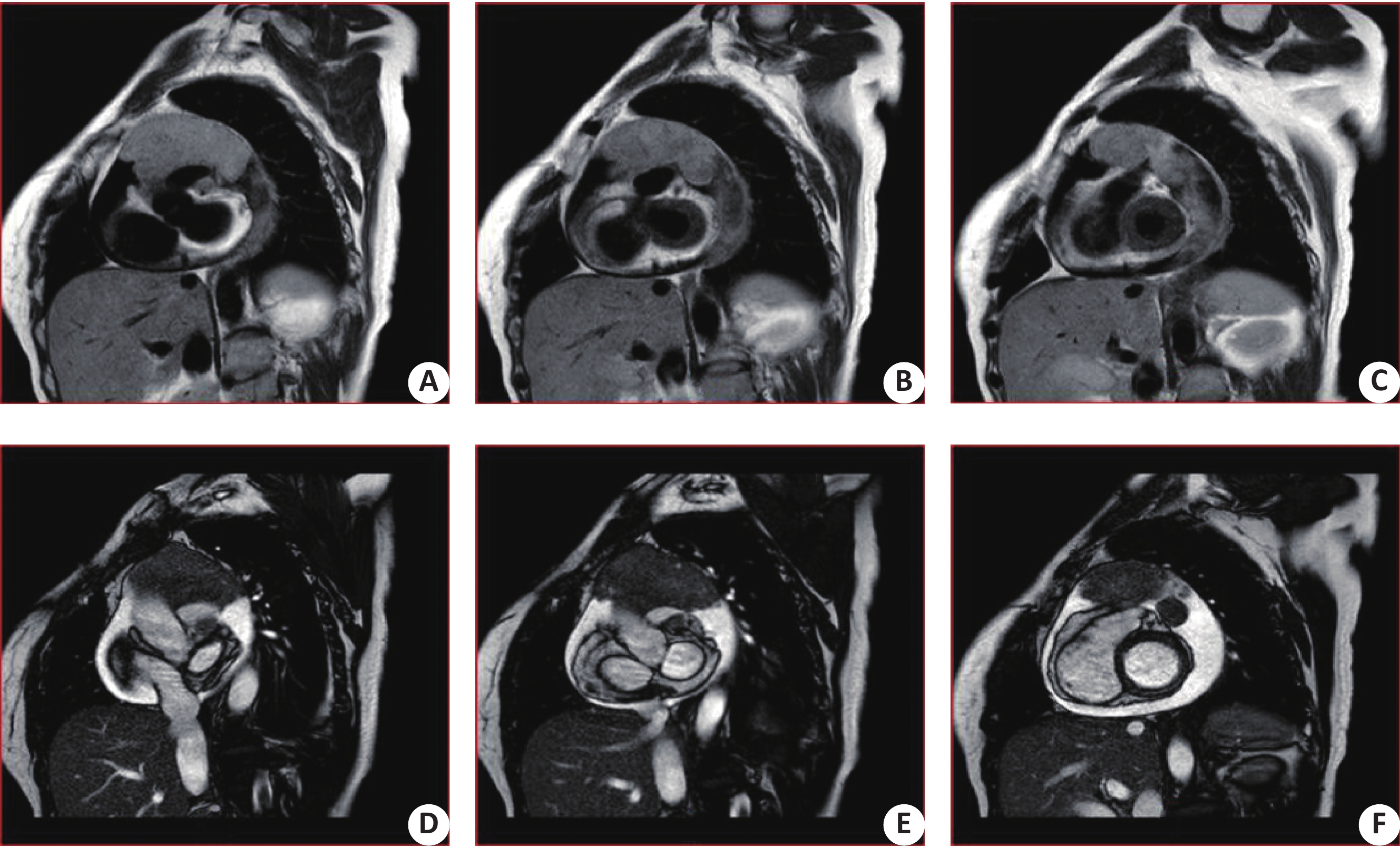
Malignant pericardial mesothelioma is a malignant tumor originating from the layer of the pericardial serosa. It is rarely in clinic and its imaging performance is lack of specificity. A case of malignant pericardial mesothelioma was diagnosed in our hospital in 2018. The imaging data are more comprehensive. The patient found abnormal cardiac shadow by X-ray examination. Further examination of CT and MRI can determine the location, morphology and related imaging features of the lesions. The imaging examination plays a key role in the clinical diagnosis of pericardial mesothelioma.
Malignant pericardial mesothelioma is a malignant tumor originating from the layer of the pericardial serosa. It is rarely in clinic and its imaging performance is lack of specificity. A case of malignant pericardial mesothelioma was diagnosed in our hospital in 2018. The imaging data are more comprehensive. The patient found abnormal cardiac shadow by X-ray examination. Further examination of CT and MRI can determine the location, morphology and related imaging features of the lesions. The imaging examination plays a key role in the clinical diagnosis of pericardial mesothelioma.
2019, 42(4): 476-479.
doi: 10.12122/j.issn.1674-4500.2019.04.13
Abstract:
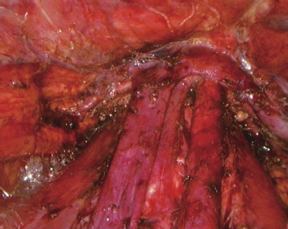
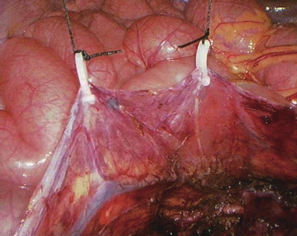
ObjectiveTo explore the method of laparoscopic bilateral retroperitoneal and pelvic lymph node dissection for testicular cancer in supine position. MethodsBilateral retroperitoneal and pelvic lymph node dissection via laparoscopy were performed in 3 cases. The literatures at home and abroad were reviewed and summarized. ResultsDuring the operation, 22 retroperitoneal lymph nodes and 10 pelvic lymph nodes were dissected, with less bleeding during the operation and returned to the ward safely after the operation. ConclusionLaparoscopic bilateral retroperitoneal and pelvic lymph node dissection through abdominal approach has a wide range, ranging from the level of the upper renal artery to the level of the lower iliac artery, with clear anatomical markers. It can achieve the same resection range as open surgery. Supine position can simultaneously remove bilateral retroperitoneal lymph nodes and pelvic lymph nodes, avoid the trouble of changing positions from left to right, and reduce the risk of operation. It can be applied to non-spermatogonial cells. Diagnosis and treatment of lymph nodes.
2019, 42(4): 480-481.
doi: 10.12122/j.issn.1674-4500.2019.04.14
Abstract:
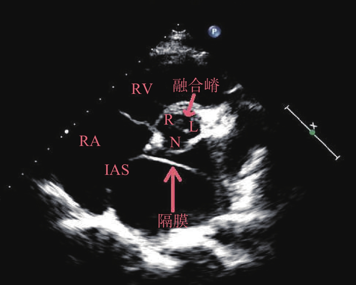
ObjectiveTo explore the application valve of echocardiography in the diagnosis of cor triatriatum (or complicated with other cardiovascular abnormalities). MethodsThe data of a case of cor triatriatum complicated with bicuspid aortic valve were retrospectively analyzed. The related literatures were analyzed. ResultsDuring the physical examination berore the college entrance examination,ultrasound showed a membrane that divided the left atrium into two chambers to form cor triatriatum. In addition, bicuspid aortic valve with right-left cusp fusion was found. ConclusionThere is usually a perforation on the septum of the atria, and those without hemodynamic significance are mostly asymptomatic. It may not need treatment. Patients with small or no access on the septum usually need surgical intervention to correct. Echocardiography is the simplest and most powerful tool in the diagnosis of cor triatriatum.
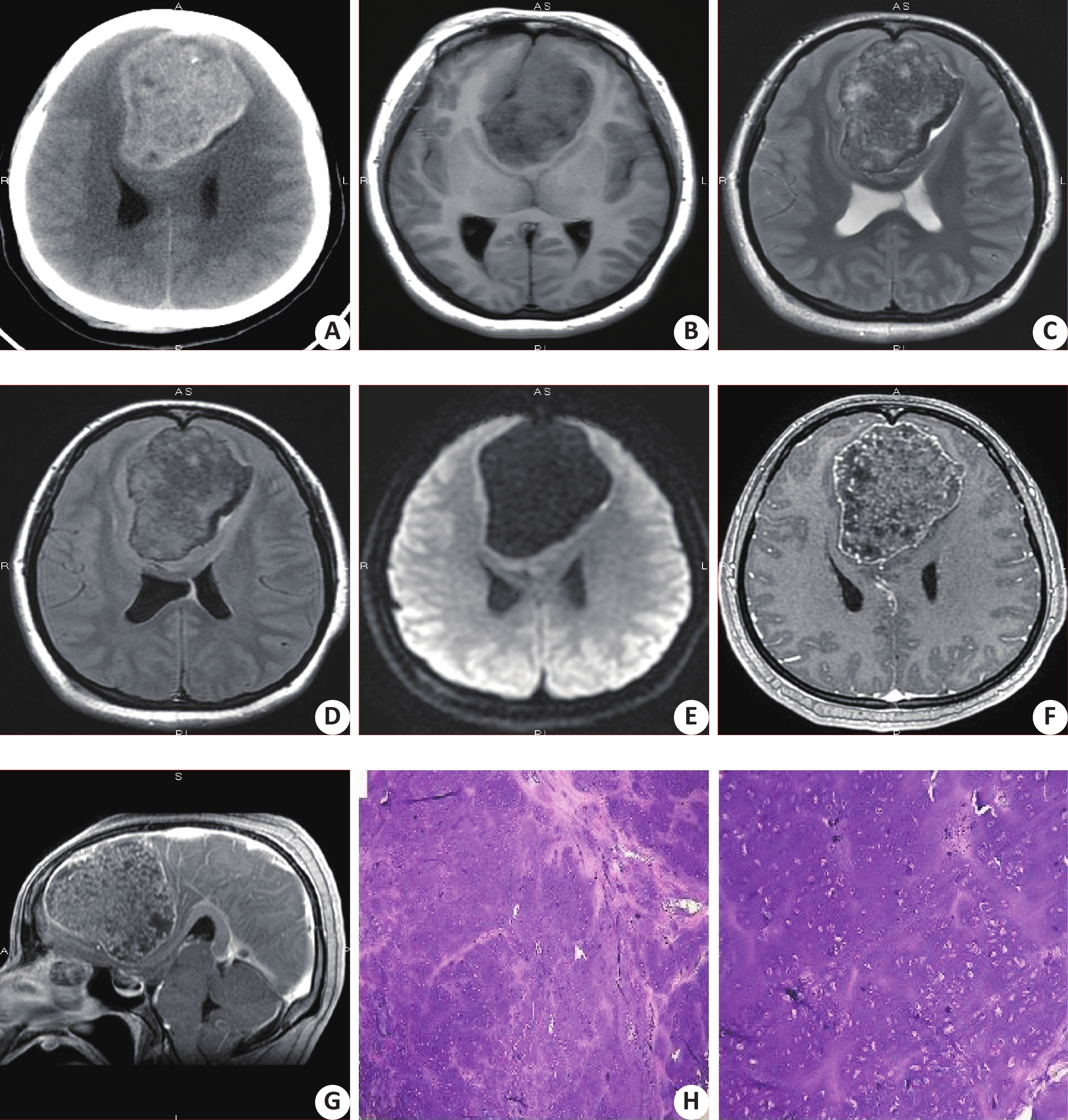
Imaging analysis of metaplastic meningioma misdiagnosed as intracranial chondrosarcom: 1 case report
2019, 42(4): 482-484.
doi: 10.12122/j.issn.1674-4500.2019.04.15
Abstract:
ObjectiveTo explore the imaging differences between the metaplasia meningioma and the typical meningioma and intracranial chondrosarcoma. MethodsA case of metaplastic meningioma misdiagnosed as intracranial chondrosarcoma was analyzed. The differences in CT and MRI and the cause of misdiagnosis were analyzed in two cases. ResultsThe characteristics of the CT and MRI of the metaplasia meningioma were not obvious. It was easy to confuse the chondrosarcoma. ConclusionThe imaging diagnosis of metaplasia meningioma is not mainly based on the tumor, but on the external brain.
2019, 42(4): 485-489.
doi: 10.12122/j.issn.1674-4500.2019.04.16
Abstract:
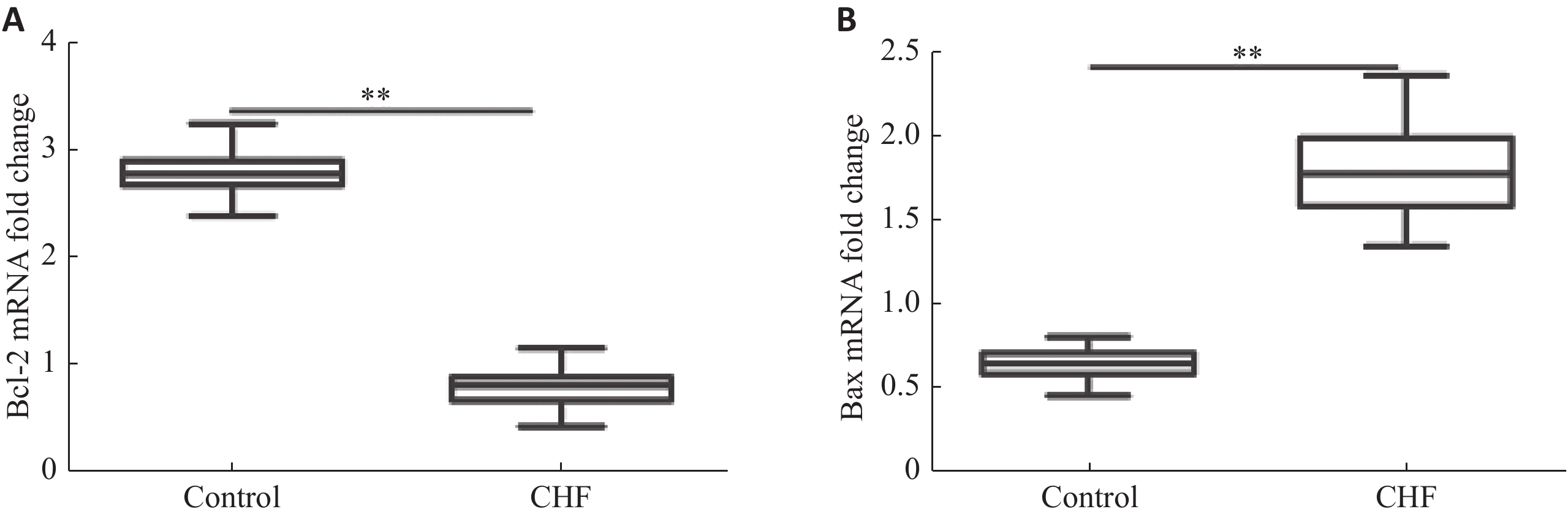
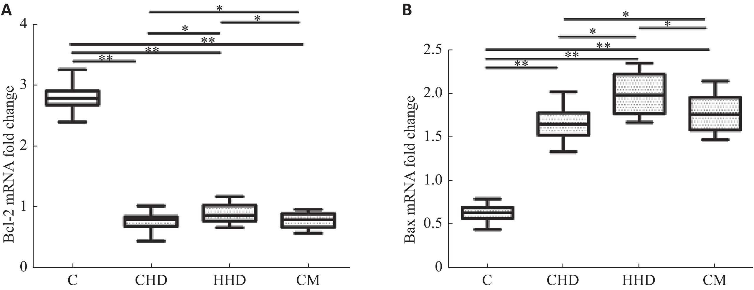
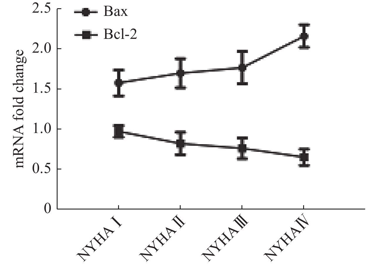
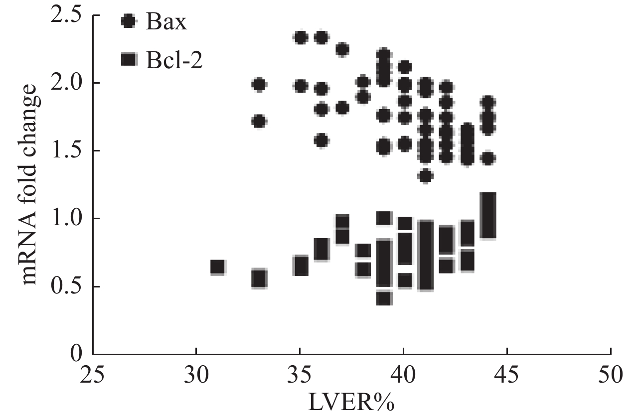
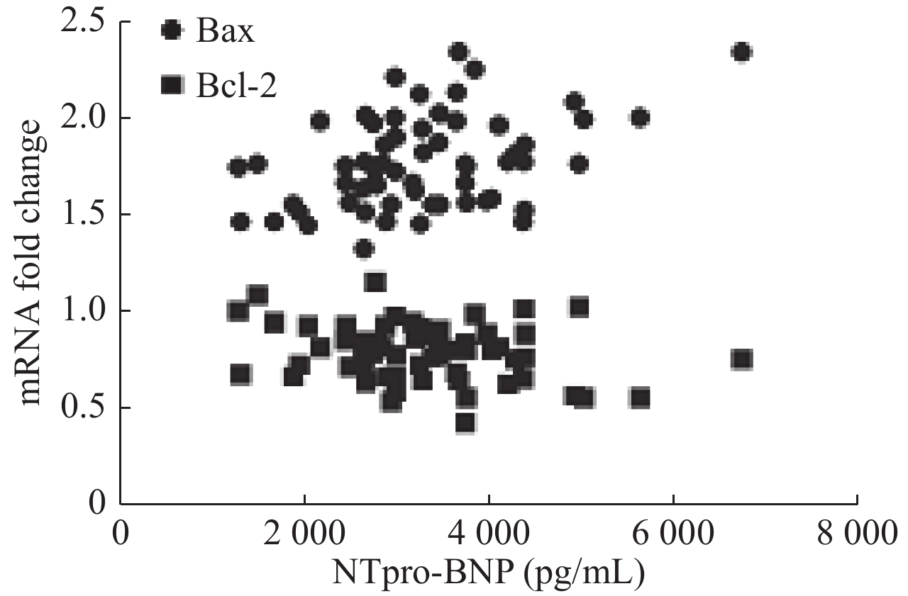
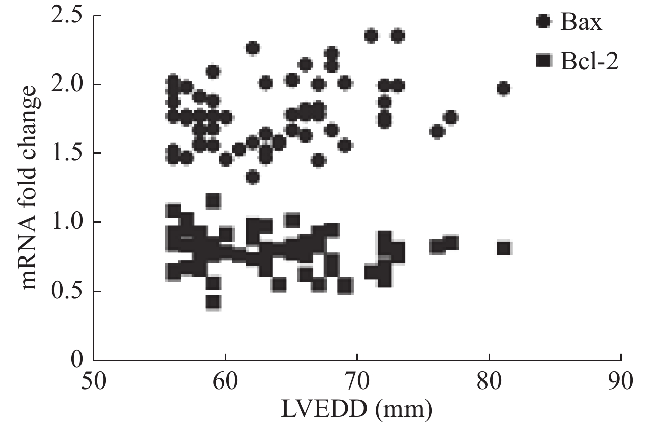
ObjectiveTo explore the relationship between mRNA level of Bax and Bcl-2 in peripheral blood mononuclear cells (PBMCs) and cardiac function and left ventricular ejection fraction. MethodsSixty patients with stable chronic heart failure and 30 healthy controls were enrolled. Monocytes were isolated and cultured from peripheral blood and mRNA were extracted. Relative content of Bax and Bcl-2 mRNA were measured by quantitative reverse transcription- polymerase chain reaction (qRT-PCR). Cardiac functions were evaluated by New York Heart Association (NYHA) functional classification system. Left ventricular ejection fraction was measured by transthoracic color doppler echocardiography. ResultsCompared with the control group, the level of Bcl-2 mRNA in PBMCs in CHF group decreased (P<0.01), and the level of Bax mRNA increased (P<0.01). The level of Bcl-2 mRNA was negatively correlated with NYHA class in CHF group (P<0.05). The level of Bax mRNA was positively correlated (P<0.05). The level of Bcl-2 mRNA in PBMCs were positively correlated with LVEF in CHF group (P<0.01. The level of Bax mRNA was negatively correlated (P<0.01). The level of Bax mRNA in PBMCs was positively correlated with the levels of NT-proBNP in CHF group (P<0.01), and the level of Bcl-2 mRNA was not correlated (P>0.05). The level of Bax mRNA in PBMCs was positively correlated with LVEDD in CHF group (P<0.05), and the level of Bcl-2 mRNA was not correlated (P>0.05). ConclusionBax and Bcl-2, a pair of apoptosis-related genes, change significantly in the expression of stable chronic heart failure. Various etiologies may lead to the common changes of these apoptosis-related genes. The mRNA of these genes is correlated with the severity of CHF. It may be one of the prognostic factors of CHF.
2019, 42(4): 490-494.
doi: 10.12122/j.issn.1674-4500.2019.04.17
Abstract:
ObjectiveTo investigate the clinicopathological characteristics of multifocal papillary thyroid carcinoma (MPTC) and the regularity of cervical lymph node metastasis, and provide evidence for the treatment of multifocal thyroid cancer. MethodsThe clinical data of 788 patients with multifocal papillary thyroid cancer who underwent surgery in the First Affiliated Hospital of Bengbu Medical College from 2012 to 2016 were analyzed retrospectively. ResultsA total of 788 MPTC patients were enrolled. The overall age distribution was 8-81 years old, with an average age of 46.28±12.49 years. There were 138 males (17.51%) and 650 females (82.49%) with an average age of 43.58±13.62 and 46.86±12.17. In addition, 287 cases (36.4%) were younger than 45 years old and 501 cases (63.6%) were younger than 45 years old. 495 patients (62.8%) had bilateral multifocal lesions and 293 patients (37.2%) had unilateral multifocal lesions. 377 cases (47.8%) with diameter >1 cm and 411 cases (52.2%) with diameter <1 cm. There were 524 cases (66.5%) with 2 lesions and 264 cases (33.5%) with more than 3 lesions. The incidence of Hashimoto's thyroiditis and tumor diameter >1 cm in bilateral multifocal group was significantly higher than that in unilateral multifocal group (P<0.01). Bilateral multiple lesions and capsular invasion were more prone to cervical lymph node metastasis, especially central lymph node metastasis (P<0.01). The lymph node metastasis rate in the central and lateral regions increased significantly with the increase of the number of cancer foci in the multifocal group (P<0.01). The risk of lymphatic metastasis in lesions larger than 1 cm was 3.805 times higher than that in lesions less than 1 cm (P<0.01). The risk of lymphatic metastasis in lesions larger than 3 was 9.848 times higher than that in lesions less than 2. ConclusionMales, patients with tumor maximum diameter >1 cm and Hashimoto's thyroiditis are prone to bilateral multifocal thyroid cancer. The proportion of bilateral multifocal thyroid cancer increases significantly with the increase of the number of cancer foci. Male, age <45 years="" old="" tumor="" diameter="">1 cm, bilateral multiple lesions, capsular invasion is more likely to occur cervical lymph node metastasis, with the increase of the number of cancer lesions, central and lateral lymph node metastasis rate increased significantly. Gender, number, size and bilateral distribution of lesions are independent risk factors for lymph node metastasis.
2019, 42(4): 495-497.
doi: 10.12122/j.issn.1674-4500.2019.04.18
Abstract:
ObjectiveTo evaluate the choice of clinical diagnosis and treatment strategies for hepatocellular carcinoma (HCC) complicated with bile duct tumor thrombi. MethodsA retrospective analysis of the surgical diagnosis and treatment of 20 cases of hepatocellular carcinoma with bile duct tumor thrombi in the hepatobiliary surgery of Hainan Hospital of PLA General Hospital from August 2016 to August 2019. ResultsSurgical operations were performed in all 20 cases. Four cases of left (right) half-hepatectomy+removal of thrombus in bile duct. Seven cases of resection of liver tumor and removal of thrombus in bile duct. Four cases radiofrequency ablation of liver tumors+removal of thrombus in bile duct. Three cases simple bile duct incision and thrombus T tube drainage, percutaneous transhepatic bile duct drainage in 2 cases. Postoperative follow-up to date,the average survival time of patients with left (right) half-hepatectomy and bile duct thrombus removal was 36.2 months. The average survival time of tumor resectionand and bile duct thrombus removal was 34.6 months. The average survival time of radiofrequency ablation and bile duct thrombus removal was 28.2 months. The average survival time of the simple-line bile duct was 13.6 months. The average survival time of percutaneous transhepatic bile duct drainage and yellowing reduction was 5.8 months. ConclusionHepatocellular carcinoma complicated with bile duct tumor thrombus, early diagnosis, active resection of tumor and removal of bile duct tumor thrombi can obtain better clinical effect.
2019, 42(4): 498-505.
doi: 10.12122/j.issn.1674-4500.2019.04.19
Abstract:
Alveolar ridge is a kind of dental support structure, which develops with the eruption of teeth. The volume and morphology can change after tooth extraction, especially the absorption of alveolar ridge in the aesthetic area of front teeth is more obvious, which may limit the later implantation or traditional restoration treatment. Application site preservation can prevent bone absorption and reduce bone loss after tooth extraction. Biological barrier membrane plays a key role in site preservation, which can prevent fibroblasts and epithelial cells from growing to the bone defect area and ensure that the bone formation process in the bone defect area is not disturbed by epithelial tissue.In this paper, the significance of site preservation, the ideal performance of the barrier membrane and the clinical application of the barrier membrane in site preservation were reviewed.
Alveolar ridge is a kind of dental support structure, which develops with the eruption of teeth. The volume and morphology can change after tooth extraction, especially the absorption of alveolar ridge in the aesthetic area of front teeth is more obvious, which may limit the later implantation or traditional restoration treatment. Application site preservation can prevent bone absorption and reduce bone loss after tooth extraction. Biological barrier membrane plays a key role in site preservation, which can prevent fibroblasts and epithelial cells from growing to the bone defect area and ensure that the bone formation process in the bone defect area is not disturbed by epithelial tissue.In this paper, the significance of site preservation, the ideal performance of the barrier membrane and the clinical application of the barrier membrane in site preservation were reviewed.
2019, 42(4): 506-509.
doi: 10.12122/j.issn.1674-4500.2019.04.20
Abstract:
Tumor is one of the most important diseases that seriously threaten children's health. But with the improvement and development of children's cancer therapy, the rate of long-term survivors was constantly improving. The number of long-term survivors is also increasing. At the same time, the long-term complications and adverse effects related to cancer treatment have attracted more and more attention. The quality of life in long-term survivors has gradually become a problem that can not be ignored. The treatment of cancer mainly includes radiation therapy, chemotherapy, surgical treatment, hematopoietic stem cell transplantation, biological therapy and so on. The effect of cancer treatment on survivors is multifaceted. In addition to the impact on physical function, there are also psychological and social impacts. The adverse effects may be gradually revealed with the prolongation of follow-up time. We must pay attention to these adverse effects, consider them comprehensively from all aspect. We should ensure the therapeutic effect, reduce the adverse effects on the body, psychology and society of the survivors as far as possible and improve the quality of life.
Tumor is one of the most important diseases that seriously threaten children's health. But with the improvement and development of children's cancer therapy, the rate of long-term survivors was constantly improving. The number of long-term survivors is also increasing. At the same time, the long-term complications and adverse effects related to cancer treatment have attracted more and more attention. The quality of life in long-term survivors has gradually become a problem that can not be ignored. The treatment of cancer mainly includes radiation therapy, chemotherapy, surgical treatment, hematopoietic stem cell transplantation, biological therapy and so on. The effect of cancer treatment on survivors is multifaceted. In addition to the impact on physical function, there are also psychological and social impacts. The adverse effects may be gradually revealed with the prolongation of follow-up time. We must pay attention to these adverse effects, consider them comprehensively from all aspect. We should ensure the therapeutic effect, reduce the adverse effects on the body, psychology and society of the survivors as far as possible and improve the quality of life.
2019, 42(4): 510-513.
doi: 10.12122/j.issn.1674-4500.2019.04.21
Abstract:
Multiple myeloma (MM) is a malignant tumor originating from plasma cells, secreting monoclonal immunoglobulin or monoclonal protein, causing damage to related organs or tissues. IgD type is a rare type of myeloma, which accounts for about 2%, and the reported rate is about 4% to 8% in China, and the prognosis is poor. Regarding the clinical characteristics and survival of IgD type MM, there is no large-scale study in China. This article reviews the clinical features, therapeutic efficacy, survival and prognosis of patients with IgD type MM. It aims to enhance our understanding of the biological characteristics of this type of MM, and help clinicians make correct diagnosis and treatment decisions and further improve the prognosis of such patients.
Multiple myeloma (MM) is a malignant tumor originating from plasma cells, secreting monoclonal immunoglobulin or monoclonal protein, causing damage to related organs or tissues. IgD type is a rare type of myeloma, which accounts for about 2%, and the reported rate is about 4% to 8% in China, and the prognosis is poor. Regarding the clinical characteristics and survival of IgD type MM, there is no large-scale study in China. This article reviews the clinical features, therapeutic efficacy, survival and prognosis of patients with IgD type MM. It aims to enhance our understanding of the biological characteristics of this type of MM, and help clinicians make correct diagnosis and treatment decisions and further improve the prognosis of such patients.
2019, 42(4): 514-517.
doi: 10.12122/j.issn.1674-4500.2019.04.22
Abstract:
Locally advanced colon cancer has poor prognosis due to non-resectable cases, palliative resection, and the increased risk of distant metastasis. Neoadjuvant therapy increases resectability of locally advanced colon cancer by reducing tumor loading and degrading stage, and improves the R0 resection rates. But the model of neoadjuvant treatment has not yet been unified. Surgery of locally advanced colon cancer follows the principle of the “en bloc” resection, with the aim of R0 resection and complete mesocolic excision as the standard procedure. The role of adjuvant chemotherapy in decreasing postoperative recurrence and distant metastasis has been fully demonstrated. This review is focused on neoadjuvant therapy, surgery and adjuvant chemotherapy for locally advanced colon cancer,in order to provide valuable guide for the selection of perioperative therapy and reach the best efficiency of individualized therapy and survival benefit for patients.
Locally advanced colon cancer has poor prognosis due to non-resectable cases, palliative resection, and the increased risk of distant metastasis. Neoadjuvant therapy increases resectability of locally advanced colon cancer by reducing tumor loading and degrading stage, and improves the R0 resection rates. But the model of neoadjuvant treatment has not yet been unified. Surgery of locally advanced colon cancer follows the principle of the “en bloc” resection, with the aim of R0 resection and complete mesocolic excision as the standard procedure. The role of adjuvant chemotherapy in decreasing postoperative recurrence and distant metastasis has been fully demonstrated. This review is focused on neoadjuvant therapy, surgery and adjuvant chemotherapy for locally advanced colon cancer,in order to provide valuable guide for the selection of perioperative therapy and reach the best efficiency of individualized therapy and survival benefit for patients.
2019, 42(4): 518-523.
doi: 10.12122/j.issn.1674-4500.2019.04.23
Abstract:
Fork head box 1 (FOXK1) is one of the members of the FOX transcription factors family that has attracted increasing attention in recent years. More and more studies have shown that FOXK1 is closely related to various tumors such as cancer of reproductive, nervous and digestive systems. Through regulating cell cycle, autophagy and mediating signal pathway, FOXK1 influences tumor invasion and metastasis. It affecting the prognosis of patients. The research of expression of FOXK1 in various tumor tissues and its mechanism provide a new direction for the diagnosis and treatment of tumors.
Fork head box 1 (FOXK1) is one of the members of the FOX transcription factors family that has attracted increasing attention in recent years. More and more studies have shown that FOXK1 is closely related to various tumors such as cancer of reproductive, nervous and digestive systems. Through regulating cell cycle, autophagy and mediating signal pathway, FOXK1 influences tumor invasion and metastasis. It affecting the prognosis of patients. The research of expression of FOXK1 in various tumor tissues and its mechanism provide a new direction for the diagnosis and treatment of tumors.
2019, 42(4): 524-527.
doi: 10.12122/j.issn.1674-4500.2019.04.24
Abstract:
Metabolic acidosis (MA) is a kind of common complication of chronic kidney disease. Serum bicarbonate (HCO3−) concentration would significantly decrease once the end-stage renal disease was diagnosed. It would worsen the condition. MA correction would contribute to the clinical therapy and the prognosis of maintenance hemodialysis (MHD) patients. Low concentration of HCO3− would accelerate the progression of kidney disease, and high concentration of HCO3− would pose the risk of cardiovascular disease. Nevertheless, the optimal clinical reference value of serum bicarbonate concentration is still unknown. The factors that should be considered in dialysis treatment are also complicated. This paper mainly reviews the factors of MA occurrence and adverse effects in MHD patients, and further discusses the relevant practical considerations in the clinical therapy of MA in MHD. We aim to provide theoretical references for MA treatment by analyzing the results of large cohort study around the world and that from wet-lab.
Metabolic acidosis (MA) is a kind of common complication of chronic kidney disease. Serum bicarbonate (HCO3−) concentration would significantly decrease once the end-stage renal disease was diagnosed. It would worsen the condition. MA correction would contribute to the clinical therapy and the prognosis of maintenance hemodialysis (MHD) patients. Low concentration of HCO3− would accelerate the progression of kidney disease, and high concentration of HCO3− would pose the risk of cardiovascular disease. Nevertheless, the optimal clinical reference value of serum bicarbonate concentration is still unknown. The factors that should be considered in dialysis treatment are also complicated. This paper mainly reviews the factors of MA occurrence and adverse effects in MHD patients, and further discusses the relevant practical considerations in the clinical therapy of MA in MHD. We aim to provide theoretical references for MA treatment by analyzing the results of large cohort study around the world and that from wet-lab.
2019, 42(4): 528-533.
doi: 10.12122/j.issn.1674-4500.2019.04.25
Abstract:
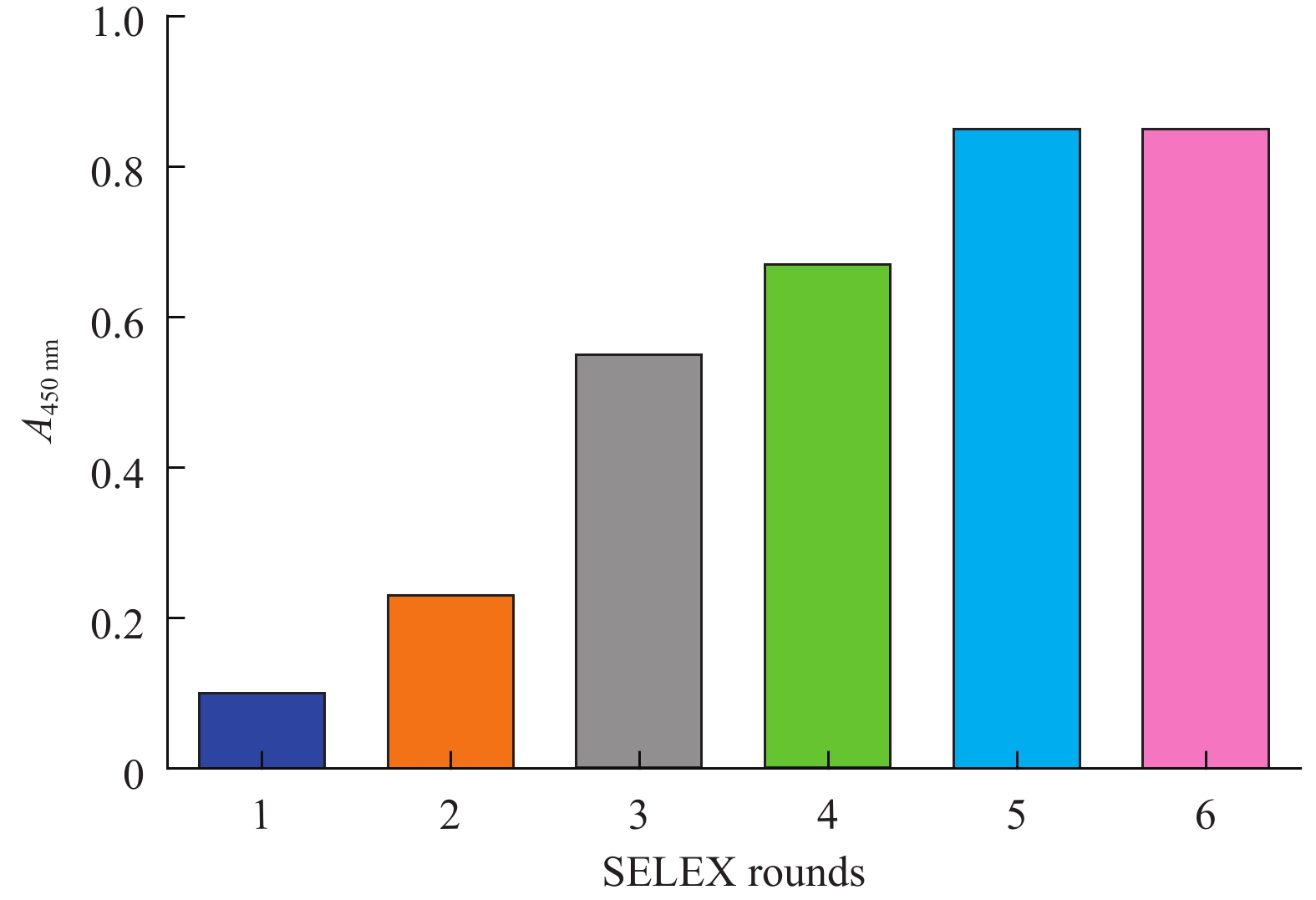
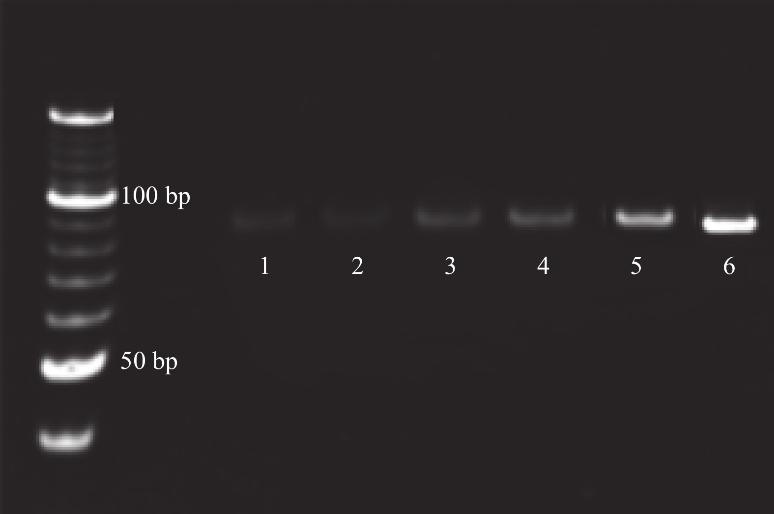
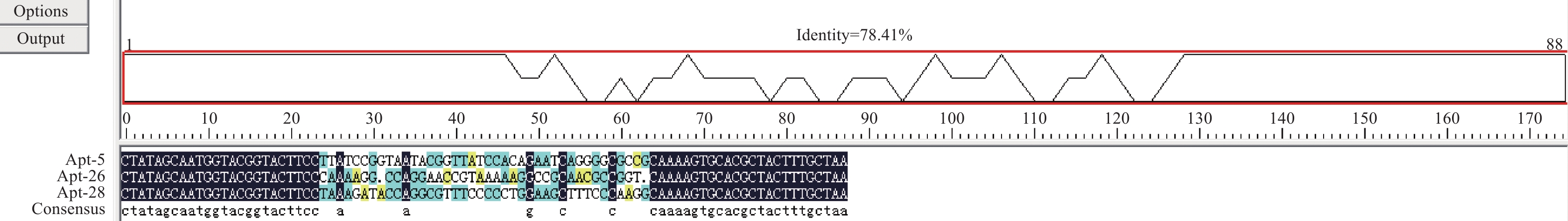
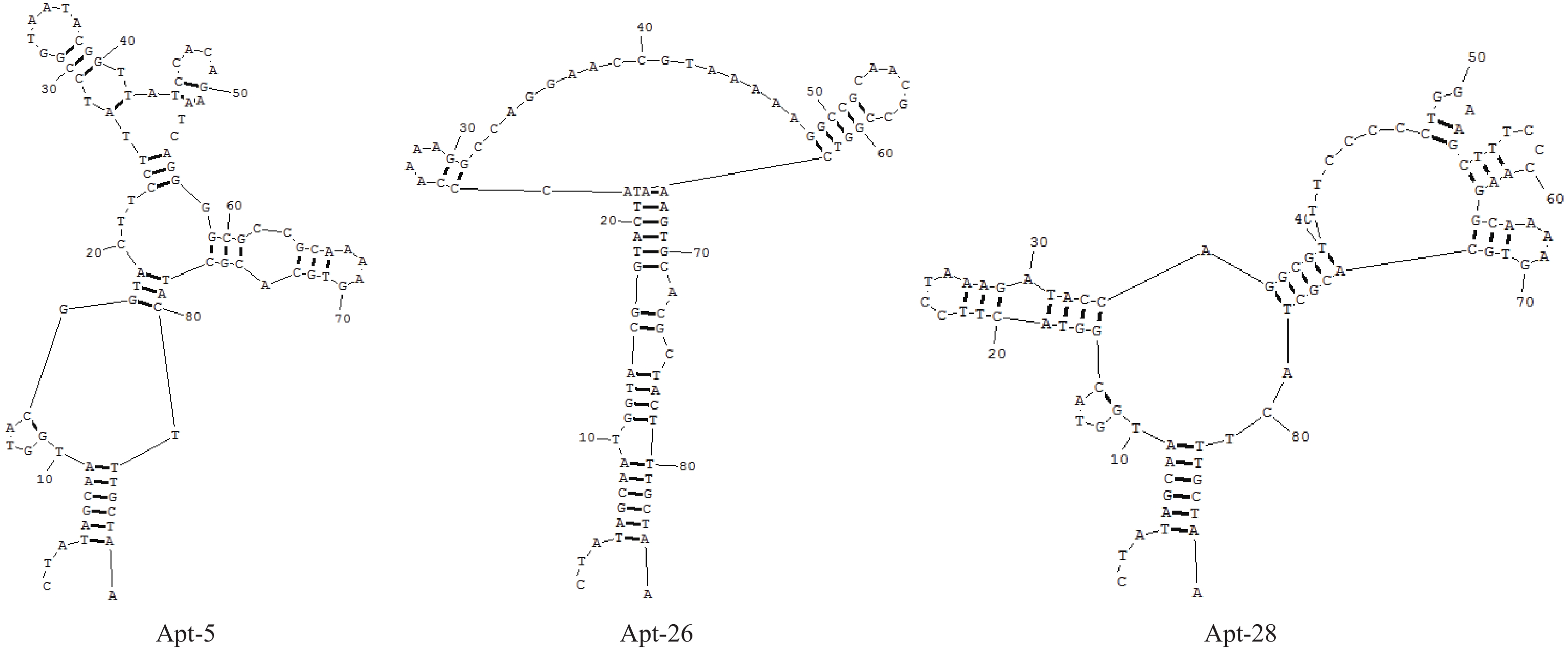
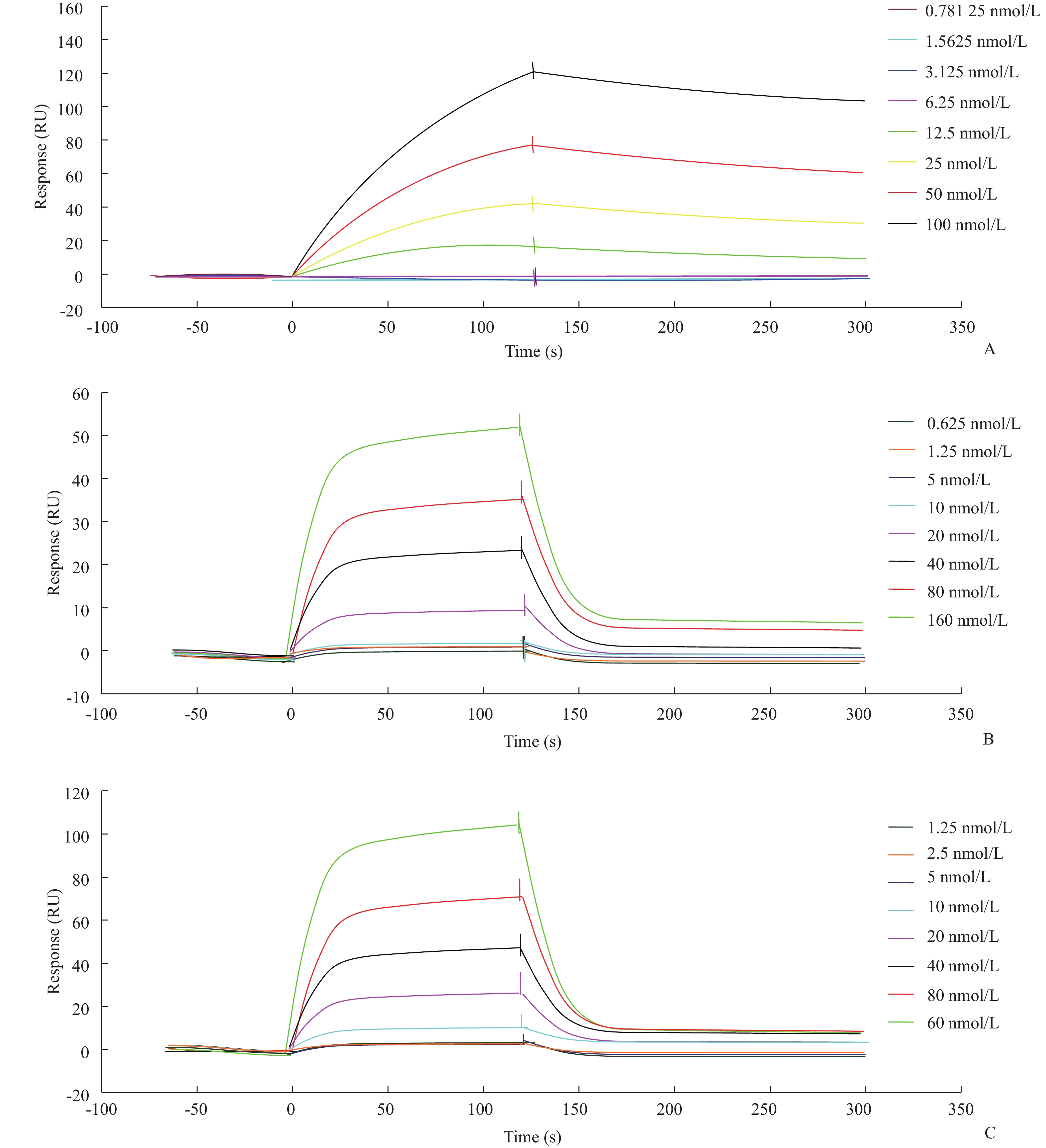
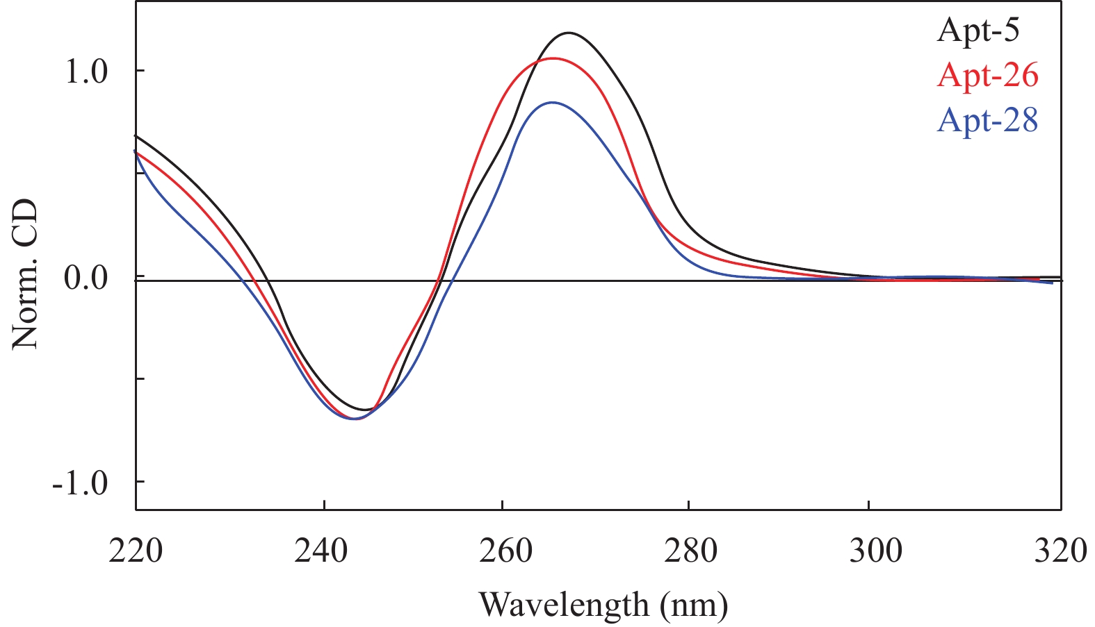
ObjectiveTo screen and identify the serum specific marker Dickkopf-1 aptamer (Apt). MethodsThe systemic evolution of ligand by exponential enrichment (SELEX) was used to select DKK1 as the target protein and carboxylated agar magnetic beads as the screening medium. A random set of random ssDNA library was selected. A nucleic acid aptamer to which it binds. Sequence analysis and secondary structure prediction of nucleic acid aptamers were carried out by bioinformatics methods. The affinity of nucleic acid aptamers was determined by surface plasmon resonance (SPR) analysis. ResultsAfter 6 rounds of SELEX screening, the affinity of the secondary ssDNA library to the DKK1 target protein was stabilized. The 6th round of screening products were amplified by PCR for high-throughput sequencing. The results of SPR assay showed that the binding dissociation constants of DKK1 aptamers and DKK1 were in the nanomolar range. The Kd value of Apt-5 was the smallest and the affinity was the highest. The affinity of Apt-26 and Apt-28 aptamers was relative weak. The predictive analysis of secondary structure indicated that the stem loop and stem loop structure were the main structural forms. The results of circular dichroism spectroscopy showed that the three candidate aptamers (Apt-5, Apt-26, Apt-28) specifically formed the G-quadruplex structure to recognize the DKK1 target protein. Conclusion The aptamer that specifically binds to the DKK1 target protein is obtained. It laids a foundation for the application of subsequent aptamers and the study of DKK1 protein function.
2019, 42(4): 534-537.
doi: 10.12122/j.issn.1674-4500.2019.04.26
Abstract:
ObjectiveTo explore the relevant factors of postoperative brain damage by analyzing the correlation between NSE and preoperative and postoperative factors. MethodsSpearman correlation method was used to analyze the correlation between neuron specific enolase NSE with various preoperative and postoperative factors. ResultThe concentration of NSE was related to intubation time (P=0.001, r=0.366), preoperative and 12 h after operation oxygenation index (P<0.01), preoperative ALT (P=0.008, r=0.193) and postoperative creatinine. ConclusionThe preoperative and 12 h after operation oxygenation index fell, hepatic insufficiency, postoperative renal dysfunction, age and cardiac dysfunction are the risk factors for brain damage after cardiac surgery. The preoperative renal insufficiency, postoperative liver function, CBP and aortic clip time are not risk factors for brain damage after cardiac surgery.
2019, 42(4): 538-543.
doi: 10.12122/j.issn.1674-4500.2019.04.27
Abstract:
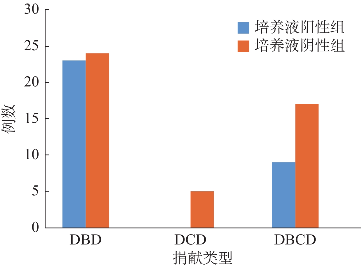
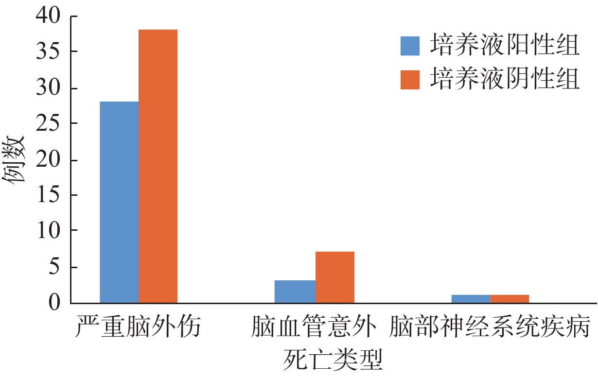
ObjectiveTo discuss the preservation solution (PS) contamination and initial experience of liver transplantation from organ donation by citizens after death and initial experience. MethodsThe 78 liver transplant recipients in a hospital in Guangzhou from March 2016 to October 2017 were divided into two groups based on the finding of the culture of PS. The positive group received the sequential therapy of antibiotics with ertapenem and imipenem for one week. The negative group stopped using imipenem. The situation of PS contamination and infection after liver transplantation and prognosis during the follow-up 3 months of the recipients were analyzed. ResultsAmong the 78 recipients, 32 PSs culture were positive (41.03%), and 33 strains of pathogens were isolated. The most common pathogenic bacteria were gram-negative bacilli (9 strains, 27.27%) and coagulase–negative staphylococci (9 strains, 27.27%). The infection rates after liver transplantation in culture positive group and culture negative group were 31.25% and 13.04%, respectively (χ2=3.837, P=0.048). The most frequent infection sites were lower respiratory tract (5 cases, 31.25%), abdominal cavity (5 cases, 31.25%) and surgical incision (4 cases, 25.00%). There was no significant difference of postoperative infection rate among patients with different CTP, MELD and surgical methods (P>0.05). One case (1.28%) was infected with the same pathogenic bacteria as PS contamination 3 weeks after liver transplantation, and died of multiple organ failure. There was no significant difference of the acute rejection rate (1, 3.13% and 2, 4.35%) and mortality (2, 6.25% and 5, 10.87%) between the two groups (P>0.05). ConclusionContamination of the PS is frequent in liver transplantation, which is the risk factor for postoperative infection of recipients. Early targeted antimicrobial treatment against pathogens cultured from PS is positive in reducing the contamination-associated infection rate after liver transplantation.
2019, 42(4): 544-547.
doi: 10.12122/j.issn.1674-4500.2019.04.28
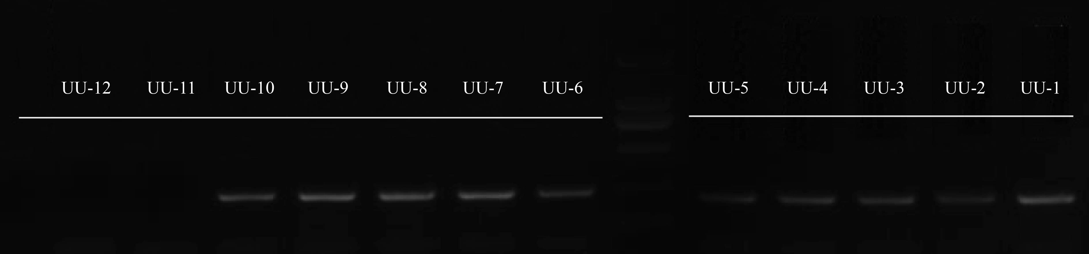
Abstract:
ObjectiveTo evaluate the performance of real time quantitative PCR methods and analyze the infection status. MethodsAccording to the official CNAS-GL039 file and product specification, performance including precision, accuracy, lower limit of detection and anti-interference were investigated. Meanwhile, the samples from outpatients and inpatients in 2018 were collected and tested. The results were statistically analyzed. ResultsThe precision was within allowed coefficient of variation (CV), the accuracy was up to 100% by compared with the golden standard (gene sequencing). The lowest detection limit was 1×10³ copies/mL. Cross-reaction between UU and common genital tract microorganisms were not present. The samples containing 4% of endogenous red blood cells did not interfere with test results. The overall positive rate of UU test was 59.8%. The detection rate in female was significantly higher than that in male (χ2=525.613, P<0 01="" among="" the="" positive="" samples="" of="" female="" patients="" the="" detection="" rate="" in="" 30="" years="" old="" was="" higher="" than="" that="" of="">30 years old (P<0.01). ConclusionThe detection method of qRT-PCR has a high detection sensitivity, good repeatability, strong anti-interference, and the lower limit of detection meets the requirements. It can be used for clinical testing. This study provides a reference basis for the diagnosis and treatment of UU infection.
2019, 42(4): 548-550.
doi: 10.12122/j.issn.1674-4500.2019.04.29
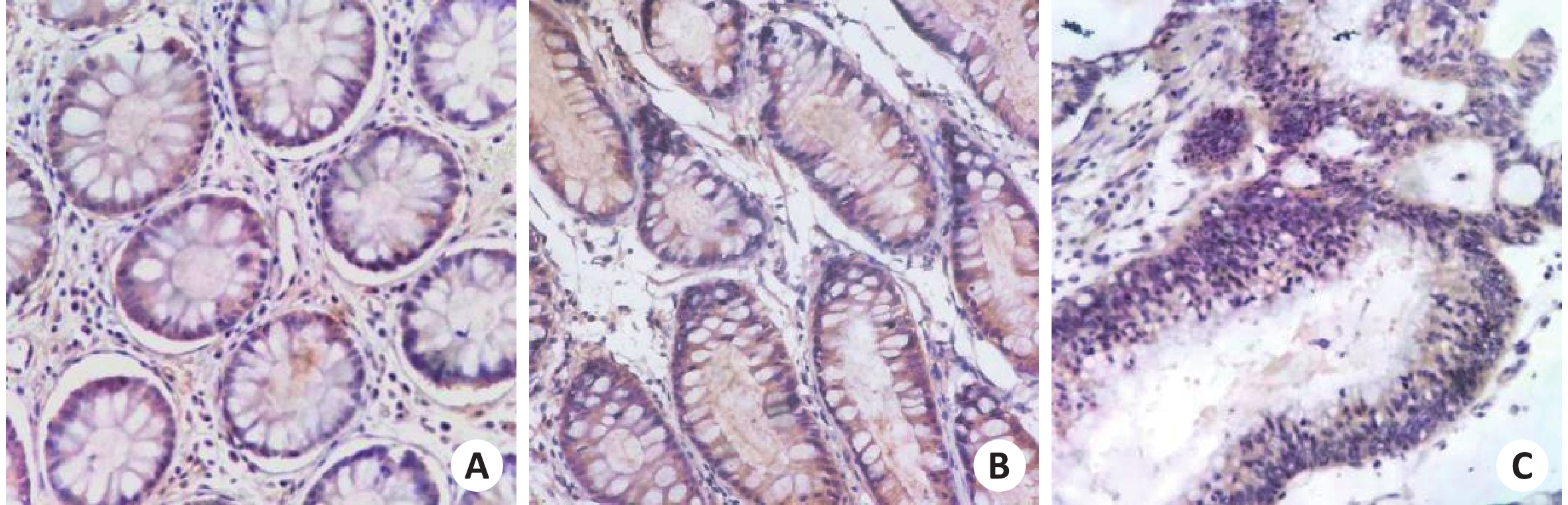
Abstract:
ObjectiveTo investigate the expression of EFNA3 in colorectal cancer preliminary to explore the relationship between EFNA3 and colorectal cancer. MethodsA total of 84 colorectal cancer cases in XiangNan University affiliated hospital were randomly selected. They were divided into 3 groups: colorectal cancer, colorectal benign tumor and normal colorectal tissue. Immunohistochemistry was used to detect the expression of EFNA3 in 3 groups. ResultsExpression intensity of EFNA3 in colorectal cancer (3.6±1.3) was lower than that in colorectum benign tumor (5.1±0.6, P<0.05) and Normal colorectum (4.9±1.3, P<0.05). Compared with the expression of EFNA3 in the tumor tissues of well-differentiated group (4.2±0.4) and poorly differentiated group (3.3±1.6), the former was lower than the latter (P<0.05). The expression of EFNA3 in the tumor tissues in the lymphatic metastasis group (3.3±1.7) was lower than not (4.13±0.28) (P<0.05). Compared with the expression of EFNA3 in the tumor tissues people at Ⅲ+Ⅳ (3.3±1.7) and Ⅰ+Ⅱ (4.13±0.28) TNM staging, the former was lower than the latter (P<0.05). ConclusionThe expression of EFNA3 is lower in Colorectal cancer. Colorectum benign tumor and Normal colorectum have no significant difference.
2019, 42(4): 551-553.
doi: 10.12122/j.issn.1674-4500.2019.04.30
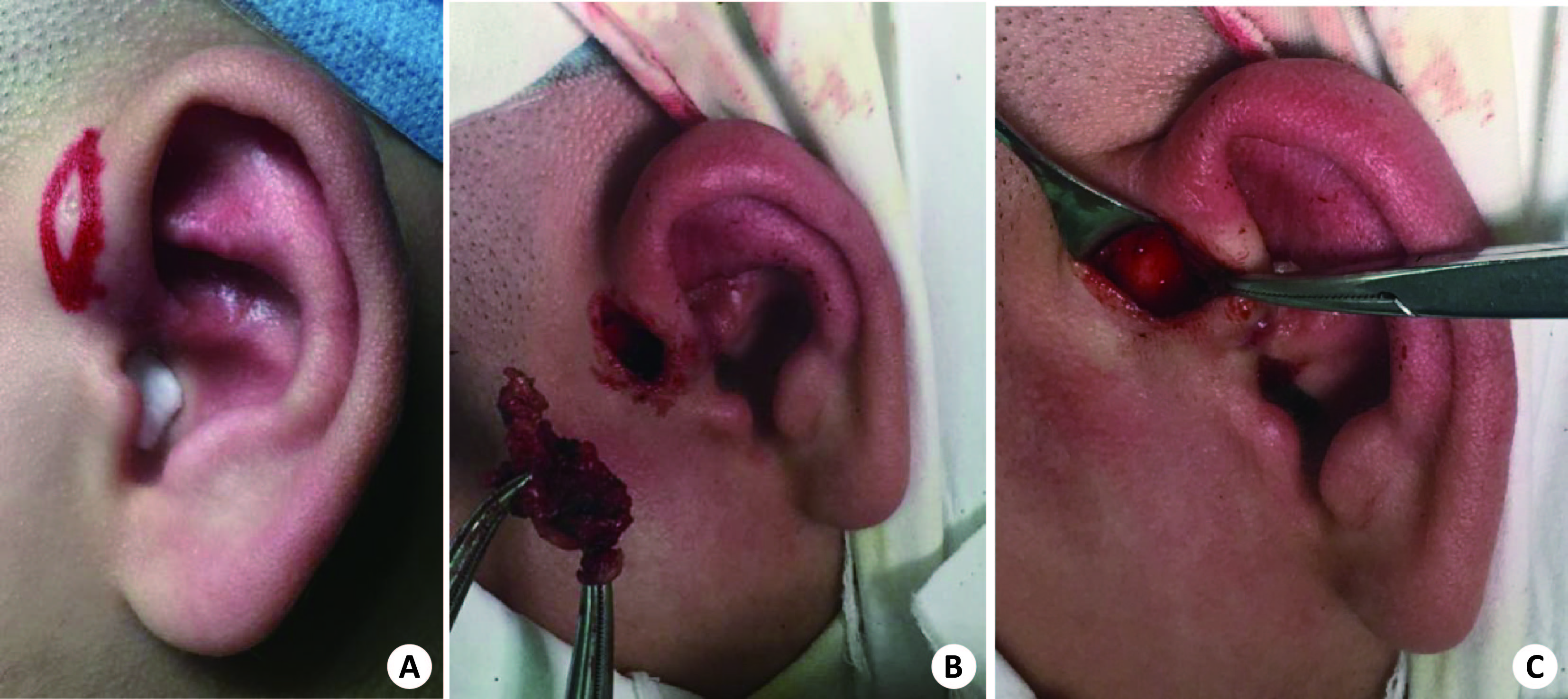
Abstract:
ObjectiveTo evaluate the effect of regional preauricular con tinuous block resection on the treatment of infectious preauricular sinus in children. MethodsRetrospective analysis of 16 cases of infectious preauricular sinus in children, preauricular sinus and scar granulation tissue were completely removed by preauricular regional continuous block resection. ResultsAll the patients had stage I healing, and no postoperative complications such as auricle deformity, pyogenic perichondritis, facial paralysis, parotid gland leakage were found. Follow-up period was 12 months to 48 months, and no recurrence was found. ConclusionFor the treatment of infectious preauricular fistula, regional continuous block resection for the treatment of infectious preauricular fistula requires no intraoperative anatomical fistula tissue, and there is no fear of staining contamination of the operative cavity. In addition, it requires simple surgical equipment, and the operation is safe and effective. It is suitable for young doctors to perform surgery and suitable for popularization in primary hospitals.