


Clinical value of 3D-CEUS detection in patients with severe uterine adhesion after hysteroscopy separation
-
摘要:
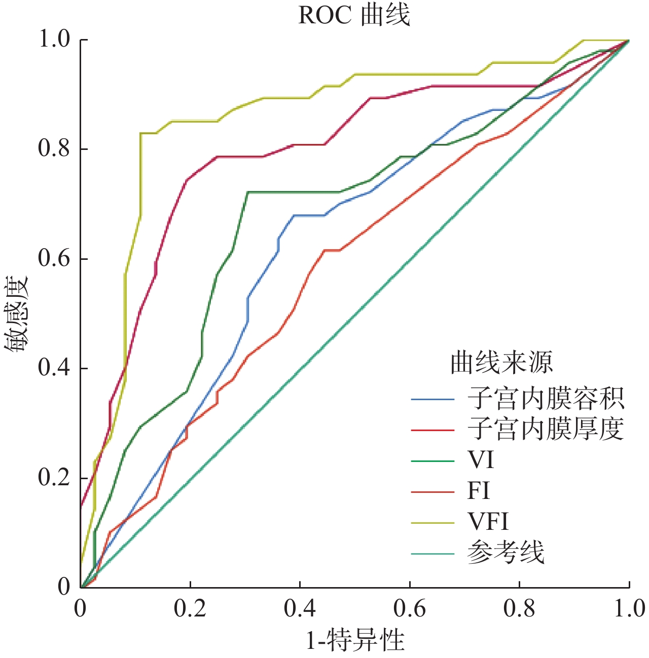
目的探讨三维超声造影(3D-CEUS)诊断宫腔镜下宫腔粘连分解术(TCRA)后重度宫腔粘连(SIUA)的价值。 方法回顾性分析83例TCRA术后IUA患者临床资料,所有患者均行宫腔镜、经阴道二维三维超声检查、3D-CEUS检查,获得子宫内膜相关参数:子宫内膜厚度、容积、血管指数、血流指数、血管化血流指数。以宫腔镜检查结果为准,分析3D-CEUS诊断TCRA术后SIUA的价值。 结果本组TCRA术后确诊SIUA 36例,SIUA三维成像表现为宫腔形态异常、狭小、边缘不规则,内膜回声不连续等。经阴道三维超声诊断TCRA术后IUA分级准确率90.36%,与宫腔镜检查结果一致性较好(Kappa=0.795,P<0.05)。SIUA患者子宫内膜厚度、容积、血管指数、血流指数、血管化血流指数低于中度组和轻度组(P<0.05),二元Logistic回归分析子宫内膜厚度、容积、血管指数、血流指数、血管化血流指数与TCRA术后SIUA发生显著相关(P<0.05)。ROC结果显示,子宫内膜厚度、血管化血流指数鉴别SIUA的效能较高,曲线下面积分别为0.794、0.856,灵敏度为80.56%、74.47%,特异度为88.89%、82.98%。 结论经阴道三维超声可清晰显示TCRA术后宫腔粘连程度和累及范围,为IUA分级提供可靠参考。3D-CEUS测量相关子宫内膜参数可作为SIUA诊断的定量指标。 Abstract:ObjectiveTo investigate the value of three-dimensional contrast-enhanced ultrasound (3D-CEUS) in the diagnosis of severe intrauterine adhesions (SIUA) after transcervical resection of adhensions (TCRA). MethodsThe clinical data of 83 patients with IUA after TCRA were retrospectively analyzed. All patients underwent hysteroscopy, transvaginal two-dimensional ultrasonography and 3D-CEUS. The endometrial thickness, volume, vascular index (VI), flow index (FI), vascularization flow index (VFI) were analyzed. Based on the results of hysteroscopy, the value of 3D-CEUS in diagnosing SIUA after TCRA was analyzed. ResultsThirty-six cases of SIUA were confirmed after TCRA. The three-dimensional imaging of SIUA showed abnormal uterine cavity shape, narrow, irregular margin and discontinuous endometrial echo. The accuracy rate of IUA classification of TCRA by transvaginal three-dimensional ultrasound was 90.36%, which was in good agreement with hysteroscopy (kappa=0.795, P<0.05). The endometrial thickness, volume, VI, FI, VFI index of SIUA patients were lower than those of moderate and mild groups (P<0.05). Dual Logistic regression analysis showed that the thickness, volume, VI, FI and VFI of endometrium were significantly correlated with SIUA after TCRA (P<0.05). The results of ROC analysis showed that endometrial thickness and VFI had a higher efficiency in identifying SIUA, with AUC of 0.794 and 0.856, sensitivity and specificity of 80.56%, 74.47%, 88.89% and 82.98%, respectively. ConclusionTransvaginal three-dimensional ultrasound can clearly show the degree and extent of intrauterine adhesions after TCRA. It provide a reliable reference for IUA classification. The measurement of endometrial parameters by 3D-CEUS can be used as a quantitative index for SIUA diagnosis. -
Key words:
- hysteroscopy /
- intrauterine adhesion /
- endometrial thickness /
- volume /
- vascular index /
- flow index /
- vascularization flow
-

图 1 SIUA超声二维成像图和三维成像图对比
A~C:SIUA超声二维成像图。A:子宫内膜线部分不连续;B:不连续区呈低回声,可见宫腔分离;C:子宫内膜厚薄不均,与周围肌层分界不清;D~F:SIUA超声三维成像图。D:内膜边缘不规则,多处回声缺失;E:子宫形态不规则;F:子宫宫腔狭小,宫角显示不清
Figure 1. Comparison of two-dimensional images and three-dimensional images in SIUA ultrasound

图 2 子宫内膜厚度、容积、VI、FI、VFI指数鉴别SIUA的ROC曲线
Figure 2. ROC curve of endometrial thickness, volume, VI、FI、VFI index SIUA.
表 1 经阴三维超声诊断IUA分级与宫腔镜结果比较(n)
Table 1. Comparison of diagnostic IUA grading and hysteroscopy results of three-dimensional ultrasonography
组别 子宫内膜 kappa P 轻度 中度 重度 轻度 17 1 0 中度 4 24 2 0.795 0.000 重度 0 1 34 合计 21 26 36  下载: 导出CSV
表 2 不同粘连程度IUA患者子宫内膜厚度、子宫容积、VI、FI、VFI差异(Mean±SD)
Table 2. The differences of endometrial thickness, uterine volume and VI、FI、VFI of patients with different degree of adhesion
组别 例数 子宫内膜 VI FI VFI 厚度(cm) 容积(cm3) 轻度 21 0.69±0.15 2.16±0.74 38.59±12.05 32.51±10.46 12.35±5.49 中度 26 0.55±0.11* 1.51±0.55* 32.51±9.75* 30.51±8.57* 10.34±5.02* 重度 36 0.41±0.09*# 0.83±0.39*# 23.35±5.24*# 26.95±6.04*# 6.34±2.54*# F 11.084 10.449 12.515 3.402 6.938 P 0.000 0.000 0.000 0.038 0.000 *P<0.05 vs轻度组;#P<0.05 vs中度组.
下载: 导出CSV
表 3 3D-CEUS子宫内膜参数与SIUA相关性
Table 3. Correlation of 3D-CEUS endometrial parameters and SIUA
指标 β S.E, Wald OR(95%CI) P 子宫内膜厚度 −0.592 0.137 18.672 0.553(0.124~0.856) 0.000 子宫内膜容积 −0.371 0.103 12.974 0.690(0.154~0.915) 0.000 VI −0.621 0.185 11.267 0.537(0.103~0.957) 0.001 FI −0.296 0.131 5.106 0.743(0.113~0.984) 0.045 VFI −0.537 0.182 8.706 0.584(0.234~0.834) 0.009
下载: 导出CSV
表 4 子宫内膜厚度、容积、VI、FI、VFI指数鉴别SIUA的效能分析
Table 4. Efficacy of endometrial thickness, volume, VI、FI、VFI index in identifying SIUA
指标 Cut-off AUC(95%CI) P 灵敏度(%) 特异度(%) 子宫内膜厚度 0.43 cm 0.794(0.694~0.893) 0.000 80.56 74.47 子宫内膜容积 1.21 cm3 0.628(0.505~0.751) 0.047 61.11 68.09 VI 27.24% 0.679(0.562~0.797) 0.005 86.21 72.34 FI 29.51 0.580(0.456~0.705) 0.211 55.56 61.70 VFI 10.02 0.856(0.767~0.945) 0.000 88.89 82.98
下载: 导出CSV
-
[1] Amin T, Saridogan E, Dooley M, et al. Morphological appearance of uterine cavity on ultrasound prior to development of intrauterine adhesions[J]. Ultrasound Obst Gyn, 2018, 51(1): 142-3. doi: 10.1002/uog.17444 [2] 管媚媚, 陈 勍, 刘畅浩, 等. 宫腔镜下宫腔粘连分离术后预防再粘连方法比较[J]. 实用妇产科杂志, 2016, 32(7): 551-3. [3] 邱梅珍, 祝彩霞, 罗海华, 等. 中重度宫腔粘连患者术后妊娠情况及其相关因素分析[J]. 中国妇幼保健, 2018, 33(14): 3131-4. [4] Kaffas A, Sigrist RMS, Fisher G, et al. Quantitative three-dimensional dynamic contrast-enhanced ultrasound imaging: first-in-human pilot study in patients with liver metastases[J]. Theranostics, 2017, 7(15): 3745-58. doi: 10.7150/thno.20329 [5] Berman JM. Intrauterine adhesions[J]. Semin Reprod Med, 2008, 26(4): 349-55. doi: 10.1055/s-0028-1082393 [6] Fedele L, Bianchi S, Dorta M, et al. Intrauterine adhesions: detection with transvaginal US[J]. Radiology, 1996, 199(3): 757-9. doi: 10.1148/radiology.199.3.8638001 [7] Zhang L, Wang M, Zhang Q, et al. Estrogen therapy before hysteroscopic adhesiolysis improves the fertility outcome in patients with intrauterine adhesions[J]. Arch Gynecol Obstet, 2019, 300(1): 933-9. [8] 程遵华, 王 丽, 项晓宇, 等. 三维超声冠状面对宫腔粘连的诊断价值[J]. 海军医学杂志, 2019, 40(4): 340-3. doi: 10.3969/j.issn.1009-0754.2019.04.019 [9] Ni J, Han B, Liang J, et al. Three-dimensional 3D ultrasound combined with power Doppler for the differential diagnosis of endometrial lesions among infertile women[J]. Int J Gynaecol Obstet, 2019, 145(2): 212-8. doi: 10.1002/ijgo.12787 [10] Kriseman M, Schutt A, Appleton J, et al. A novel ultrasound-guided Technique for hysteroscopic adhesiolysis in high-risk patients[J]. J Ultrasound Med, 2019, 38(5): 1383-7. doi: 10.1002/jum.14815 [11] 李明明, 邓艳蕾, 钟少卫, 等. 二维、三维超声综合评分法诊断宫腔粘连的临床价值[J]. 中国妇幼保健, 2018, 33(23): 5604-7. [12] Lowe C, Abbas A, Rogers S, et al. Three-dimensional contrast-enhanced ultrasound improves endoleak detection and classification after endovascular aneurysm repair[J]. J Vasc Surg, 2017, 65(5): 1453-9. doi: 10.1016/j.jvs.2016.10.082 [13] Zheng Q, Zhang JC, Wang Z, et al. Assessment of angiogenesis in rabbit orthotropic liver tumors using three-dimensional dynamic contrast-enhanced ultrasound compared with two-dimensional DCE-US[J]. Jpn J Radiol, 2019, 37(10): 701-9. doi: 10.1007/s11604-019-00861-z [14] 刘 鑫, 马黛群. 经阴道三维超声对宫腔粘连患者宫腔容积和血流变化的临床诊断价值研究[J]. 现代医学, 2018, 46(3): 259-63. [15] 季晓媛, 凌秀凤. 宫腔粘连的研究进展[J]. 中国妇幼健康研究, 2015, 26(5): 1097-9. doi: 10.3969/j.issn.1673-5293.2015.05.070 [16] 方道昶, 李文念. B超检测子宫内膜厚度预测稽留流产术后宫腔粘连的价值分析[J]. 影响研究与医学应用, 2018, 2(9): 175-6. [17] 李雪凤, 闫雅妮, 冯艳霞, 等. 经阴道超声对宫腔粘连患者宫腔容积和血流变化的临床评价及诊断价值[J]. 河北医科大学学报, 2017, 38(9): 1072-5. doi: 10.3969/j.issn.1007-3205.2017.09.019 [18] 李明明, 邓艳蕾, 钟少卫, 等. 经阴道三维超声评估宫腔粘连患者内膜容积和血流变化[J]. 中国妇幼保健, 2018, 33(20): 4786-8. [19] Liu MJ, Liu ZF, Yin WH, et al. Application of transvaginal three-dimensional power Doppler ultrasound in benign and malignant endometrial diseases[J]. Medicine (Baltimore), 2019, 98(46): e17965-73. doi: 10.1097/MD.0000000000017965 -

 点击查看大图

计量
- 文章访问数: 988
- HTML全文浏览量: 433
- PDF下载量: 9
- 被引次数: 0