


Clinical characteristics and CT findings of COVID-19
-
摘要:
目的探讨COVID-19的临床特征和CT表现特点。 方法回顾性分析我院核酸检测确诊的13例COVID-19患者的临床和CT资料。CT重点观察病灶的密度、数目、分布、位置和形态,以及有无胸膜增厚、胸腔积液、纵膈淋巴结肿大或其他伴随征象。 结果确诊患者包括男性9例,女性4例,年龄31~67岁(49±12岁)。常见临床症状包括发热(8例),干咳或咳嗽(3例),伴有腹泻、恶心呕吐(1例)。2例出现外周淋巴细胞计数减低,5例出现C-反应蛋白增加。CT表现:病灶多发10例,双肺下叶背段或后基底段累及者共11例,以周围性分布(位于胸膜下或叶间裂胸膜下区)为主12例,沿支气管树分布10例,形态以团片状和斑片状为主10例,伴网格状影9例,密度以磨玻璃密度病灶为主11例,病灶临近支气管血管束增粗9例,伴有胸膜增厚或牵拉变形(包括叶间裂扭曲)8例,无胸腔积液和纵膈淋巴结增大。 结论COVID-19临床特征常表现为发热和C-反应蛋白的增加,影像表现为肺部外周性磨玻璃影,胸部高分辨率CT扫描能早期发现COVID-19患者的肺部改变。 Abstract:ObjectiveTo investigate the clinical characteristics and CT features of novel coronavirus pneumonia (COVID-19). MethodsThe clinical and CT data of 13 cases of COVID-19 diagnosed by nucleic acid test in our hospital were retrospectively analyzed. The CT features focused on the density, number, distribution, location and morphology of the lesions, as well as pleural thickening, pleural effusion, mediastinal lymph node enlargement or other accompanying signs. ResultsThere were 9 males and 4 females, aged 31-67 years, with an average age of 49±12 years. Common clinical symptoms included fever (8/13), dry cough or cough (3/13), accompanied by diarrhea, nausea and vomiting in 1 case (1/13). Lymphocyte count in peripheral blood decreased in 2 cases (2/13) and CRP increased in 5 cases (5/13). 10 cases (10/12) have multiple lesions, 11 cases (11/12) involve the dorsal and posterior basal segment of the lower lobe bilateral lung. Peripheral distribution (located in subpleural or subpleural area of interlobular fissure) was dominant in 12 cases (12/12), and distribution along the bronchial tree in 10 cases (10/12), There were 10 cases (10/12) with mass and patchy morphology, 9 cases (9/12) with grid shadow. The density was mainly ground-glass opacity lesions (11/12 cases), thickening of the adjacent bronchial bundle was observed in 9 cases (9/12), Pleural thickening or traction deformation (including interlobular fissure distortion) was observed in 8 cases (8/12). ConclusionThe clinical features of COVID-19 are fever and increased CRP. The imaging features is peripheral GGO, and HRCT of the chest can detect the pulmonary changes of COVID-19 patients at an early stage. -
Key words:
- COVID-19 /
- clinical characteristics /
- CT
-
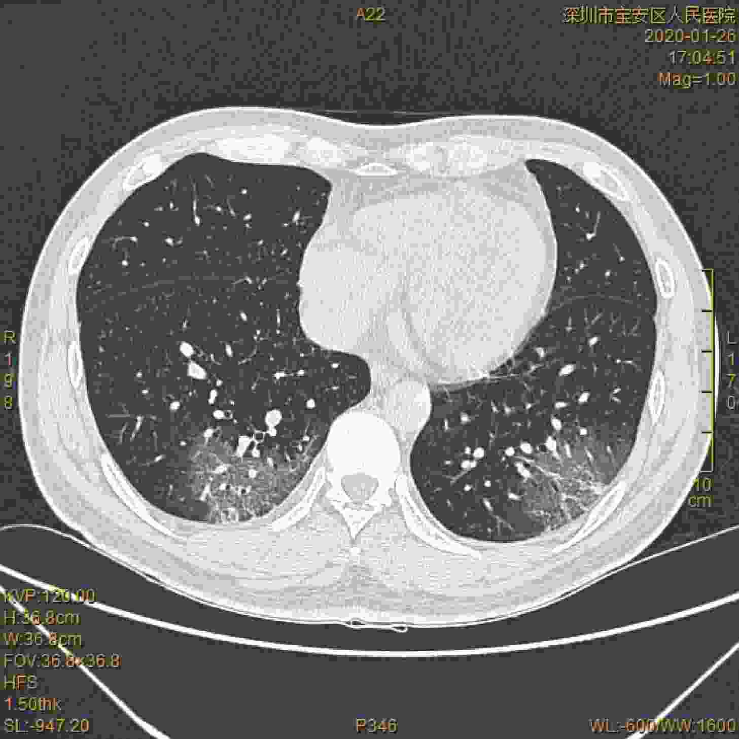
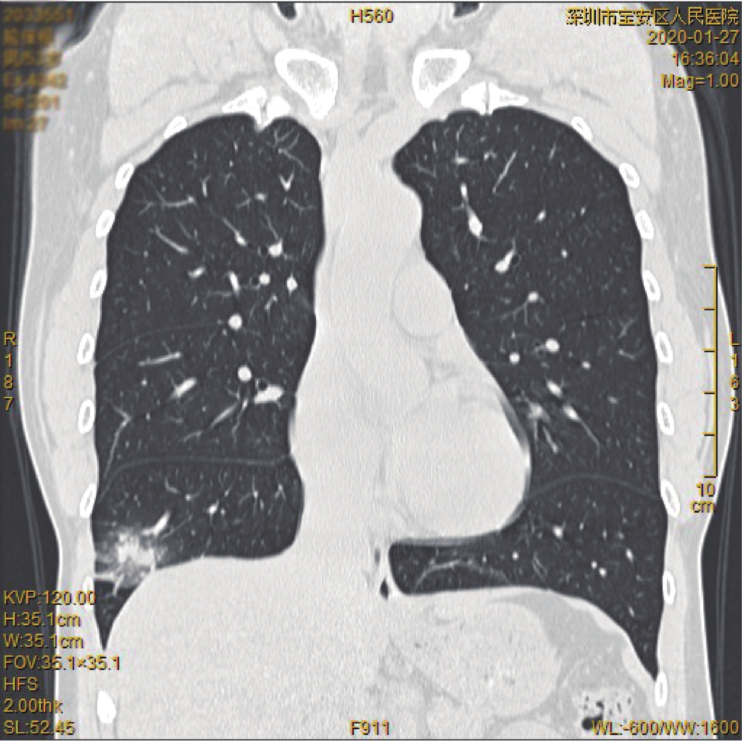
图 1 双肺下叶后部胸膜下区多发磨玻璃样密度影,右侧可见病灶支气管血管束增粗
Figure 1. Multiple glass-like density shadows in subpleural area of posterior sublobar of both lungs, thickening of bronchial vascular bundle in right side
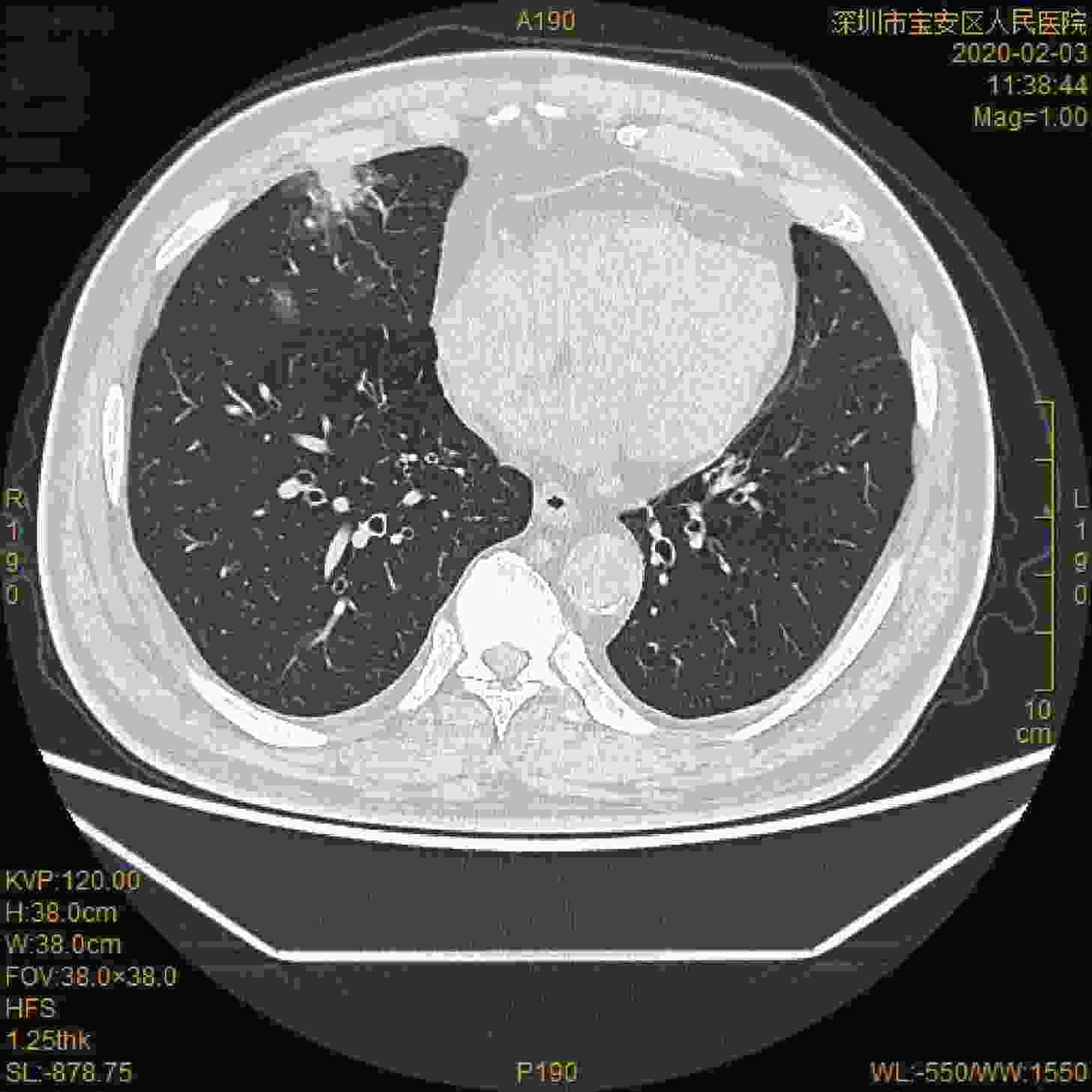
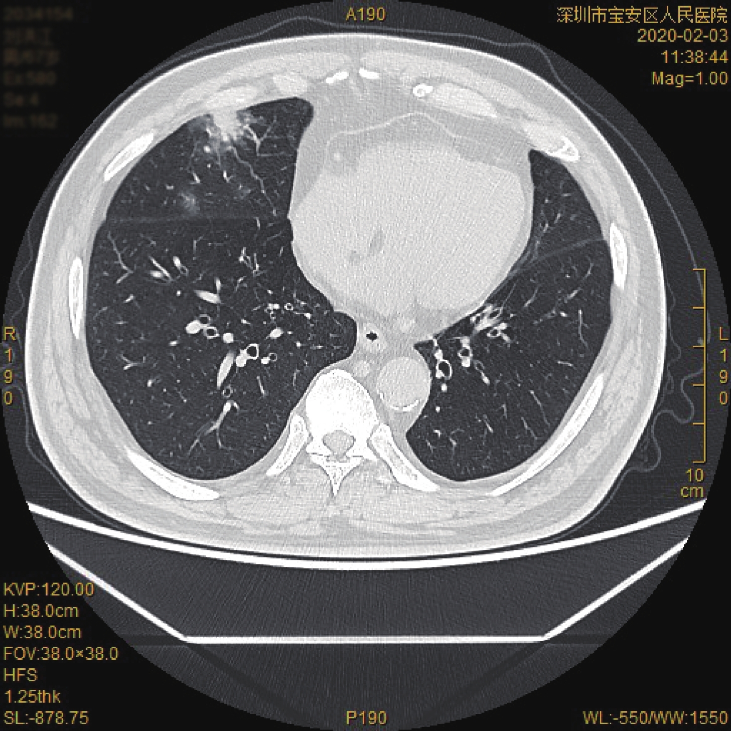
图 2 右肺中叶内多发斑片状磨玻璃样密度影和磨玻璃与实性混杂高密度影
Figure 2. Multiple variegated glass-like density shadow and glass-grinding high density shadow in middle lobe of right lung
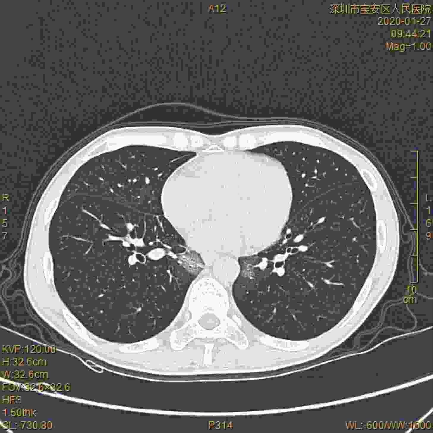
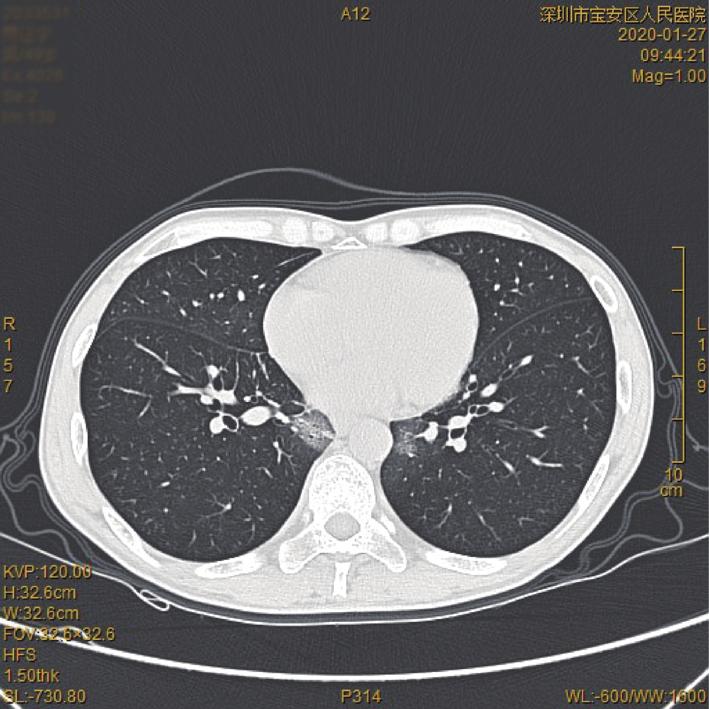
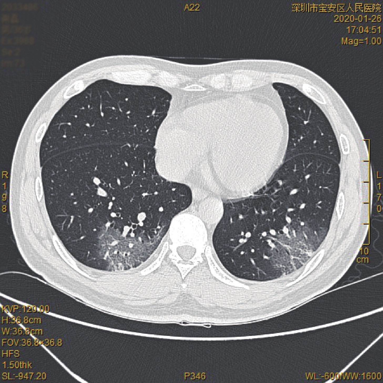
图 3 双肺下叶纵膈旁胸膜下区小团片状磨玻璃样密度影,内部见细小网格状改变
Figure 3. Small patches glass-like density shadow in subphrenic subphrenic pleural area of lower lobe of both lungs
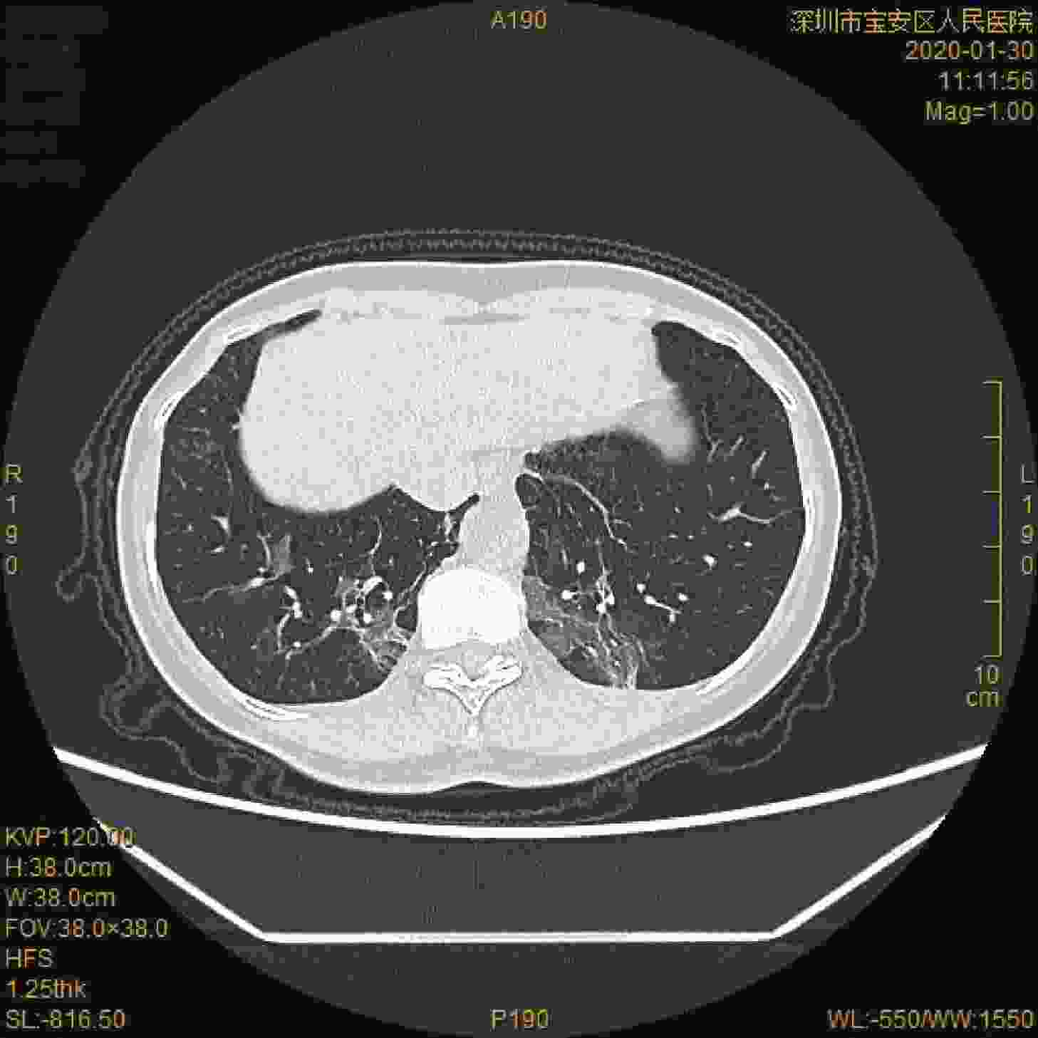
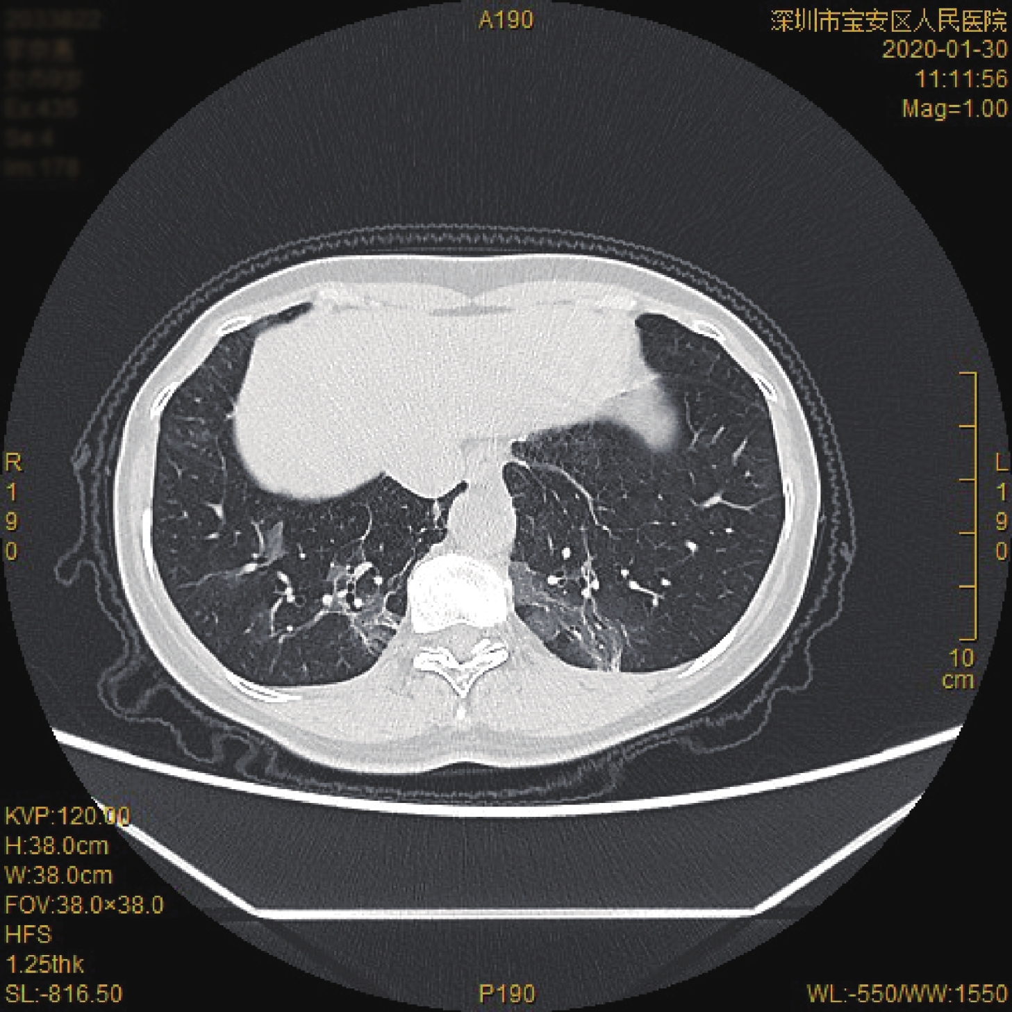
图 4 双肺下叶磨玻璃样及条索状高密度影
Figure 4. Glass-like and striped high density shadow of lower lobe of both lungs
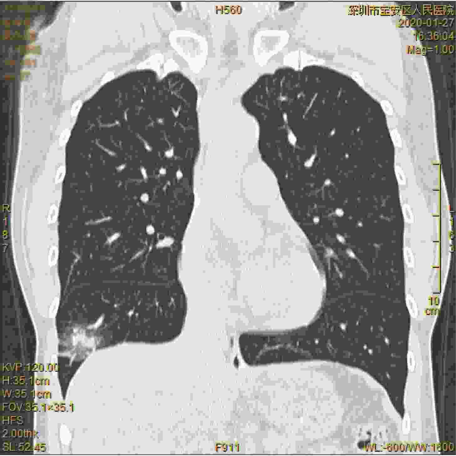
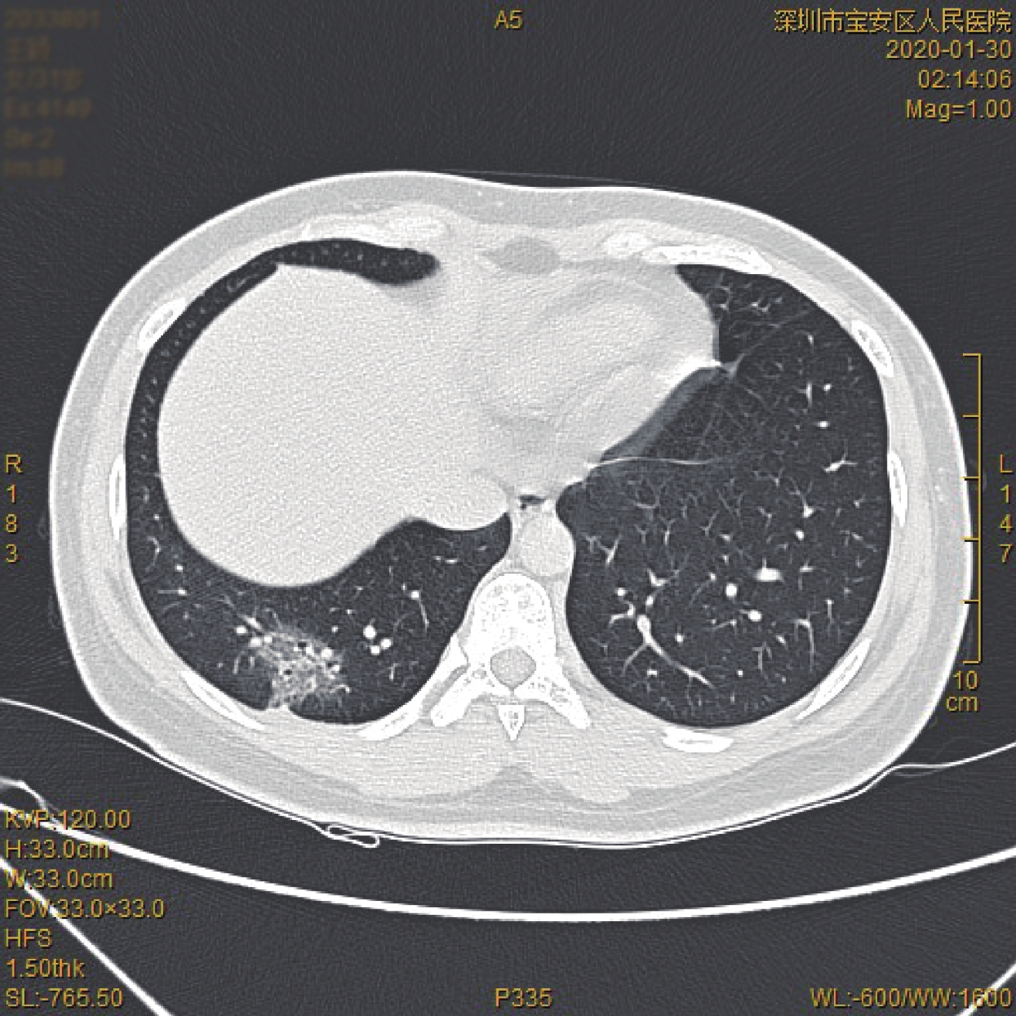
图 5 右肺下叶磨玻璃与实性混杂高密度影,临近支气管血管束增粗
Figure 5. Grind glass and solid hybrid high density shadow in inferior lobe of right lung, thickening of adjacent bronchial blood vessels
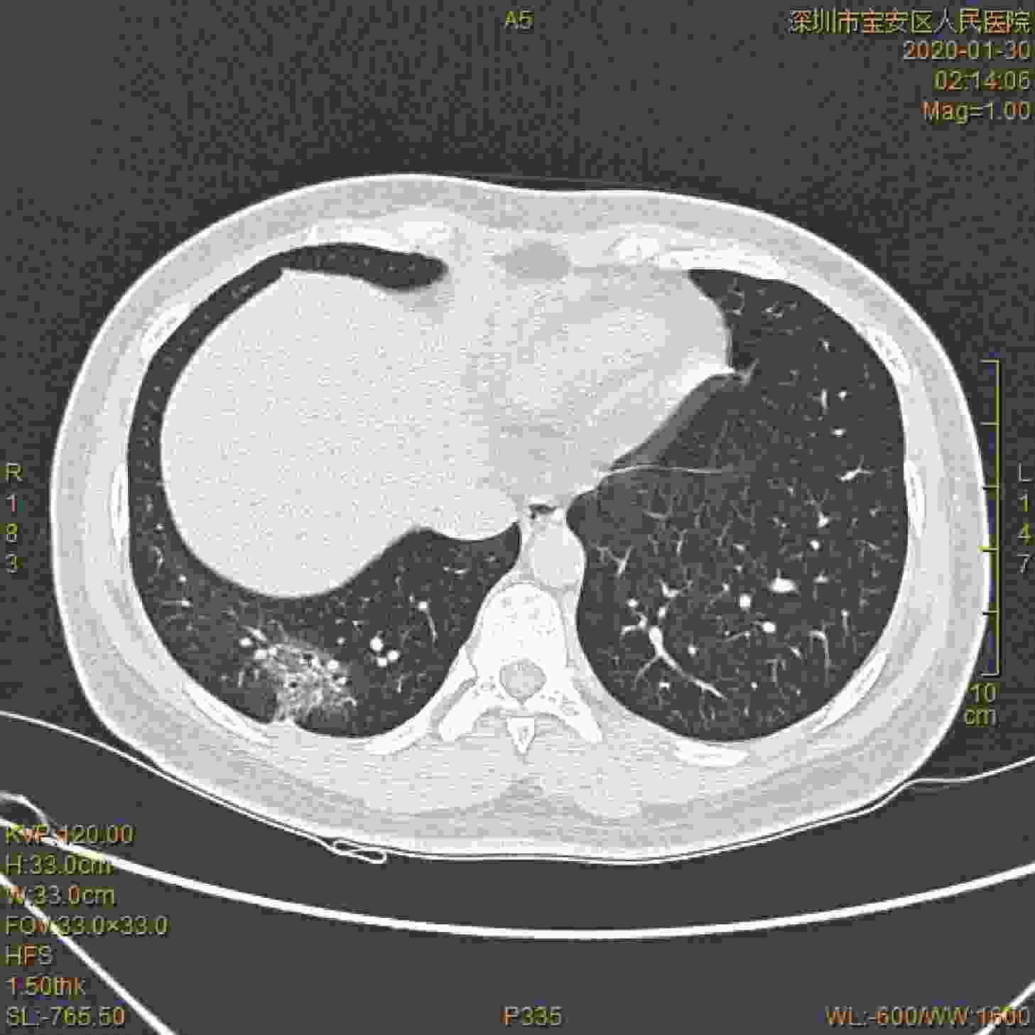
图 6 右肺下叶胸膜下去团片状高密度影,内部见网状小叶间隔增厚,临近胸膜可见增厚牵拉改变
Figure 6. Right inferior lobe pleura subcluster lamellar high density shadow,thickening of mesophyll septum
表 1 COVID-19患者一般资料、临床表现和外周血检查
Table 1. General data, clinical manifestation and peripheral blood examination of patients with COVID-19
编号 一般资料 临床症状 外周血检查 性别 年龄 接触史 发热 干咳 其他 淋巴细胞减低 C-反应蛋白增加 1 男 36 有 有 无 无 无 有 2 女 32 有 有 无 无 无 无 3 男 49 有 无 无 无 有 无 4 男 53 有 无 无 无 无 无 5 男 35 有 有 有 无 无 无 6 女 31 有 有 无 无 无 有 7 男 54 有 有 无 无 无 有 8 女 59 有 有 有 无 有 无 9 男 61 有 有 有 无 无 有 10 男 48 有 有 无 无 无 有 11 男 67 有 有 无 腹泻 无 无 12 男 63 有 无 无 无 无 无 13 女 51 有 无 无 无 无 无  下载: 导出CSV
-
[1] Novel Coronavirus(2019-nCoV) Situation Report–22 [EB/OL]. https://www.who.int/docs/default-source/coronaviruse/situation-reports/20200211-sitrep-22-ncov.pdf?sfvrsn=fb6d49b1_2, 2020. [2] 新冠状病毒诊疗方案(试行第六版) [EB/OL]. http://www.nhc.gov.cn/xcs/zhengcwj/202002/8334a8326dd94d329df351d7da8aefc2/files/b218cfeb1bc54639af227f922bf6b817.pdf, 2020. [3] Jasper C, Yuan S, Kok K, et al. A familial cluster of pneumonia associated with the 2019 novel coronavirus indicating person-to-person transmission: a study of a family cluster[J]. The Lancet, 2020, 395(10223): 514-23. doi: 10.1016/S0140-6736(20)30154-9 [4] Lu R, Zhao X, Li J, et al. Genomic characterisation and epidemiology of 2019 novel coronavirus: implications for virus origins and receptor binding[J]. The Lancet, 2020, 395(10224): 565-74. doi: 10.1016/S0140-6736(20)30251-8 [5] Lai CC, Shih TP, Ko WC, et al. Severe acute respiratory syndrome coronavirus 2 (SARS-CoV-2) and corona virus disease-2019 (COVID-19-19): the epidemic and the challenges[J]. Int J Antimicrob Agents, 2020: https://doi.org/10.1016/j.ijantimicag.2020.105924. [6] Phan LT, Nguyen TV, Luong QC, et al. Importation and human-to-human transmission of a novel coronavirus in vietnam[J]. N Engl J Med, 2020: https://doi.org/10.1056/NEJMc2001272. [7] Li Q, Guan X, Wu P, et al. Early transmission dynamics in Wuhan, China, of novel coronavirus-Infected pneumonia[J]. N Engl J Med, 2020: https://doi.org/10.1056/NEJMoa2001316. [8] Zhou P, Yang X, Wang X, et al. A pneumonia outbreak associated with a new coronavirus of probable bat origin[J]. Nature, 2020: https://doi.org/10.1038/s41586-020-2012-7. [9] 中国疾病预防控制中心新型冠状病毒肺炎应急响应机制流行病学组. 新型冠状病毒肺炎流行病学特征分析[J]. 中华流行病学杂志, 2020, 41(2): 145-51. doi: 10.3760/cma.j.issn.0254-6450.2020.02.003 [10] Huang C, Wang Y, Li X, et al. Clinical features of patients infected with 2019 novel coronavirus in Wuhan, China[J]. The Lancet, 2020, 395(10223): 497-506. doi: 10.1016/S0140-6736(20)30183-5 [11] Wang C, Horby P, Hayden F, et al. A novel coronavirus outbreak of global health concern[J]. The Lancet,, 2020, 395(10223): 470-3. doi: 10.1016/S0140-6736(20)30185-9 [12] Lin C, Ding Y, Xie B, et al. Asymptomatic novel coronavirus pneumonia patient outside WuHan: the value of CT images in the course of the disease[J]. Clinical Imaging, 2020, 63(2020): 7-9. [13] Chen N, Zhou M, Dong X, et al. Epidemiological and clinical characteristics of 99 cases of 2019 novel coronavirus pneumonia in Wuhan, China: a descriptive study[J]. The Lancet, 2020, 395(10223): 507-13. doi: 10.1016/S0140-6736(20)30211-7 [14] Xu Z, Shi L, Wang Y, et al. Pathological findings of COVID-19-19 associated with acute respiratory distress syndrome[J]. Lancet Respir Med, 2020: https://doi.org/10.1016/S2213-2600(20)30076-X. [15] Wrapp D, Wang N, Corbett KS, et al. Cryo-EM structure of the 2019-nCoV spike in the prefusion conformation[J]. Science, 2020: https://doi.org/10.1126/science.abb2507. -

 点击查看大图
计量
- 文章访问数: 2454
- HTML全文浏览量: 914
- PDF下载量: 49
- 被引次数: 0