


Application of 3D high resolution magnetic resonance imaging in simple focal cortical dysplasia with children
-
摘要:
目的利用3D高分辨磁共振成像来分析儿童单纯型局灶性皮质发育不良(FCD)的影像学特征。 方法回顾分析山东大学齐鲁儿童医院2016年3月~2018年10月经病理学证实的57例单纯型FCD患儿的MRI资料。其中男性33例,女性24例,年龄6月~11.5岁。分析其主要MRI阳性征象、各种征象在不同类型FCD及各亚型中出现的频率,然后比较各种征象在不同类型FCD及各亚型中检出率是否有差异。 结果Ⅰ型FCD主要MRI征象为局灶性灰白质分界模糊(轻度)(n=9,39.13%)、局灶性皮层变薄(n=9,39.13%)及节段性脑叶萎缩与局部白质体积缩小(n=8,34.78%),上述3种征象的检出率高于Ⅱ型FCD,差异有统计学意义(P<0.05)。Ⅱ型主要MRI征象有:局灶性灰白质分界模糊(较明显)(n=29,85.29%)、局灶性皮层增厚(n=25,73.53%)、transmantle征(n=16,47.06%)、灰/白质内信号异常(n=17,50.00%;n=24,70.59%)及异常脑沟/回形态(n=13,38.24%),上述征象检出率高于Ⅰ型患儿,差异有统计学意义(P<0.05)。FCDⅡb型中局灶性灰白质分界模糊(较明显)、局灶性皮层增厚、transmantle征(自皮层下向脑室方向延伸类似锥形信号)及白质信号异常的检出率(100.00%、95.00%、80.00%、90.00%)高于FCDⅡa型,差异有统计学意义(P<0.05);FCDⅠ型各亚型的MRI征象检出率差异无统计学意义(P>0.05)。 结论高分辨成像能够在术前判断FCD类型,对FCDⅡ型各亚型也具有鉴别价值,这对指导其术前方案的制定具有重要意义。 Abstract:ObjectiveTo explore simplex focal cortical dysplasia (FCD) imaging characteristics with children by 3D high resolution magnetic resonance imaging. MethodsWe retrospectively analyzed MRI data of 57 children (33 males, 24 females, 6 months to 11.5 years old ) with simple FCD confirmed by pathology from March 2016 to October 2018 in Qilu Children's Hospital of Shandong University. The main MRI positive signs and the frequency of various signs in different types of FCD and subtypes were analyzed. The differences in the detection rates of various signs in different types of FCD and subtypes were compared. ResultsThe main MRI signs of type Ⅰ FCD were focal gray-white matter blurring (mild) (n=9, 39.13%), focal cortex thinning (n=9, 39.13%), segmental cerebral lobe atrophy, and local white matter volume reduction (n=8, 34.78%). The detection rate of the three signs was significantly higher than that of FCD Ⅱ(P<0.05). The main MRI signs of type Ⅱ were blurred gray matter boundary (obvious) (n=29, 85.29%), focal cortex thickening (n=25, 73.53%), transmantle sign (n=16, 47.06%), abnormal signal in gray-white matter(n=17, 50.00%; n=24 cases, 70.59%), abnormal sulci and morphology (n=13, 38.24%). The detection rate of the above signs was significantly higher than that of children with type I(P<0.05). The detection rate of focal gray-white matter in FCDⅡb type was blurred (more obvious). The focal cortical thickening, transmantle sign (like a cone signal extending from the subcortex to the ventricle), and abnormal white matter signal detection rates(100.00%, 95.00%, 80.00%, 90.00% respectively)were significantly higher than that in FCD Ⅱa (P < 0.05). Ther difference in the detection rate of MRI signs of type FCD Ⅰwas not significant (P>0.05). ConclusionHigh-resolution imaging could determine the type of FCD before surgery. It has a discriminating value for each subtype of FCD Ⅱ, which is significant in guiding the formulation of its preoperative plan. -

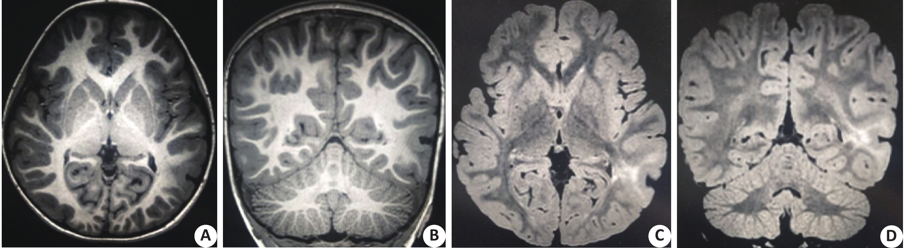
图 1 患者男,3岁,癫痫频繁发作2年余,术后病理学证实为FCDⅡb型
A,B: T1WI显示左枕叶可见明显局灶性灰白质分界模糊及相应皮质增厚;C,D: T2WI-FLAIR显示病变部位灰白质内均可见异常高信号,并可见自皮层下向脑室方向延伸的锥形高信号灶.
Figure 1. The patient, male ,3 years old, frequent for more than 2 years, postoperative pathology confirmed FCDⅡb type
表 1 其它相关MRI序列扫描参数
Table 1. Additional MRI sequence scan parameters
序列 TR(ms) TE(ms) NSA FOV(mm×mm) 矩阵 层厚(mm) 间距(mm) TSE-T1WI 638.0 7.1 2 260×260 280×280 5 1 TSE-T2WI 2 600.0 94.0 2 260×260 280×280 5 1 T2WI-FLAIR 12 000.0 130.0 2 260×260 280×280 5 1  下载: 导出CSV
表 2 不同MRI征象在两型FCD中出现频率的对比[n(%)]
Table 2. Comparison of the frequency of different MRI signs in two types of FCD
类型 局灶性灰白质
分界模糊(轻度)局灶性灰白质
分界模糊(较明显)局灶性
皮层增厚局灶性
皮层变薄transmantle征 灰质信号
异常白质信号
异常节段性脑叶萎缩与
局部白质体积缩小异常脑沟
、回形态Ⅰ型 9(39.13) 10(43.48) 7(30.43) 9(39.13) 0(0.00) 5(21.74) 9(39.13) 8(34.78) 1(4.35) Ⅱ型 2(5.88) 29(85.29) 25(73.53) 4(11.76) 16(47.06) 17(50.00) 24(70.59) 2(5.88) 13(38.24) χ2 7.720* 11.103* 10.348* 4.385# — 4.623* 5.569* 6.049# 6.772# P 0.005 0.001 0.001 0.036 0.000 0.032 0.018 0.014 0.009 *采用Pearson卡方检验法;#采用连续校正卡方检验法;—:采用Fisher’s精确检验法.
下载: 导出CSV
表 3 不同MRI征象在FCDⅡa、Ⅱb型中出现频率的对比[n(%)]
Table 3. The comparison of frequency of different MRI signs in FCDⅡa、Ⅱb type
类型 局灶性灰白质
分界模糊(轻度)局灶性灰白质
分界模糊(较明显)局灶性
皮层增厚局灶性
皮层变薄transmantle征 灰质信号
异常白质信号
异常节段性脑叶萎缩与
局部白质体积缩小异常脑沟
、回形态Ⅱa型 2(14.29) 9(64.29) 6(42.86) 3(21.43) 0(0.00) 6(42.86) 6(42.86) 1(7.14) 4(28.57) Ⅱb型 0(0.00) 20(100.00) 19(95.00) 1(5.00) 16(80.00) 11(55.00) 18(90.00) 1(5.00) 9(45.00) P 0.162 0.007 0.001 0.283 0.000 0.728 0.006 1.000 0.477 采用Fisher’s精确检验法.
下载: 导出CSV
表 4 不同MRI征象在FCDⅠa、Ⅰb、Ⅰc型中出现频率的对比[n(%)]
Table 4. The comparison of frequency of different MRI signs in FCDⅠa、Ⅰb、Ⅰc type
类型 局灶性灰白质
分界模糊(轻度)局灶性灰白质
分界模糊(较明显)局灶性
皮层增厚局灶性
皮层变薄transmantle征 灰质信号
异常白质信号
异常节段性脑叶萎缩与
局部白质体积缩小异常脑沟
、回形态Ⅰa型 2(33.3) 1(16.7) 2(33.3) 2(33.3) 0(0.0) 0(0.0) 0(0.0) 1(16.7) 0(0.0) Ⅰb型 4(44.4) 4(44.4) 3(33.3) 5(55.6) 0(0.0) 3(33.3) 2(22.2) 4(44.4) 0(0.0) Ⅰc型 3(37.5) 7(87.5)* 2(25.0) 2(25.0) 0(0.0) 2(25.0) 7(87.5)* 3(37.5) 1(12.5) P >0.05 0.026 >0.05 >0.05 >0.05 >0.05 0.005 >0.05 >0.05 采用Fisher’s精确检验法,每种征象进行两两比较。*P<0.05 vs Ⅰa型.
下载: 导出CSV
-
[1] Choi SA, Kim KJ. The Surgical and cognitive outcomes of focal cortical dysplasia[J]. J Korean Neurosurg Soc, 2019, 62(3): 321-7. doi: 10.3340/jkns.2019.0005 [2] Bender B, Rona S, Focke N, et al. MR-imaging of focal cortical dysplasia[J]. Rofo, 2014, 186(11): 987-90. doi: 10.1055/s-0034-1369334 [3] Ahmed R, Rubinger L, Go C, et al. Utility of additional dedicated high-resolution 3 T MRI in children with medically refractory focal epilepsy[J]. Epilepsy Res, 2018, 143(2018): 113-9. [4] Blümcke I, Thom M, Aronica E, et al. The clinicopathologic spectrum of focal cortical dysplasias: a consensus classification proposed by an ad hoc Task Force of the ILAE diagnostic methods commission[J]. Epilepsia, 2011, 52(1): 158-74. doi: 10.1111/j.1528-1167.2010.02777.x [5] Jayalakshmi S, Nanda SK, Vooturi S, et al. Focal cortical dysplasia and refractory epilepsy: role of multimodality imaging and outcome of surgery[J]. Am J Neuroradiol, 2019, 40(5): 892-8. doi: 10.3174/ajnr.A6041 [6] Sacino MF, Ho CY, Whitehead MT, et al. Resective surgery for focal cortical dysplasia in children: a comparative analysis of the utility of intraoperative magnetic resonance imaging (iMRI)[J]. Childs Nerv Syst, 2018, 32(6): 1101-7. [7] Battal B, Ince S, Akgun V, et al. Malformations of cortical development: 3 T magnetic resonance imaging features[J]. World J Radiol, 2019, 7(10): 329-35. [8] Kim SH, Choi J. Pathological Classification of focal cortical dysplasia: personal comments for well understanding FCD classification[J]. J Korean Neurosurg Soc, 2019, 62(3): 288-95. doi: 10.3340/jkns.2019.0025 [9] Veersema TJ, Ferrier CH, van Eijsden P, et al. Seven tesla MRI improves detection of focal cortical dysplasia in patients with refractory focal epilepsy[J]. Epilepsia Open, 2017, 2(2): 162-71. doi: 10.1002/epi4.12041 [10] Zucca I, Milesi G, Medici V, et al. Type II focal cortical dysplasia: Ex vivo 7T magnetic resonance imaging abnormalities and histopathological comparisons[J]. Ann Neurol, 2016, 79(1): 42-58. doi: 10.1002/ana.24541 [11] Hong SJ, Bernhardt BC, Caldairou B, et al. Multimodal MRI profiling of focal cortical dysplasia type II[J]. Neurology, 2017, 88(8): 734-42. doi: 10.1212/WNL.0000000000003632 [12] Chassoux F, Landré E, Mellerio C, et al. Type II focal cortical dysplasia: electroclinical phenotype and surgical outcomerelated to imaging[J]. Epilepsia, 2012, 53(2): 349-58. doi: 10.1111/j.1528-1167.2011.03363.x [13] Mata-Mbemba D, Iimura Y, Hazrati LN, et al. MRI, Magnetoencephalography and surgical outcome of oligodendrocytosis versus focal cortical dysplasia type I[J]. Am J Neuroradiol, 2018, 39(12): 2371-7. doi: 10.3174/ajnr.A5877 [14] Taylor DC, Falconer MA, Bruton CJ, et al. Focal dysplasia of the cerebral cortex in epilepsy[J]. J Neurol Neurosurg Psychiatry, 1971, 34(4): 369-87. doi: 10.1136/jnnp.34.4.369 [15] Jin B, Krishnan B, Adler S, et al. Automated detection of focal cortical dysplasia type II with surface-basedmagnetic resonance imaging postprocessing and machine learning[J]. Epilepsia, 2018, 59(5): 982-92. doi: 10.1111/epi.14064 [16] Guye M, Bartolomei F, Ranjeva JP. Malformations of cortical development: the role of 7- tesla magnetic resonance imaging in diagnosis[J]. Rev Neurol(Paris), 2019, 175(3): 157-62. doi: 10.1016/j.neurol.2019.01.393 [17] Bourdillon P, Rheims S, Catenoix H, et al. Malformations of cortical development: new surgical advances[J]. Rev Neurol (Paris), 2019, 175(3): 183-8. doi: 10.1016/j.neurol.2019.01.392 [18] Colon AJ, van Osch MJ, Buijs M, et al. Detection superiority of 7 T MRI protocol in patients with epilepsy and suspected focal cortical dysplasia[J]. Acta Neurol Belg, 2016, 116(3): 259-69. doi: 10.1007/s13760-016-0662-x -

 点击查看大图
计量
- 文章访问数: 824
- HTML全文浏览量: 325
- PDF下载量: 5
- 被引次数: 0