


Experimental study of lymph node imaging by MR contrast enhanced contrast agent
-
摘要:
目的研究大分子阳性亲淋巴对比剂二氨基乙基乙二醇醚-DTPA酰胺共聚物钆配合物(Gd-poly-DTPA-EOEA)增强MR淋巴造影对肿瘤转移、炎性增生淋巴结显像的鉴别诊断价值。 方法本研究共14只新西兰大白兔,其中7只于后肢注射完全型免疫佐剂,建立腘窝淋巴结的炎性增生组模型,另7只于后肢肌肉内接种VX2瘤,建议腘窝淋巴结肿瘤转移模型,对侧正常腘窝淋巴结作为对照。实验兔在接种前后行2次MR淋巴造影检查。图像3D重建后,选取兔腘窝淋巴结中直径最大者测量其各观察时间点的信噪比。采用成组设计t检验比较每一观察时间点的正常对照侧、炎性增生侧、肿瘤转移侧淋巴结之间的信噪比差异,并与病理检查结果相对照。 结果1只兔接种肿瘤未成功,其他接种均良好。全部模型兔均完成二次MR淋巴造影检查。在MR淋巴造影图像上,炎性增生组在各扫描时间点,炎性侧与对照侧腘窝淋巴结信噪比之间差异均无统计学意义(P>0.05)。肿瘤转移组在各扫描时间点,肿瘤侧与对照侧腘窝淋巴结信噪比之间差异均有统计学意义(P<0.01)。进一步比较炎性侧和肿瘤侧腘窝淋巴结,在各扫描时间点,炎性侧与肿瘤侧腘窝淋巴结信噪比差异有统计学意义(P<0.01)。 结论基于大分子阳性亲淋巴对比剂Gd-poly-DTPA-EOEA增强的MR淋巴造影是特异性地鉴别良、恶性淋巴结的敏感检查方法。 Abstract:ObjectiveTo explore the differential diagnostic value of macromolecular positive lymphophilic contrast agent diaminoglycol ether DTPA amide copolymer gadolinium complex (Gd-poly-DTPA-EOEA) enhanced MR lymphography (MRL) in lymph node imaging of tumor metastasis and inflammatory hyperplasia. MethodsA total of 14 New Zealand white rabbits. Seven rabbits were injected with complete immune adjuvant at the hind limb to establish an inflammatory hyperplasia group model of popliteal fossa lymph node. The Other 7 were inoculated with VX2 tumor in the posterior limb muscle, suggesting the popliteal fossa lymph node metastasis model and the opposite normal popliteal fossa lymph node as the control. Two magnetic resonance lymphography were performed before and after inoculation. After 3D reconstruction, the signal to noise ratio (SNR) of the largest rabbit popliteal fossa nodes was measured. The SNR differences between normal control side, inflammatory hyperplasia side and metastatic side lymph nodes at each observation time point were compared with the results of pathological examination. ResultOne rabbit was not successfully inoculated with tumor, and other inoculations were good. All the model rabbits completed the secondary MR lymphography. On the MR lymphangiography image, there was no significant difference between the inflammatory side and the control side popliteal fossa nodes SNR at all scanning time points in the inflammatory hyperplasia group (P>0.05). In the tumor metastasis group, there were significant differences between the tumor side and the control side popliteal fossa lymph node SNR at each scanning time point (P<0.01). Further comparison was made between the inflammatory side and the tumor side popliteal fossa lymph nodes. The difference between the inflammatory side and the tumor side popliteal fossa lymph nodes SNR was significant at each scanning time point (P<0.01). ConclusionMR lymphography (MRL) based on macromolecular positive lymphophilic contrast medium (Gd-poly-DTPA-EOEA) enhancement is a sensitive method for differentiating benign from malignant lymph nodes. -
Key words:
- magnetic resonance imaging /
- contrast media /
- lymphography /
- neoplasm metastasis /
- inflammation
-
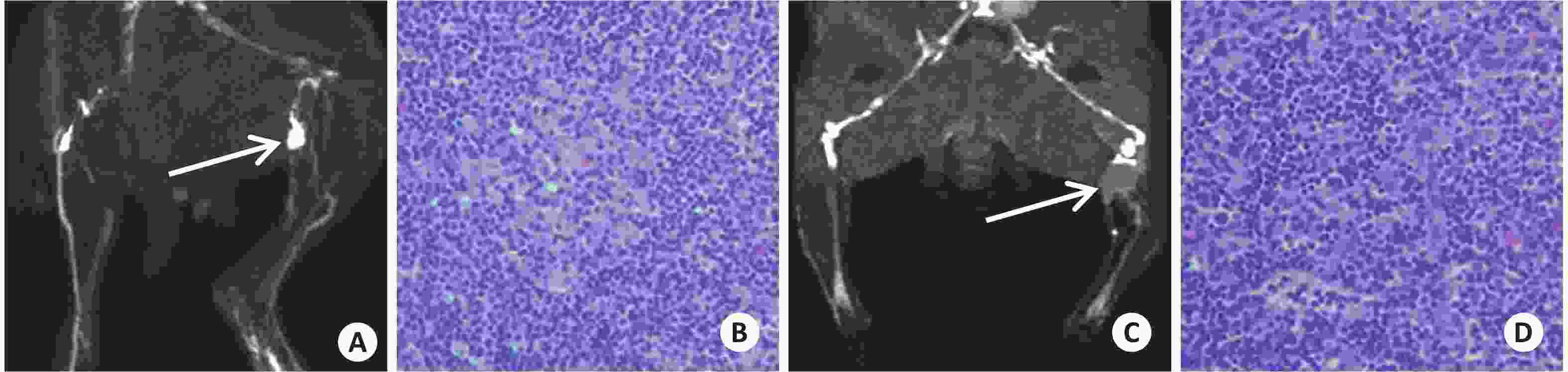
图 1 炎性侧与肿瘤侧腘窝淋巴结信噪与病理对照
A: 炎性增生组, 兔左侧足部淋巴管扩张, 腘窝淋巴结较对侧肿大; B: 炎性增生组病理切片, 显示淋巴结内可见大量炎性细胞浸润(HE, ×400); C: VX2肿瘤转移组, 兔左侧腘窝可见充盈缺损区(箭); D: VX2肿瘤转移组, 淋巴结内可见大量肿瘤细胞浸润(HE, ×400).
表 1 MR淋巴成像扫描各时间点病变侧与正常对照侧腘窝淋巴结信噪比统计(Mean±SD)
组别 淋巴结(n) 扫描时间点(min) 5 15 30 60 90 120 炎性增生组 炎性侧 7 10.78±0.16 19.01±0.31 29.79±0.16 27.97±0.56 24.77±0.14 22.85±0.39 对照侧 7 10.89±0.17 18.87±0.47 29.96±0.64 27.96±0.27 24.85±0.44 22.94±0.40 P 0.239 0.542 0.490 0.962 0.634 0.435 肿瘤转移组 肿瘤侧 6 4.15±0.12 5.94±0.12 6.93±0.09 6.54±0.11 5.84±0.18 5.89±0.11 对照侧 6 11.33±0.19 18.29±0.70 31.87±0.50 27.63±0.23 24.58±0.15 23.07±0.30 P 0.000 0.000 0.000 0.000 0.000 0.000 炎症与肿瘤 肿瘤侧 6 4.15±0.12 5.94±0.12 6.93±0.09 6.54±0.11 5.84±0.18 5.89±0.11 炎性侧 7 10.78±0.16 19.01±0.31 29.79±0.16 27.97±0.56 24.77±0.14 22.85±0.39 P 0.000 0.000 0.000 0.000 0.000 0.000  下载: 导出CSV
-
[1] Moskovic E, Fernando I, Blake P, et al. Lymphography--current role in oncology[J]. Br J Radiol, 1991, 64(761): 422-7. doi: 10.1259/0007-1285-64-761-422 [2] Akduman EI, Momtahen AJ, Balci NC, et al. Comparison between malignant and benign abdominal lymph nodes on diffusion-weighted imaging[J]. Acad Radiol, 2008, 15(5): 641-6. doi: 10.1016/j.acra.2007.12.023 [3] Misselwitz B. Mr contrast agents in lymph node imaging[J]. Eur J Radiol, 2006, 58(3): 375-82. doi: 10.1016/j.ejrad.2005.12.044 [4] Som PM. Detection of metastasis in cervical lymph nodes: CT and Mr criteria and differential diagnosis[J]. Am J Roentgenol, 1992, 158(5): 961-9. doi: 10.2214/ajr.158.5.1566697 [5] 周正扬, 朱 斌, 俞海平, 等. 间质磁共振淋巴造影的实验研究[J]. 南京大学学报:自然科学版, 2005, 21(2): 120-4. [6] 周正扬, 朱 斌, 俞海平, 等. 间质磁共振淋巴造影的技术方法及其实验研究[J]. 实用放射学杂志, 2005, 21(11): 1121-3, 1230. doi: 10.3969/j.issn.1002-1671.2005.11.001 [7] Zhou Z, Guo J, Yu H, et al. Comparison of Gd[DTPA-bis (2-aminoethoxy) ethane] polymeric contrast agent with gadodiamide injection for interstitial Mr lymphography: experimental study with rabbits[J]. J Magn Reson Imag, 2005, 22(3): 361-7. doi: 10.1002/jmri.20395 [8] 周正扬, 朱 斌, 俞海平, 等. 亲淋巴磁共振造影剂的合成、表征和性能研究[J]. 现代生物医学进展, 2009, 9(7): 1300-3. [9] 周正扬, 俞海平, 陈君坤, 等. 阳性亲淋巴对比剂增强MR淋巴成像对淋巴结病变的诊断价值[J]. 中华放射学杂志, 2010, 44(4): 434-9. doi: 10.3760/cma.j.issn.1005-1201.2010.04.025 [10] Harisinghani MG, Barentsz J, Hahn PF, et al. Noninvasive detection of clinically occult lymph-node metastases in prostate cancer[J]. N Engl J Med, 2003, 348(25): 2491-9. [11] Bellin MF, Lebleu L, Meric JB. Evaluation of retroperitoneal and pelvic lymph node metastases with MRI and Mr lymphangiography[J]. Abdom Imag, 2003, 28(2): 155-63. doi: 10.1007/s00261-001-0182-9 [12] Herborn CU, Vogt FM, Lauenstein TC, et al. Assessment of normal, inflammatory, and tumor-bearing lymph nodes with contrast-enhanced interstitial magnetic resonance lymphography: preliminary results in rabbits[J]. J Magn Reson Imag, 2003, 18(3): 328-35. doi: 10.1002/jmri.10357 [13] Misselwitz B, Platzek J, Weinmann HJ. Early Mr lymphography with gadofluorine M in rabbits[J]. Radiology, 2004, 231(3): 682-8. doi: 10.1148/radiol.2313021000 [14] Tsuda N, Tsuji T, Kato N. Interstitial magnetic resonance lymphography using gadolinium-ethoxybenzyl-diethylenetriamine pentaacetic acid in rabbits with lymph node metastasis[J]. Invest Radiol, 2005, 40(5): 306-12. doi: 10.1097/01.rli.0000160606.54347.15 [15] Weissleder R, Heautot JF, Schaffer BK, et al. Mr lymphography: study of a high-efficiency lymphotrophic agent[J]. Radiology, 1994, 191(1): 225-30. doi: 10.1148/radiology.191.1.8134576 [16] Yan GP, Xu W, Yang L, et al. Dextran Gadolinium complexes as contrast agents for magnetic resonance imaging to sentinel lymph nodes[J]. Pharm Res, 2010, 27(9): 1884-92. doi: 10.1007/s11095-010-0187-6 [17] 方 婧, 洪 颖. 磁共振间质淋巴造影术在宫颈癌前哨淋巴结检测中的临床价值[J]. 实用医学杂志, 2010, 34(17): 3262-4. doi: 10.3969/j.issn.1006-5725.2010.17.085 [18] 齐云平, 曲 坚. 亚甲蓝和纳米炭对子宫内膜癌前哨淋巴结的识别价值[J]. 山东医药, 2011, 51(13): 21-3. doi: 10.3969/j.issn.1002-266X.2011.13.011 [19] 谭红娜, 彭卫军, 杨犇龙, 等. MR淋巴管造影显示VX2兔乳腺癌前哨淋巴结[J]. 中国医学计算机成像杂志, 2012, 25(1): 67-71. doi: 10.3969/j.issn.1006-5741.2012.01.017 [20] Shen NA, Xu X, Sha Y, et al. Indirect computed tomography lymphography identifies lymph node metastasis in rabbit pyriform sinus VX2 carcinoma[J]. Oncol Lett, 2015, 9(4): 1802-6. doi: 10.3892/ol.2015.2899 [21] Wang P, Xie X, Wang J, et al. Ultra-small superparamagnetic Iron oxide mediated magnetic hyperthermia in treatment of neck lymph node metastasis in rabbit pyriform sinus VX2 carcinoma[J]. Tumour Biol, 2015, 36(10): 8035-40. doi: 10.1007/s13277-015-3538-4 [22] 周明祎, 王晓彬, 王纯雁, 等. 前哨淋巴结在子宫内膜癌中应用的研究进展[J]. 现代妇产科进展, 2016, 18(9): 703-5. [23] Yang Y, Zhou J, Shi X, et al. Long-term observation of indirect lymphography using gadolinium-loaded polyethylenimine-entrapped gold nanoparticles as a dual mode CT/Mr contrast agent for rabbit lingual sentinel lymph node identification[J]. Acta Otolaryngol, 2017, 137(2): 207-14. doi: 10.1080/00016489.2016.1222550 [24] Yang Y, Zhou B, Zhou J, et al. Assessment of lingual sentinel lymph nodes metastases using dual-modal indirect CT/Mr lymphography with gold-gadolinium-based nanoprobes in a tongue VX2 carcinoma model[J]. Acta Otolaryngol, 2018, 138(8): 727-33. doi: 10.1080/00016489.2018.1441544 -

 点击查看大图

图(1) / 表(1)
计量
- 文章访问数: 827
- HTML全文浏览量: 386
- PDF下载量: 88
- 被引次数: 0