


Application and clinical analysis of MRI in the TORCH of children
-
摘要:
目的探讨MRI在儿童先天性TORCH综合征中的应用价值。 方法回顾性分析经证实的36例TORCH患儿的影像资料及临床资料,包括男19例、女17例,年龄5 d~3岁,所有患儿均行头颅MRI扫描。 结果弥漫性囊性脑软化20例及穿通囊肿7例;27例脑实质不同程度钙化,呈条状及斑点状,以及少许脑回样融合性钙化,主要位于室管膜下及脑室周围;小头畸形,平滑脑11例,巨脑回畸形15例,小脑发育不全3例,脑裂畸形6例;积水性无脑畸形3例;脑实质脱髓鞘及胶质增生13例;脑积水17例,硬膜下积液9例及弥漫性脑萎缩21例。 结论MRI能够对TORCH综合征中的各种征象进行直观显示,为本病的一种首选影像学检查。 Abstract:ObjectiveTo explore the value of MRI in the TORCH of children. MethodsThe imaging and clinical data of 36 children confirmed TORCH were retrospectively analyzed. Nineteen boys and 17 girls were included, ranging in age from 5 d to 3 years old. All the children underwent head scan of MRI. ResultsDiffuse cystic encephalomalacia accounted for 20 cases and penetrating cyst were 7 cases. There were 27 cases with varying degrees of calcification in the brain parenchyma, which were striated and spotted. A few cases with ileocentric fusion calcification, mainly located in the subependymal and periventricular areas. Microcephaly, smooth brain accounted for 11 cases, giant gyrus malformation 15 cases, cerebellar hypoplasia 3 cases, cleft brain malformation 6 cases,hydroanencephaly 3 cases, demyelination and gliosis of brain parenchyma 13 cases. There were 17 cases of hydrocephalus, 9 cases of subdural effusion and 21 cases of diffuse cerebral atrophy. ConclusionMRI could visually display various signs in TORCH syndrome. It is a preferred imaging examination for this disease. -
Key words:
- children /
- apriority /
- TORCH /
- magnetic resonance imaging /
- clinic
-
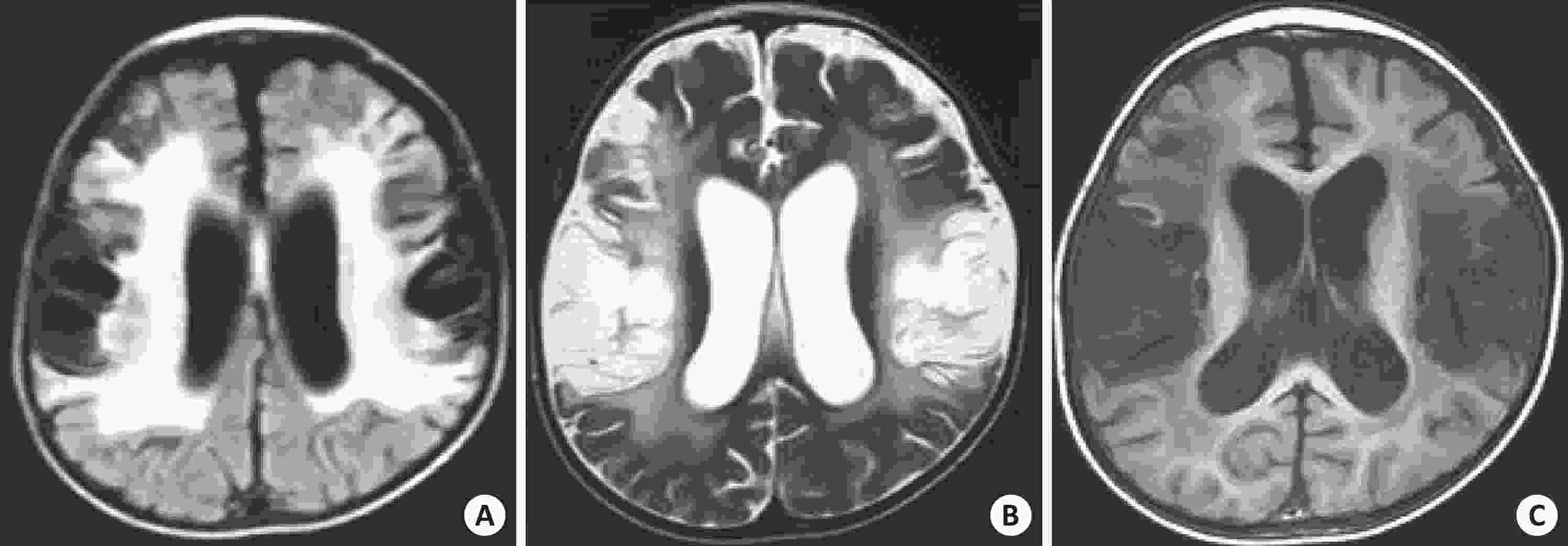
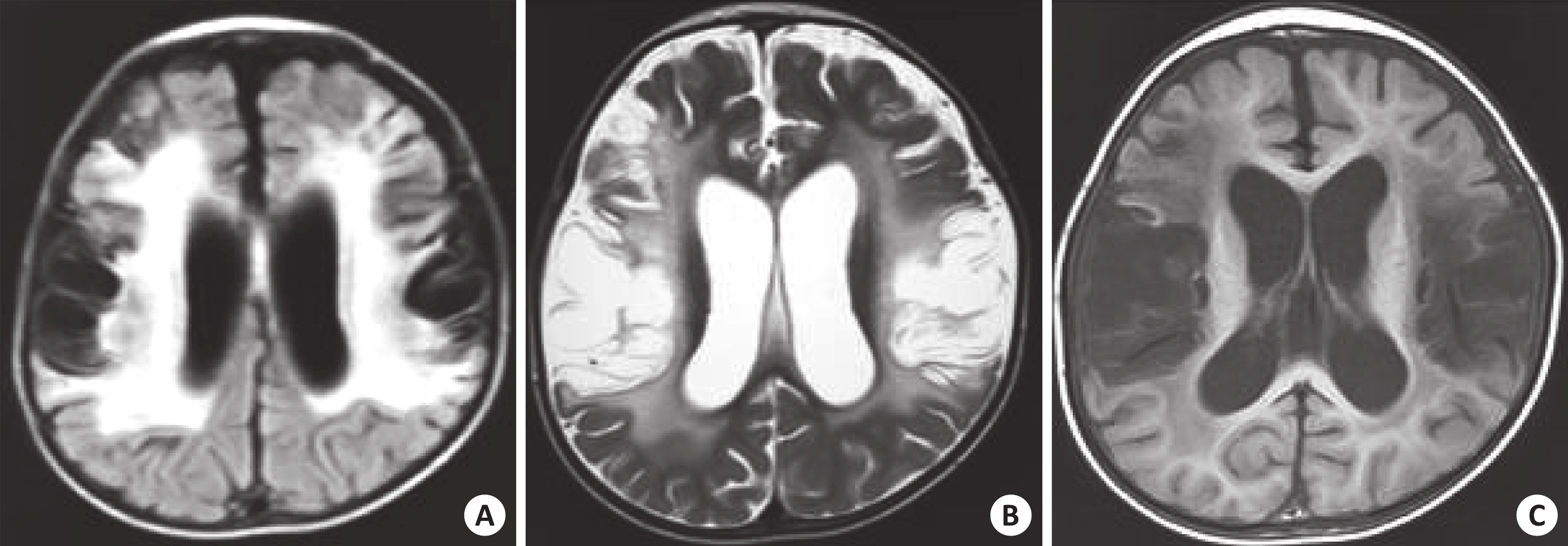
图 1 巨细胞病毒感染
男孩, 1岁7月; A: T2WI-FLAIR; B: T2WI; C: T1WI. 示脑实质内弥漫性多囊性脑软化, 脑萎缩, 并胶质增生, 胼胝体萎缩.
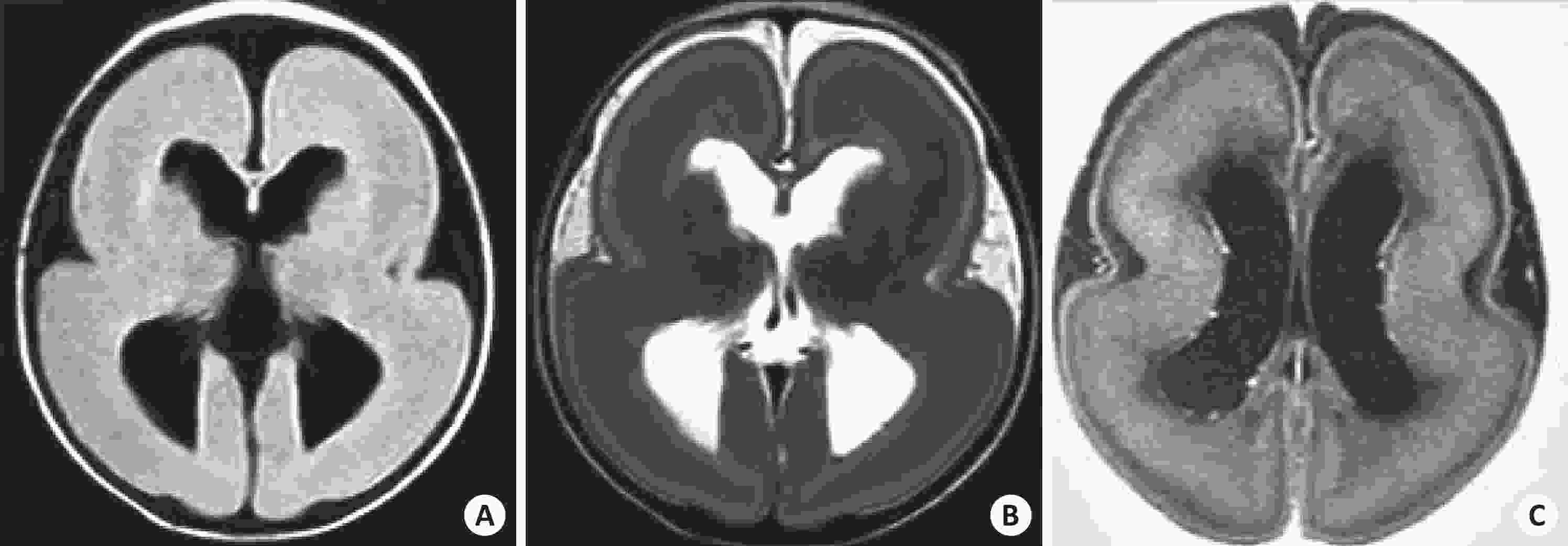
图 2 弓形体感染
男孩, 1月; A: T2WI-FLAIR; B: T2WI; C: T1WI. 双侧侧脑室体周, 三角区多发点状短T1, 短T2信号影, 同时伴无脑回畸形.
-
[1] Cofre F, Delpiano L, Labraña Y, et al. TORCH syndrome: rational approach of pre and post natal diagnosis and treatment, recommendations of the Advisory Committee on Neonatal Infections Sociedad Chilena de Infectología[J]. Rev Chilena Infectol, 2016, 33(2): 191-216. [2] Rasti S, Ghasemi FS, Abdoli A, et al. ToRCH “co-infections” are associated with increased risk of abortion in pregnant women[J]. Congenit Anom (Kyoto), 2016, 56(2): 73-8. doi: 10.1111/cga.12138 [3] Neu N, Duchon J, Zachariah P. TORCH infections[J]. Clin Perinatol, 2015, 42(1): 77-103. doi: 10.1016/j.clp.2014.11.001 [4] Chou YL, Hsieh KH, Perng CL, et al. High level antibodies to TORCH in the IVIG preparation from Taiwanese[J]. J Chin Med Assoc, 2019, 82(6): 510-4. doi: 10.1097/JCMA.0000000000000043 [5] Schwartz DA. The origins and emergence of Zika virus, the newest TORCH infection: what's old is new again[J]. Arch Pathol Lab Med, 2017, 141(1): 18-25. doi: 10.5858/arpa.2016-0429-ED [6] Suh CH, Kim HS, Jung SC, et al. MRI as a diagnostic biomarker for differentiating primary central nervous system lymphoma from glioblastoma: a systematic review and meta-analysis[J]. J Magn Reson Imag, 2019, 50(2): 560-72. doi: 10.1002/jmri.26602 [7] Forbes K. MRI brain white matter change: spectrum of change - how we can grade[J]. J Coll Physicians Edinb, 2017, 47(3): 271-5. [8] Sacks G, Sargent I, Redman C. An innate view of human preg Nancy[J]. Immunol Today, 1999, 20(3): 114-8. doi: 10.1016/S0167-5699(98)01393-0 [9] 张增俊, 黄明霞, 毕晓辰, 等. TORCH综合征的脑部CT表现[J]. 实用放射学杂志, 2010, 26(5): 724-5, 735. doi: 10.3969/j.issn.1002-1671.2010.05.031 [10] Halawa S, Mcdermott L, Donati M, et al. TORCH screening in pregnancy. Where are we now? An audit of use in a tertiary level centre[J]. J Obstet Gynaecol, 2014, 34(4): 309-12. doi: 10.3109/01443615.2013.872609 [11] 任德麟. TORCH感染对妊娠胎儿的影响[J]. 中国医师杂志, 1999, 1(1): 32-4. [12] Prasoona KR, Srinadh B, Sunitha T, et al. Seroprevalence and influence of torch infections in high risk pregnant women: a large study from South India[J]. J Obstet Gynaecol India, 2015, 65(5): 301-9. doi: 10.1007/s13224-014-0615-3 [13] Kishore J, Misra R, Paisal A, et al. Adverse reproductive outcome induced by parvovirus B19 and TORCH infections in women with high-risk pregnancy[J]. J Infect Dev Ctries, 2011, 5(12): 868-73. doi: 10.3855/jidc.1533 [14] 彭 丽, 孟繁峥, 霍淑芳. 新生儿TORCH感染的临床研究[J]. 中国实验诊断学, 2002, 6(2): 90-1. doi: 10.3969/j.issn.1007-4287.2002.02.010 [15] 李建超. 先天性TORCH感染35例临床分析[J]. 新生儿科杂志, 2000, 15(2): 57-8. [16] Basílio-Queirós D, Venturini L, Laib Sampaio K, et al. Fast and efficient titration of human cytomegalovirus stocks with a self-excisable bacterial artificial chromosomes cassette by flow cytometry[J]. Hum Gene Ther Methods, 2019, 30(4): 122-6. doi: 10.1089/hgtb.2019.084 [17] 李 欣, 李明村. 先天性TORCH感染脑CT表现[J]. 中华放射学杂志, 1997, 31(3): 160-3. doi: 10.3760/j.issn:1005-1201.1997.03.004 [18] 孙国强. 实用儿科放射诊断学[M]. 北京: 人民军医出版社, 2011: 79-82. [19] Levine D, Jani JC, Castro-Aragon I, et al. How does imaging of congenital Zika compare with imaging of other TORCH infections[J]. Radiology, 2017, 285(3): 744-61. doi: 10.1148/radiol.2017171238 [20] Teele RL, Hernanz-Schulman M, Sotrel A. Echogenic vasculature in the basal ganglia of neonates: a sonographic sign of vasculopathy[J]. Radiology, 1988, 169(2): 423-7. doi: 10.1148/radiology.169.2.2845473 [21] Post Y, Clevers H. Defining adult stem cell function at its simplest: the ability to replace lost cells through mitosis[J]. Cell Stem Cell, 2019, 25(2): 174-83. doi: 10.1016/j.stem.2019.07.002 [22] Kao YC, Peng SS, Weng WC, et al. Evaluation of white matter changes in agyria-pachygyria complex using diffusion tensor imaging[J]. J Child Neurol, 2011, 26(4): 433-9. doi: 10.1177/0883073810382452 -

 下载:
下载:
 点击查看大图
图(3)
计量
- 文章访问数: 1000
- HTML全文浏览量: 495
- PDF下载量: 8
- 被引次数: 0