


Cerenkov luminescence imaging of microRNA expression in tumor using an HSV1-tk reporter gene
-
摘要:
目的构建一种基于I型单纯疱疹病毒胸苷激酶(HSV1-tk)的新型报告基因体系,用于对肿瘤内微小核糖核酸(miR-21)的表达进行契伦科夫光学成像。 方法将细胞巨病毒(CMV)启动子基因、HSV1-tk基因以及可被miR-21互补结合的三联miR-21靶基因序列,串联构建成报告基因(CMV-HSV1-tk-3×miR-21t),即将CMV-HSV1-tk-3×miR-21t序列连接到pcDNA3.1质粒载体中并转染A549细胞。向稳定表达上述报告基因的A549细胞(A549T细胞)加入9-[4-[18F]氟-3(羟甲基)丁基]鸟嘌呤([18F] FHBG)孵育;或采用可互补结合miR-21的梯度剂量反义寡聚miR-21(Anti-miR-21)处理A549T细胞后,加入[18F]FHBG孵育。分别对上述细胞进行契伦科夫光学成像和放射性γ计数。另构建A549T裸鼠皮下移植瘤模型,分为2组分别瘤内注射Anti-miR- 21与对照混合RNA,然后分别经尾静脉注射[18F]FHBG后进行活体契伦科夫光学成像。 结果A549T细胞摄取[18F]FHBG后,其光学信号强度、γ计数分别与细胞数量之间呈线性正相关(R2=0.9962、0.9807);加入Anti-miR-21的剂量与光学信号强度、γ计数之间分别呈剂量依赖性正相关(P < 0.05);A549T细胞皮下瘤模型成像结果显示,瘤内注射Anti-miR-21与对照RNA的移植瘤对比,肿瘤组织信号更高且视觉对比显著。 结论基于microRNA调控的示踪剂摄取相关报告基因体系,本研究成功构建了一种用于对肿瘤内miR-21表达进行契伦科夫光学成像的新方法。 -
关键词:
- I型单纯疱疹病毒胸苷激酶 /
- 报告基因 /
- 微小核糖核酸 /
- 契伦科夫光学 /
- 分子影像
Abstract:ObjectiveTo establish a novel reporter gene system based on the herpes simplex virus type-1 thymidine kinase (HSV1-tk) gene for the Cerenkov optical imaging of microRNA (miR-21) expression inside tumors. MethodsThe cytomegalovirus (CMV) promoter gene, HSV1-tk gene, and the triple complementary target sequences for miR-21 were constructed in series to form the MV-HSV1-tk-3×miR-21t reporter gene. The CMV-HSV1-tk-3×miR-21t gene was inserted into the pcDNA3.1 vector and transfected to the A549 cell line. The transfected cells which can stably express the reporter gene aforementioned (A549T cells) were incubated with 9-[4-[18F]fluoro-3-(hydroxymethyl)butyl)guanine ([18F]FHBG), or treated with antisense miR-21 ribonucleic acid (Anti-miR-21) in gratitude dose and then incubated with [18F]FHBG. The Cerenkov optical signal and γ radioactive counts per minute were acquired, respectively. Except that, we set up subcutaneous A549T xenograft models using athymic nude mice and divide them into two groups: one group was intratumorally injected with Anti-miR-21, and another group was the control injected with the control of RNA mixture only. Then these two groups were administrated with [18F]FHBG and in vivo scanned in Cerenkov modality. ResultsAfter uptaking [18F]FHBG, the A549T cells can transmit marked optical signal and γ radioactivity in a linear positive correlation with the number of cells(R2=0.9962, 0.9807). The intensity of the optical signal and γ radioactivity is dose-dependently correlated with the Anti-miR-21 added into the A549T cell culture(P < 0.05). The in vivo Cerenkov images of the subcutaneous xenograft model show that compared to the group intratumorally injected with control RNA, the signal from the xenograft of the group injected with Anti-miR-21 is visually higher with a sharp contrast. ConclusionA novel reporter gene system employing the regulation on tracer uptake via microRNA for the Cerenkov imaging of endogenous miR-21 expression has been established successively. -

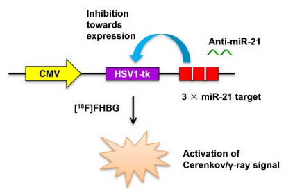
图 1 CMV-HSV1-tk-3×miR-21t/[18F]FHBG报告基因成像体系原理示意图.
细胞内源性miR-21可互补结合到HSV1-tk mRNA下游3’-端非翻译区的miR-21靶序列,抑制HSV1-tk mRNA表达,而外源加入Anti-miR-21后可抑制内源性miR-21的功能; Anti-miR-21: 反义寡聚miR-21; CMV: 细胞巨病毒启动子基因; HSV1-tk: I型单纯疱疹病毒胸苷激酶基因; 3×miR- 21 target: 三联miR-21互补结合靶序列; [18F]FHBG: 9-[4-[18F]氟-3-(羟甲基)丁基]鸟嘌呤.
Figure 1. Schematic diagram of the CMV-HSV1- tk- 3 × miR- 21t/[18F] FHBG reporter gene system.

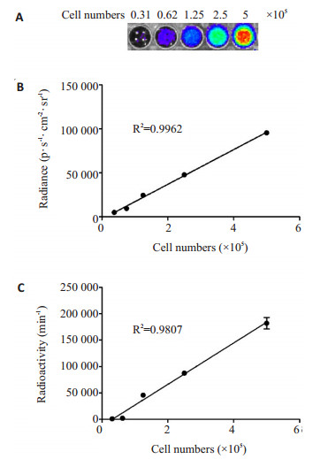
图 2 稳定表达报告基因载体的梯度数量A549T细胞与[18F]FHBG共孵育120 min后的成像信号.
A: 细胞的典型契伦科夫光学影像; B: 契伦科夫光学信号强度与细胞数量之间的线性相关性; C: 放射性γ计数率与细胞数量之间的线性相关性.
Figure 2. The signals transmitted by the A549T cells in gratitude number stably expressing the reporter gene vector after the incubation with[18F]FHBG for 120 min.

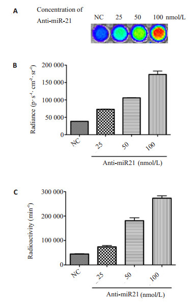
图 3 加入梯度剂量反义寡聚miR-21处理的稳定表达报告基因载体的A549T细胞与[18F]FHBG共孵育120 min后的成像信号
Anti-miR-21: 反义寡聚miR-21; NC: 阴性对照; A: 细胞的典型契伦科夫光学图像; B: 细胞的契伦科夫光学信号强度(P < 0.05); C: 细胞的放射性γ计数率(P < 0.05).
Figure 3. The signals transmitted by the A549T cells stably expressing the reporter gene vector after the treatment with antisense oligomeric miR- 21 in gratitude concentration and the subsqeuent incubation with[18F]FHBG for 120 min.

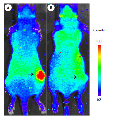
图 4 稳定表达报告基因载体的A549T细胞皮下移植瘤模型小鼠在注射[18F]FHBG 2 h后的典型活体契伦科夫光学图像.
箭头所指为移植瘤体; A: 瘤内注射In vivo-jetPEI载体包裹AntimiR-21 (n=5); B: 瘤内注射In vivo-jetPEI载体包裹对照RNA (n=5).
Figure 4. The typical in vivo Cerenkov images of subcutaneous A549T xenograft model stably expressing the reporter gene vector at 2 h after the injection of[18F]FHBG.
-
[1] Oh SW, Hwang DW, Lee DS. In vivo monitoring of microRNA biogenesis using reporter gene imaging[J]. Theranostics, 2013, 3 (12): 1004-11. doi: 10.7150/thno.4580 [2] Li M, Wang Y, Liu M, et al. Multimodality reporter gene imaging: Construction strategies and application[J]. Theranostics, 2018, 8 (11): 2954-73. doi: 10.7150/thno.24108 [3] Cao LQ, Yang XW, Chen YB, et al. Exosomal miR-21 regulates the TETs/PTENp1/PTEN pathway to promote hepatocellular carcinoma growth[J]. Mol Cancer, 2019, 18(1): 148. doi: 10.1186/s12943-019-1075-2 [4] 王红, 魏福兰, 王春玲. miR-21研究进展[J]. 口腔生物医学, 2018, 9 (2): 106-9. https://www.cnki.com.cn/Article/CJFDTOTAL-KQSW201802011.htm [5] Yang WD, Qin WW, Hu ZH, et al. Comparison of Cerenkov Luminescence Imaging (CLI) and gamma camera imaging for visualization of let-7 expression in lung adenocarcinoma A549 Cells [J]. Nucl Med Biol, 2012, 39(7): 948-53. doi: 10.1016/j.nucmedbio.2012.05.004 [6] Dubey P. Reporter Gene Imaging - Methods and Protocols[M]. New York: Springer-Humana Press, 2018. [7] Liu H, Ren G, Liu S, et al. Optical imaging of reporter gene expression using a positron-emission-tomography probe[J]. J Biomed Opt, 2010, 15(6): 1-3. [8] 杨卫东, 田捷, 汪静. MicroRNAs靶向肿瘤分子显像[J]. 中华核医学与分子影像杂志, 2014, 34(4): 335-8. doi: 10.3760/cma.j.issn.2095-2848.2014.04.018 [9] Baril P, Ezzine S, Pichon C. Monitoring the spatiotemporal activities of miRNAs in small animal models using molecular imaging modalities[J]. Int J Mol Sci, 2015, 16(3): 4947-72. doi: 10.3390/ijms16034947 [10] Ying ZM, Wu Z, Tu B, et al. Genetically encoded fluorescent RNA sensor for ratiometric imaging of MicroRNA in living tumor cells [J]. J Am Chem Soc, 2017, 139(29): 9779-82. doi: 10.1021/jacs.7b04527 [11] Simion V, Sobilo J, Clemoncon R, et al. Positive radionuclide imaging of miRNA expression using RILES and the human sodium iodide symporter as reporter gene is feasible and supports a protective role of miRNA-23a in response to muscular atrophy[J]. PLoS One, 2017, 12(5): e0177492. doi: 10.1371/journal.pone.0177492 [12] Wang F, Song X, Li X, et al. Noninvasive visualization of microRNA-16 in the chemoresistance of gastric cancer using a dual reporter gene imaging system[J]. PLoS One, 2013, 8(4): e61792. doi: 10.1371/journal.pone.0061792 [13] Shaikh FA, Kurtys E, Kubassova O, et al. Reporter gene imaging and its role in imaging-based drug development[J]. Drug Discov Today, 2020, 25(3): 582-92. doi: 10.1016/j.drudis.2019.12.010 [14] Jo MH, Jeong MS, Ko HY, et al. Radioisotope imaging of microRNA-9-regulating neurogenesis using sodium iodide sympoter [J]. Biomaterials, 2013, 34(20): 4803-9. doi: 10.1016/j.biomaterials.2013.03.032 [15] 杨柳, 李彦鹏, 吴彬彬, 等. 优化18F-FHBG自动化合成方法及其质量评估[J]. 中国医学影像技术, 2019, 35(3): 417-22. https://www.cnki.com.cn/Article/CJFDTOTAL-ZYXX201903037.htm [16] Mäkilä J, Kiviniemi A, Saanijoki T, et al. Noninvasive and quantitative monitoring of the distributions and kinetics of MicroRNA-targeting molecules in vivo by positron emission tomography[J]. Mol Pharm, 2019, 16(4): 1507-15. doi: 10.1021/acs.molpharmaceut.8b01169 [17] Song Y, Xu Z, Wang F. Genetically encoded reporter genes for MicroRNA imaging in living cells and animals[J]. Mol Ther Nucleic Acids, 2020, 21: 555-67. doi: 10.1016/j.omtn.2020.06.021 [18] Liu J, Zheng T, Tian Y. Functionalized h-BN nanosheets as a theranostic platform for SERS real-time monitoring of MicroRNA and photodynamic therapy[J]. Angew Chem Int Ed Engl, 2019, 58 (23): 7757-61. doi: 10.1002/anie.201902776 [19] Qu A, Sun M, Xu L, et al. Quantitative zeptomolar imaging of miRNA cancer markers with nanoparticle assemblies[J]. PNAS, 2019, 116(9): 3391-400. doi: 10.1073/pnas.1810764116 [20] Fu ZL, Wu YD, Ren CN, et al. Cancer cell-targeted nanoprobe for multilayer imaging of diverse biomarkers and precise photodynamic therapy[J]. Chem Commun Camb Engl, 2020, 56(96): 15208-11. doi: 10.1039/D0CC06305C [21] Wu H, Chen TT, Wang XN, et al. RNA imaging in living mice enabled by an in vivo hybridization chain reaction circuit with a tripartite DNA probe[J]. Chem Sci, 2020, 11(1): 62-9. doi: 10.1039/C9SC03469B [22] Wang F, Zhang B, Zhou L, et al. Imaging dendrimer-grafted graphene oxide mediated anti-miR-21 delivery with an activatable luciferase reporter[J]. ACS Appl Mater Interfaces, 2016, 8(14): 9014-21. doi: 10.1021/acsami.6b02662 [23] Liu H, Ren G, Miao Z, et al. Molecular optical imaging with radioactive probes[J]. PLoS One, 2010, 5(3): e9470. doi: 10.1371/journal.pone.0009470 -

 下载:
下载:
 点击查看大图
计量
- 文章访问数: 322
- HTML全文浏览量: 178
- PDF下载量: 5
- 被引次数: 0