x

ISSN 1674-4500 CN 44-1630/R

Journal of Molecular Imaging (ISSN 1674-4500; CN 44-1630/R) is a national official journal managed by the editorial department of Journal of Southern Medical University. It is a monthly journal and a Chinese Science and Technology Core Journal (Statistical Source Journal of Chinese Science and Technology Papers), with the latest impact factor of  1.162.

It has been the Chinese Core Journals of Science and Technology (the Source of Scientific Papers Statistical Journal of China), China Academic Journal Comprehensive Evaluation Database (CAJCED), Chinese Core Journals (selection) Database", "China Academic Journals (cd-rom version)", "China Science and Technology Periodical Citation Report", Ulrichsweb (USA).


The main column of Journal:  Basic research, clinical research, technology and Methods, etc. We does not charge any fee to review manuscripts. The publication time of the manuscript shall be within 6 months.

Address:  1838 North Guangzhou Dadao, Guangzhou City, Guangdong Province, China

PostCode: 510515 PhoneNo:020-62789575,61648177,61648176

Articles in press have been peer-reviewed and accepted, which are not yet assigned to volumes /issues, but are citable by Digital Object Identifier (DOI).
Display Method:
Display Method:
Clinical Research
Application value of amide proton transfer combined with quantitative susceptibility mapping in the diagnosis of Parkinson's disease
HOU Pengfei, HAN Xue, WANG Hong, JU Chao, ZHANG Fulan, HAO Lu
2024, 47(3): 225-230.   doi: 10.12122/j.issn.1674-4500.2024.03.01
[Abstract](1) [FullText HTML](2) [PDF 1315KB](1)
Abstract:
  Objective  To explore the clinical application value of amide proton transfer (APT) and quantitative susceptibility mapping (QSM) for the diagnosis of Parkinson's disease (PD).  Methods  A total of 38 patients who were diagnosed with PD in the Second Affiliated Hospital of Xinjiang Medical University from June 2022 to June 2023 were included as the PD group, and 22 matched healthy control volunteers were recruited during the same period. QSM and APT images of all subjects were collected. The magnetic susceptibility value (MSV) and magnetization transfer asymmetry (MTRasym) of the substantia nigra region were obtained. The prediction probability of the combined diagnosis of two kinds of parameters was modeled by Logistics regression, and the diagnostic efficiency of the single imaging technology and the combined diagnosis of two kinds of imaging technology was compared by the ROC curve.  Results  Compared with the healthy control group, the average MSV value of substantia nigra in the PD group was significantly higher, while the average MTRasym value of substantia nigra in the PD group was significantly lower (P < 0.001). MSV value of substantia nigra on the more heavily affected side was higher than that on the less lightly affected side(P < 0.001), and MTRasym value of substantia nigra on the more heavily affected side was lower than that on the less lightly affected side (P < 0.05). Using the average values of bilateral substantia nigra, the area under the ROC curve of APT, QSM and QSM combined with APT were 0.812, 0.873, 0.897. While using the values of the more affected side, the area under the ROC curve of QSM combined with APT was 0.928.  Conclusion  Both QSM and APT have high diagnostic efficacy for PD. QSM combined with APT has higher diagnostic efficacy for PD compared with QSM or APT alone and can provide an imaging basis for accurate diagnosis of PD.
Value of DCE-MRI in the evaluation of preoperative core needle biopsy diagnosis of ductal carcinoma in situ and postoperative pathology upgrade to invasive ductal carcinoma
LI Rurui, ZENG Hui, FENG Jieying, LAI Wenjia, CHEN Weiguo
2024, 47(3): 231-236.   doi: 10.12122/j.issn.1674-4500.2024.03.02
[Abstract](0) [FullText HTML](0) [PDF 1476KB](0)
Abstract:
  Objective  To explore the evaluation value of dynamic contrast-enhanced magnetic resonance imaging (DCE-MRI) in breast cancer by analyzing the MRI features of cases with ductal carcinoma in situ (DCIS) and upgraded to invasive ductal carcinoma (IDC) after surgery diagnosed by preoperative core needle biopsy (CNB).  Methods  The data of 104 patients with breast DCIS (including microinvasion) diagnosed by CNB admitted to the Sixth Affiliated Hospital of South China University of Technology from January 2012 to December 2022 were retrospectively analyzed. All patients underwent breast DCE-MRI. The DCE-MRI features of postoperative non-upgraded DCIS and postoperative upgraded IDC were compared, including size, morphology, edge, and internal enhancement pattern.  Results  Among 104 cases with DCIS by CNB before surgery, 64 remained as DCIS postoperatively (including microinvasion) and 40 were postoperatively upgraded to IDC, with an upgrading ratio (pathological underestimation) of approximately 38.5%. In the non-upgraded DCIS group, there were 18 cases of mass lesions and 46 cases of non-mass lesions; in the upgraded IDC group, there were 19 cases of mass lesions and 21 cases of non-mass lesions with statistically significant differences between the two groups (P < 0.05). In terms of DCE-MRI features, when the lesions appeared as non-mass enhancement, the non-upgraded DCIS mainly showed segmental distribution and heterogeneous enhancement, while the upgraded IDC mainly showed regional distribution and clustered enhancement. When the lesions appeared as mass enhancement, the non-upgraded DCIS group had clear margins, while the the upgraded IDC mostly had spiculated margins. Concerning the time- signal strength curve classification, the non-upgraded DCIS mainly showed plateau curves followed by persistent enhancing curves, while the the upgraded IDC mainly exhibited outflow curves followed by plateau curves, with statistically significant differences between the two groups (P < 0.05).  Conclusion  Breast DCE-MRI has potential value in determining postoperative pathological upgrade of DCIS confirmed by CNB, which is conducive to the formulation of targeted treatment plans in clinical practice.
Relationship between and FNA-Tg and the size of metastatic lymph nodes from papillary thyroid carcinoma
CHEN Yilai, CHEN Yudong, ZHAN Weiwei, ZHOU Wei
2024, 47(3): 237-241.   doi: 10.12122/j.issn.1674-4500.2024.03.03
[Abstract](1) [FullText HTML](0) [PDF 1108KB](0)
Abstract:
  Objective  To analyze the relationship between and the value of FNA-Tg and the size of metastatic lymph nodes from papillary thyroid carcinoma (PTC).  Methods  The clinical data of 96 patients with suspected cervical lymph node metastasis or recurrence of thyroid papillary carcinoma in Ruijin Hospital affiliated to Medical College of Shanghai Jiaotong University from April 2018 to January 2019 were collected. A total of 136 suspicious cervical lymph nodes from 96 PTC patients were prospectively included, and the lymph nodes were divided into benign and malignant according to the results of aspiration cytology or paraffin pathology. The long and short axes of each lymph node on the largest section were measured by ultrasound, each suspected lymph node was aspirated with a 22-gauge needle, then FNA-Tg was measured. ECL analyzer (Cobas E602, Roche, Switzerland) and ELEXSYS TG Ⅱ kit were used to determine the Tg value of FNA-Tg samples. Serological examination of thyroid function was performed in all patients within 2 weeks before FNA. The relationship between metastatic lymph node size and FNA-Tg in PTC patients was analyzed.  Results  Among the 136 lymph nodes, 89 (65.44%) were diagnosed as metastatic lymph nodes and 47 (34.56%) were benign. The level of FNA-Tg in metastatic lymph nodes was significantly higher than that in non-metastatic lymph nodes (median 631.550 ng/mL vs 0.056 ng/mL). The cut-off value of FNA-Tg and FNA-Tg/sTg in the diagnosis of metastatic lymph nodes was 2.71 ng/mL and 6.50 ng/mL separately. There was no significant correlation between the size of lymph nodes and FNA-Tg (P>0.05).  Conclusion  The FNA-Tg level of metastatic lymph nodes in PTC patients is significantly higher than that of non-metastatic lymph nodes. The size of lymph nodes alone can not predict the level of FNA-Tg.
A quantitative study of magnetic resonance ESWAN sequence on the establishment of a hierarchy of cerebral microbleeds lesions
SONG Shiqian, LÜ Yueqin, ZHANG Qiongfang, RUAN Wenle, ZHANG Yinping, ZENG Wei, CHEN Wei, ZHANG Hui
2024, 47(3): 242-248.   doi: 10.12122/j.issn.1674-4500.2024.03.04
[Abstract](2) [FullText HTML](1) [PDF 1676KB](0)
Abstract:
  Objective  To enhance the grading criteria for cerebral microbleeds (CMBs) imaging diagnosis by applying the phase value (PV) of magnetic resonance enhanced gradient echo GRE T2-star weighted angiography (ESWAN) sequences.  Methods  A retrospective analysis was conducted on 100 patients with cerebral small vessel disease (CSVD) presenting CMBs and 29 healthy volunteers without any abnormal brain imaging findings or underlying diseases, who underwent ESWAN sequence scans at Xiangya Hospital of Central South University from May 2021 to May 2022. The CSVD group was divided into CMBs group, lacunar infarction (LI) group (CMBs with LI only), white matter lesions (WML) group (CMBs with WML only), and LI + WML group based on the lesion diagnosis results. LI confined to two brain regions and with fewer than three lesions was defined as scattered LI, otherwise as multiple LI; Fazekas grade Ⅰ was defined as mild WML, otherwise as severe WML. Hospital post-processing software was used to outline and measure the PV of each CMBs lesion in the CSVD group and the red nucleus and substantia nigra in the healthy group, analyzing the differences in PV values between CMBs lesions and the healthy red nucleus and substantia nigra, and the relationship between LI, WML lesions occurrence and PV values of CMBs.  Results  Statistical significance was found in the differences between the average PV values of CMBs lesions in various brain regions of the CSVD group and the red nucleus and substantia nigra of the healthy group (F=65.599, P < 0.001); Differences in average PV values of CMBs were statistically significant across different degrees of WML lesions and numbers of LI lesions (P < 0.05), showing a negative correlation (P=0.027, 0.047). Based on the degree of impact, preliminary definitions were established: Grade Ⅰ CMBs as PV values >-0.74, grade Ⅱ CMBs as PV values between -0.81 and -0.74, grade Ⅲ CMBs as PV values between -0.84 and -0.81, grade Ⅳ CMBs as PV values between -0.89 and -0.84, and grade Ⅴ CMBs as PV values < -0.89.  Conclusion  CMBs lesions and the severity of LI and WML lesions mutually influence each other; PV values from magnetic resonance ESWAN sequences can quantitatively reflect the severity of CMBs lesions. Based on this, a grading system for CMBs lesions is established, helping to replace the currently more subjective manual counting assessment method. This aids in the precise detection of early CMBs lesions and rapid diagnosis of CSVD at its initial stages.
Value of high-resolution CT radiomics model in differentiating glandular precursor lesions and minimally invasive adenocarcinoma presenting as subcentimeter pure ground glass nodules
XU Zhenyu, YANG Yunjun, DUAN Rui, GUO Li, XU Zhifeng
2024, 47(3): 249-255.   doi: 10.12122/j.issn.1674-4500.2024.03.05
[Abstract](1) [FullText HTML](0) [PDF 1739KB](1)
Abstract:
  Objective  To assess the efficacy of a radiomics model rooted in high-resolution CT imaging for the differentiation of precursor glandular lesions and minimally invasive adenocarcinoma (MIA) manifesting as subcentimeter pure ground-glass nodules (pGGN).  Methods  A total of 68 patients (75 pulmonary nodules) with subcentimeter pGGN confirmed by surgical pathology from July 2020 to April 2022 were retrospectively analyzed, including 6 atypical adenomatous hyperplasia (AAH), 26 adenocarcinoma in situ (AIS) and 43 MIA. According to the pathological type, the patients were divided into precursor glandular lesions group (AAH+AIS) and minimally invasive group (MIA), including 54 cases in the training group (60 pGGN) and 14 cases in the validation group (15 pGGN). Clinical data (age, gender), CT qualitative parameters (margin, spiculation, lobulation, air bronchogram, internal vseesl sign, bubblen, pleural attachment) and quantitative parameters (longest diameter, shortest diameter, average CT value, maximum CT value, minimum CT value) were collected. Manual segmentation of each pGGN was performed using ITK-SNAP software, and image features were extracted using AK software. Statistical analyses included univariate and multivariate methods to identify significant differences between the two subgroups in the training group. We used these analyses to create imaging radiomics models, clinical models, and combined models through multivariate Logistic regression. The prediction efficiency of each model was compared by ROC curve and the area under the curve (AUC), and Delong's test was used to compare whether there were significant differences among the models. The calibration curve and the decision curve analysis were used to evaluate the calibration and clinical application of the combined model, and Hosmer-Lemeshow test was used to analyze the fitting degree between the predicted value and the observed value of the combined model.  Results  The combined model had highest diagnostic efficiency in both the training group and the text group (AUC=0.857, 95% CI: 0.764-0.951, P < 0.0001 in the training group; AUC=0.84, 95% CI: 0.592-1.000, P=0.0071 in the text group), which was higher than the radiomics model (AUC=0.835, 95% CI: 0.735-0.935, P < 0.0001 in the training group; AUC=0.82, 95% CI: 0.563-1.000, P=0.0145 in the text group) and clinical model (AUC=0.764, 95% CI: 0.636-0.864, P < 0.0001 in the training group; AUC=0.63, 95% CI: 0.347~0.913, P=0.3677 in the text group). Furthermore, the combined model demonstrated a commendable degree of consistency between its predicted values and actual observations in both the training and text group.  Conclusion  The combined model based on CT radiomics and clinical features is helpful to distinguish precursor glandular lesions and MIA which presenting as subcentimeter pure ground glass nodules before operation, and improve the level of diagnosis, treatment and management of pulmonary nodules.
A preliminary study of the correlation between ultrasound detection of multi site visceral fat and cerebral ischemic stroke
TANG Min, YAN Tianhui, YANG Qian, SUN Hui, ZHANG Xia
2024, 47(3): 256-263.   doi: 10.12122/j.issn.1674-4500.2024.03.06
[Abstract](1) [FullText HTML](1) [PDF 2728KB](0)
Abstract:
  Objective  To evaluate the association between visceral fat in multi sites and cerebral ischemic stroke (CIS) by applying ultrasound detection, identify relevant risk factors, and provide a new perspective for the early diagnosis and prevention of CIS.  Methods  Eighty-nine patients diagnosed with CIS in the neurology department of Yijishan Hospital, Wannan Medical College as well as 90 healthy volunteers from September 2022 to September 2023 were included, as stroke group and control group respectively. Ultrasound technology was used to measure visceral fat parameters in multi sites, and compared between groups.  Results  The differences of Chinese visceral adiposity index, intima- media thickness (IMT), epicardial adipose tissue (EAT), preperitoneal fat thickness, posterior right periphery fat thickness, visceral adipose tissue and left or right renal fat sac thickness between the two groups were statistically significant (P < 0.05). The OR value of EAT was 2.258 (1.805-2.834), which was significantly higher than other indicators. After adjusting for multiple variables, multivariate Logistic regression analysis showed that IMT (OR=1.004, P=0.013) and EAT (OR=4.040, P < 0.001) had a statistically significant impact on CIS. ROC curve analysis determined that the AUC values of IMT and EAT were 0.708, 0.882, respectively, which had high sensitivity, and the critical values were 1275 μm and 17.54 mm, respectively.  Conclusion  Ultrasound measurement of multi sites visceral fat parameters is related to CIS, and IMT and EAT are independent influencing factors of CIS. The higher the IMT and EAT values, the higher the risk of developing CIS, with EAT having higher diagnostic efficacy.
Influence of exposure to a closed and isolated occupational environment on seafarers brain function by resting-state fMRI
CHU Zhezhe, ZHANG JianPing, CAI Zhiyuan, CHEN Shuanghong
2024, 47(3): 264-270.   doi: 10.12122/j.issn.1674-4500.2024.03.07
[Abstract](0) [FullText HTML](0) [PDF 1726KB](0)
Abstract:
  Objective  To investigate the effects of marine closed isolation environment and exposure years on seafarers' brain function, and to provide evidence for preventing occupational environmental exposure to seafarers' brain function injury.  Methods  In August 2023, 30 male professional seafarers who have been engaged in marine operations for a long time were recruited as seafarer group, aged 19~29 years old and with 1~11 years of working experience. Resting-state functional brain MRI was performed on seafarers using the Magneton Skyra 3.0 T MRI scanner in Sanya People's Hospital. A total of 30 subjects with no experience of working at sea were matched as control group, aged 18~28 years old. The Magneton Skyra 3.0 T resting-state magnetic resonance data for the control group were downloaded from the OpenNeuro public database. The resting state ALFF, fALFF and ReHo indexes of the two groups were calculated. The two-sample t-test was used to compare the differences of ALFF, fALFF and ReHo between the two groups, and the correlation between ALFF, fALFF and ReHo values and the working years and years of at sea was further analyzed by partial correlation analysis.  Results  Compared with control group, the ALFF values of the right postcentral gyrus and right cerebellar foot area 1 were higher in the seafarer group, and the ALFF values of the left olfactory cortex were lower in the seafarer; The fALFF values of left median cingulate and paracingulate gyrus and right superior temporal gyrus were higher in the seafarer group compared with the control group, and the fALFF values of right middle occipital gyrus, left inferior parietal angular gyrus, left precentral gyrus and right precentral gyrus were lower in the seafarer group; The ReHo values of the right parahippocampal gyrus were higher in the seafarer group, and the ReHo values of the right inferior temporal gyrus, the right orbital middle frontal gyrus, the left inferior parietal angular gyrus, the left angular gyrus, the right middle temporal gyrus and the left middle temporal gyrus were lower in the seafarer group. All the above differences were statistically significant (P < 0.05), corrected by GRF, with voxel levels P < 0.001 and cluster levels P < 0.05. In the seafarer group, there was a weak positive correlation between the ALFF values of the right posterior central gyrus and the working years (r=0.369, P=0.049), and the values of fALFF of the right middle occipital gyrus was weak negative correlated with the working years (r=-0.370, P=0.048).  Conclusion  Long- term exposure to occupational closed isolation environment had an effect on the brain function activities of seafarers in multiple brain regions, and there was a weak positive correlation between the ALFF values of the right postcentral gyrus and the working years, and the fALFF values of the right middle occipital gyrus was weak negative correlated with the working years.
Intermediate value of grayscale ultrasound image-based radiomics in discriminating the pathological grade of bladder urothelial carcinoma
WANG Dan, REN Ruimin, REN Wen, CHEN Xiubin, YAO Fucheng, XUE Jiping
2024, 47(3): 271-276.   doi: 10.12122/j.issn.1674-4500.2024.03.08
[Abstract](1) [FullText HTML](1) [PDF 1872KB](0)
Abstract:
  Objective   To discriminate the pathological grade of bladder urothelial carcinoma through grayscale ultrasound image- based radiomics analysis.   Methods   A retrospective analysis was conducted on 153 patients with bladder urothelial carcinoma confirmed by pathology in our hospital from April 2016 to May 2023. The grayscale ultrasound images were manually delineated to outline the tumor region of interest and extract radiomics features. LASSO feature selection was utilized for dimensionality reduction, followed by modeling with three machine learning methods to identify the best model. The performance of the models was evaluated using ROC curves, and the goodness-of-fit was assessed using the Hosmer-Lemeshow test and calibration curve. Furthermore, decision curve analysis was conducted to explore the clinical utility of the model.   Results   Among the three machine learning models, the Support vector machine model exhibited the best performance, with an AUC of 0.858 (95% CI: 0.787-0.928) on the training set and 0.832 (95% CI: 0.708-0.936) on the test set. The calibration curve demonstrated good consistency. The decision curve analysis also showed a high net benefit.   Conclusion   Grayscale ultrasound image- based radiomics has preoperative diagnostic value in distinguishing the pathological grading of bladder urothelial carcinoma, which contributes to precise clinical diagnosis and treatment.
Value of MRI diagnosis of degenerative knee joint lesions and its imaging features
LI Xiaojun, LI Xuebing, CHEN Yidi, YAO Peng, YANG Zao, HU Chunyang, GONG Yan
2024, 47(3): 277-281.   doi: 10.12122/j.issn.1674-4500.2024.03.09
[Abstract](2) [FullText HTML](1) [PDF 1161KB](1)
Abstract:
  Objective   To explore the changes in MRI imaging features of degenerative lesions of the knee joint and analyze the diagnostic value of MRI quantitative indicators for it.   Methods   A total of 162 patients with suspected degenerative lesions of the knee joint who were admitted to Minda Hospital, Hubei University for Nationalities from February 2019 to April 2023 were selected. Before examination, patients rested for 30 min before undergoing MRI scans, and measured the T1, T2, T2* values of the medial and lateral condyles, as well as the patellar fat fraction (FF) values. Knee arthroscopy was performed within 1 week after MRI scanning, and the patients were divided into observation group (n=93) and control group (n=69) according to whether the microscopic findings were consistent with degenerative lesions of the knee joint. The MRI indexes of the two groups were compared, and the diagnostic value of each index for degenerative lesions of the knee joint was analyzed by ROC curve.   Results   The detection rate of articular cartilage degeneration by routine MRI scanning was only 66.67%, and the detection rate of meniscus degeneration was 61.53%. The detection rates of hyperostosis at the joint margin, joint cavity stenosis, and joint effusion were 89.47%, 92.86%, and 93.33%, respectively. The medial condylar T1 value, medial condylar T2 value, medial condylar T2* value, lateral condylar T1 value, lateral condylar T2 value, lateral condylar T2* value and FF (%) of the patients in the observation group were higher than those in the control group, and the difference was statistically significant (P < 0.05). ROC analysis results showed that the cutoff values of medial condylar T1, medial condylar T2, medial condylar T2*, lateral condylar T1, lateral condylar T2, lateral condylar T2* and FF for the diagnosis of degenerative lesions of the knee joint were 1018.92, 39.27, 22.34, 994.71, 39.91, 22.74, 79.18%, respectively, and the AUCs were 0.911, 0.892, 0.619, 0.813, 0.904, 0.917, 0.791 (P < 0.05).   Conclusion   Routine MRI scanning has a low detection rate for articular cartilage degeneration and meniscus degeneration. The values of medial condyle T1, medial condyle T2, medial condyle T2*, lateral condyle T1, lateral condyle T2, lateral condyle T2* and FF (%) can be used for the diagnosis of degenerative lesions of the knee joint.
Effect of different reconstruction algorithms for ultra-low dose chest CT on quantitative detection of pneumonia and image quality
CHEN Weiting, MA Guangming, HE Liyu, YANG Lu, JIN Chenwang
2024, 47(3): 282-288.   doi: 10.12122/j.issn.1674-4500.2024.03.10
[Abstract](2) [FullText HTML](0) [PDF 1849KB](1)
Abstract:
  Objective   To investigate the effect of different image reconstruction algorithms, including filtered back projection, adaptive statistical iterative reconstruction V and deep learning image reconstruction (DLIR) on AI- assisted quantitative analysis of pneumonia (uAI-Discover-NCP) and image quality under ultra-low dose chest CT scanning conditions.   Methods   Fourty-three patients undergoing follow-up for pneumonia from July to December 2023 at the Affiliated Hospital of Shaanxi University of Chinese Medicine were included in this study. Each patient underwent personalized ultra-low dose CT scanning. The raw data were reconstructed using filtered back projection, 40% intensity adaptive statistical iterative reconstruction V, various intensities of DLIR (DLIR-M, DLIR-H), and DLIR-H with additional E2 edge enhancement (DLIR-H+E2), resulting in five groups of images. Measurements were taken for CT values and noise values in the ROIs-air, lung tissue, thoracic aorta, left subscapularis muscle, and thoracic vertebra 10. The signal-to-noise ratio was calculated. Two physicians subjectively rated the overall quality and pneumonia presentation of the five groups of images on a 5-point scale. The images were independently analyzed using CT image-assisted pneumonia quantitative analysis software, recording pneumonia index, pneumonia volume and its percentage, and pneumonia quality and its percentage. Repeated measures ANOVA or Friedman's rank-sum test were used to compare quantitative parameters and subjective scores among groups.   Results   There was no significant difference in CT values of the lung parenchyma, thoracic aorta, left subscapularis muscle, and thoracic vertebra 10 across the five image groups (P>0.05). However, differences in noise values and signal-to-noise ratio among tissues were statistically significant (P < 0.05), with the DLIR- H group demonstrating the lowest image noise and highest signal- to- noise ratio, significantly outperforming the others four groups (P < 0.05). The consistency of subjective scoring by the two physicians was high (Kappa= 0.811- 0.894). There was a significant difference in overall image quality and pneumonia presentation scores across the five groups (P < 0.001), with the highest scores in the DLIR-H and DLIR-H+E2 groups, although the difference between these groups was not significant (P>0.05). There was no significant difference in the overall variation of the five pneumonia quantitative parameters among the groups (P>0.05).   Conclusion   Under ultra-low dose scanning conditions, AI-assisted quantitative analysis of pneumonia is not affected by the reconstruction algorithm. Compared to filtered back projection and 40% adaptive statistical iterative reconstruction V, high- intensity DLIR significantly reduces image noise and noticeably improves image quality, offering substantial clinical diagnostic advantages.
Application and prognostic value of indocyanine green combined with highly sensitive fluorescence image-guided surgery system in breast-conserving surgery of breast cancer
XU Hua, ZHANG Chuanqiang, CHEN Yufan, ZHOU Daili, LI Xiaowei
2024, 47(3): 289-293.   doi: 10.12122/j.issn.1674-4500.2024.03.11
[Abstract](1) [FullText HTML](0) [PDF 1093KB](0)
Abstract:
  Objective   To explore the application value of indocyanine green (ICG) combined with highly sensitive fluorescence image- guided surgery system in breast- conserving surgery for breast cancer.   Methods   A total of 91 patients with axillary lymph node-negative primary breast cancer who came to Affiliated Jiangsu Shengze Hospital of Nanjing Medical University for treatment from May 2017 to October 2020 were selected. According to the different methods of ICG intraoperative localization, the patients were divided into control group (preoperative metal guide wire localization guidance, n=33) and observation group (ICG localization guidance, n=58). The operation time, whether the surgical margin was clean, the width of the resection margin, and the tumor size (average volume) of the two groups were recorded, postoperative serum tumor index CA15- 3 and carcinoembryonic antigen, and the prognosis survival without local recurrence, disease- free survival rate and overall survival rate were observed.   Results   Compared with the control group, there were no significant differences between observation groups in positive margins, tumor resection volume, operation time, serum CA15-3 and carcinoembryonic antigen levels after operation, disease-free survival rate and overall survival rate (P>0.05), while the number of tumor margins ≤2 mm and survival rate without local recurrence increased, and the difference between groups was statistically significant (P < 0.05).   Conclusion   ICG combined with highly sensitive fluorescence image-guided surgery system can effectively reduce the width of breast cancer surgical margins, and the patients'prognosis is good.
Value of ultrasonography combined with model in predicting ipsilateral central cervical lymph node metastasis in papillary thyroid carcinoma
ZHOU Yuhao, HUANG Lihua, WEN Ge
2024, 47(3): 294-303.   doi: 10.12122/j.issn.1674-4500.2024.03.12
[Abstract](0) [FullText HTML](0) [PDF 2213KB](0)
Abstract:
  Objective   To investigate the value of ultrasound combined model in predicting ipsilateral central cervical lymph node metastasis (CLNM) in papillary thyroid carcinoma (PTC).   Methods   The clinical data and preoperative two-dimensional ultrasound images of 137 patients with pathologically confirmed PTC in Nanfang Hospital of Southern Medical University from January 2021 to July 2021 were retrospectively analyzed, and they were were divided into metastatic group (n=65) and non-metastatic group (n=72) by postoperative pathological results. All patients underwent prophylactic central lymph node dissection. The lesions were delineated manually in the ultrasound images, and the texture features were derived from the processed ultrasound images. Then ICC, statistical screening, correlation coefficient screening and LASSO method were used, and the non-0 features filtered by LASSO were used as input to build the image feature model. 137 patients' clinically effective information was used to construct the same clinical feature model as the image feature model. A combined model was constructed by combining imaging features with clinical features.   Results   Among the image feature models, the ExtraTrees model has the best performance, and the AUC of the training set and the test set are 0.895 and 0.836 respectively. The optimal model for clinical features is also the ExtraTrees model, with AUC of 0.843 and 0.701 in the training and test sets, respectively. The combined model has the best predictive ability, with AUC of 0.900 and 0.854 for the training set and test set, respectively.   Conclusion   The combined model combining imaging features and clinical features has a good ability to predict CLNM in the ipsilateral central region of PTC, and it can provide a non-invasive and effective method for clinical decision-making.
Application of compressed sensing- mDIXON, spectrally attenuated inversion recovery and short-time of inversion recovery techniques in lumbar MR scanning
LIU Peng, NIU Meichen, LIU Hong
2024, 47(3): 304-310.   doi: 10.12122/j.issn.1674-4500.2024.03.13
[Abstract](1) [FullText HTML](0) [PDF 1285KB](0)
Abstract:
  Objective   To study the application value of compressed sensing (CS) combined with mDIXON technique, spectrally attenuated inversion recovery (SPAIR) technique and short- time of inversion recovery (STIR) technique in lumbar MR by suppressing fat.   Methods   A total of 60 patients with lumbar diseases who underwent MR examination in Fuyang People's Hospital from September 7, 2023 to January 9, 2024 were selected for sagittal T2WI scans with techniques of CS-mDIXON, SPAIR and STIR. The image quality of the three groups were subjectively evaluated by two doctors using 5-point system. A region of interest was placed in the median position and relevant air area of the L2 and L5 vertebral bodies and L1/2 and L5/S1 intervertebral discs. The signal-to-noise ratio (SNR) and contrast-to-noise ratio (CNR) were calculated.   Results   The subjective scores were consistent (Kappa>0.75). CS-mDIXON technique has better scores than SPAIR technique and STIR technique (P < 0.05); At the level of L2 and L5, the signal value and noise value of the three groups increased in turn, and the differences were statistically significant (P < 0.001). The SNR and CNR values of CS-mDIXON technique were higher than those of SPAIR and STIR technique, and the differences were statistically significant (P < 0.001); At the level of L1/2 and L5/S1, the signal intensity values of CS- mDIXON technique were lower than those of the latter two groups, and the differences were statistically significant (P < 0.001). The noise values of the three groups increased in turn, while the SNR and CNR values of the three groups decreased in turn, and the differences were statistically significant (P < 0.001).   Conclusion   In lumbar MR Scanning, CSmDIXON technique has a better effect on fat suppression and reduces the scanning time, and it can be the best choice for the scan of lumbar MR by fat suppressing sequence.
Comparation of image quality in the different exposure modes of mammography
LIN Yuanbi, XU Weimin, QIN Genggeng, CHEN Weiguo, HUANG Anhong, ZHOU Meijuan, DING Zhenhua
2024, 47(3): 311-314.   doi: 10.12122/j.issn.1674-4500.2024.03.14
[Abstract](1) [FullText HTML](1) [PDF 1125KB](0)
Abstract:
  Objective  To investigate the quality of image and the radiation dose in different exposure mode by mammography, so as to summarize the most suitable exposure mode and exposure parameters in different patients.  Methods  Retrospective analysis clinical data of 90 female mammography patients and 12 phantoms (MPW- 01) in Nanfang Hospital from May to December 2020 were collected, dividing into two groups as patients group and phantoms group. The technical parameters, average glandular dose and image quality score with high contrast mode (CNT), standard mode (STD) and low dose mode (DOSE) of the two groups were compared and summarize the most suitable exposure mode and exposure parameters in different patients were summarized.  Results  The image quality of patients at different exposure mode was no significant difference, but the DOSE mode have the lowest dosed (0.99±0.18 mGy); secondly STD mode increased about 22.2% (1.21±0.18 mGy), and finally CNT mode increased about 108% (2.06±0.42 mGy). Phantoms in different exposure mode, have the lowest dosed in DOSE mode (about 0.9 mGy); secondly STD mode increased about 50% (1.35 ± 0.01 mGy); and finally CNT mode increased about 127% (2.05±0.01 mGy). The different exposure modes of phantoms was statistically different between in fiber and mass lesions. CNT mode was the best mode which can display the two kinds of lesions, the second was the STD mode, and the last one was DOSE mode.  Conclusion  On the premise of guarantee the quality of image, the optimal exposure combination can be selected as much as possible based on the glandular condition of the subject, in order to reduce the radiation dose of client, reinspection rate and recall rate.
Application value of artificial intelligence in the diagnosis of fresh rib fractures by physicians
LI Ling, ZHENG Shuangshuang, LIU Li
2024, 47(3): 315-320.   doi: 10.12122/j.issn.1674-4500.2024.03.15
[Abstract](2) [FullText HTML](0) [PDF 1289KB](4)
Abstract:
  Objective  To investigate the difference of detection rate of fresh rib fracture lesions between radiology residents and attending physicians by artificial intelligence (AI) software and the consistency evaluation before and after application of AI, and to evaluate the improvement of efficiency of doctors at all levels in diagnosing fresh rib fractures after applying AI.  Methods  A total of 300 patients with acute chest trauma underwent chest CT scan, 152 of which were confirmed to be rib fracture. 6 physicians were divided into resident physician group and attending physician group, with three physicians in each. 300 randomly assigned CT images were reviewed independently. After the washout interval of 4 weeks, the physicians combined with AI read the film for second time. Chi-square test was used to compare the difference in the detection rate of fresh rib fracture lesions and different types of lesions between the two groups, and to evaluate the difference in consistency, sensitivity and specificity before and after the application of AI in each group.  Results  After applying AI, the detection rate of all fresh rib fractures, complete fractures and incomplete fractures by residents and attending physicians was higher than that by physicians alone, and the difference was statistically significant (P<0.001). The Kappa values and Phi coefficients of all rib fractures and incomplete fractures were significantly improved by residents + AI and attending physicians + AI, and the improvement of incomplete fractures was the most significant. The sensitivity of fresh rib fractures detected by residents+AI, attending physicians+AI were significantly different from that by residents and attending physicians alone (P<0.001), there was no significant difference in specificity.  Conclusion  AI can effectively improve the detection efficiency of fresh rib fractures among physicians of different levels, and improve the consistency and sensitivity among physicians of different levels.
Clinical and CT imaging features of Mycobacterium kansasii pulmonary disease
LIU Zengwei, CHEN Pinru, LI Huiru, CHEN Hua, XIE Zhien, FANG Weijun
2024, 47(3): 321-326.   doi: 10.12122/j.issn.1674-4500.2024.03.16
[Abstract](1) [FullText HTML](1) [PDF 1388KB](0)
Abstract:
  Objective  To explore the clinical and CT features of patients with Mycobacterium kansasii pulmonary disease, so as to improve the understanding, diagnosis and treatment of this disease.  Methods  A retrospective analysis was conducted on the clinical and CT imaging data of 66 patients with Mycobacterium kansasii pulmonary disease (kansasii group) admitted to Guangzhou Chest Hospital from January 2021 to December 2022. An additional 80 patients with active pulmonary tuberculosis (tuberculosis group) were selected and compared in terms of age, gender, and CT features between the two groups.  Results  The main symptoms of Mycobacterium kansasii pulmonary disease are cough, sputum, hemoptysis, chest tightness, fever, and chest pain. The resistance rates of Mycobacterium kansasii to commonly used anti-mycobacterium drugs were 1.5% for rifampicin, 3.0% for Bedaquiline, 4.5% for ethambutol and clofazimine, 9.1% for protionamide, 12.1% for levofloxacin, 15.2% for moxifloxacin, 25.8% for linezolid, and 95.5% for isoniazid, amikacin, and capreomycin. The positive rate of γ-interferon release assays (IGRAs) in patients with Mycobacterium kansasii pulmonary disease was 32.4% (12/37). The main CT findings include lobular central nodules, cavities, bronchiectasis, ground- glass opacities, fibrous cord opacities, consolidation and lung volume decreased. The proportion of lesions with a distribution range of less than 3 lobes in the kansasii group was higher than that in the tuberculosis group, while the proportion of lung volume decreased, consolidation, and pleurisy was lower than that in the tuberculosis group (P<0.05). The proportion of solitary cavities, cavities distributed in the upper right lung, and the largest cavity located outside field of the lung in the kansasii group was higher than that in the tuberculosis group, while the wall thickness of the largest cavity was lower than that in the tuberculosis group (P<0.05).  Conclusion  The CT manifestations of Mycobacterium kansasii pulmonary disease have certain characteristics and are of great value in distinguishing it from active pulmonary tuberculosis.
Clinical value of multi-slice spiral CT for infant umbilical hernia
YAO Liangfeng, YOU Ahua, OUYANG Xin, ZHANG Jing, CUI Yunneng
2024, 47(3): 327-330.   doi: 10.12122/j.issn.1674-4500.2024.03.17
[Abstract](1) [FullText HTML](1) [PDF 1064KB](0)
Abstract:
  Objective  To investigate the clinical value of multi-slice spiral CT (MSCT) in the diagnosis of umbilical hernia in infants.  Methods  Data of 10 infants with umbilical hernia who were clinically diagnosed and underwent MSCT examination in Foshan Women and Children Hospital from February 2015 to August 2023 were retrospectively analyzed, including 6 male patients and 4 female patients, with an age at 0-7 months and a median age at 2 months. The imaging features of umbilical hernia were analyzed on CT images, and the width of the hernia ring and the hernia-neck ratio were measured. Additionally, the effect of the treatment was tracked.  Results  All 10 infants showed umbilical protrusion and abdominal wall muscle interruption. The contents of umbilical hernia were either inflatable intestinal tube or fat, or a mixture of soft tissues. The size of the hernia ring ranged from 3.1-33.2 mm, with a median value of 14.9 mm. The hernia-neck ratio ranged from 0.18-2.06, with a median value of 1.49. One infant, who shared the biggest hernia ring/hernia-neck-ratio underwent umbilical hernia repair surgery, while nine infants received conservative treatment, all of which achieved good therapeutic effect.  Conclusion  MSCT can provide objective imaging evidence for evaluating umbilical hernia in infants, and has potential advantages in the diagnosis and treatment options of umbilical hernia.
Review
Advances in MRI research on the brain structure and function in chronic obstructive pulmonary disease
LI Kun, LI Ning, YANG Jianya, ZHANG Jingyi, ZHANG Xu, LI Suyun
2024, 47(3): 331-335.   doi: 10.12122/j.issn.1674-4500.2024.03.18
[Abstract](5) [FullText HTML](0) [PDF 1072KB](0)
Abstract:
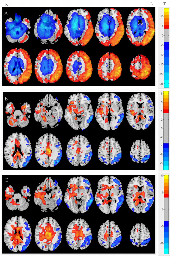
Chronic obstructive pulmonary disease can not only involve the lungs but also cause systemic adverse reactions, and extrapulmonary effects can aggravate its severity, in which brain damage is gradually gaining attention from researchers. In recent years, neurological imaging technology has made great progress, especially based on voxel morphometry, diffusion tensor imaging, magnetic resonance spectroscopy and functional magnetic resonance imaging, which can not only non-invasively observe and detect the morphology and structure of the body, but also provide microcosmic information such as functional metabolism. This article reviews the characteristics, possible mechanism and research status of brain structural and functional changes in patients with chronic obstructive pulmonary disease from the perspective of MRI multi-parameter imaging, with a view to the more extensive and mature application of MRI in the field of neuroscience and the study of central nervous system diseases. Thus, it provides a new method and important information for further study and study of the pathophysiological mechanism of chronic obstructive pulmonary disease, clinical diagnosis and treatment and prognosis of the disease.
Research progress of dual-energy CT in the diagnosis of thyroid nodules
ZHAO Zihan, CHEN Yingmin
2024, 47(3): 336-340.   doi: 10.12122/j.issn.1674-4500.2024.03.19
[Abstract](5) [FullText HTML](3) [PDF 1950KB](2)
Abstract:
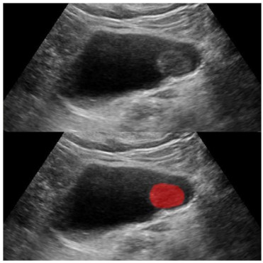
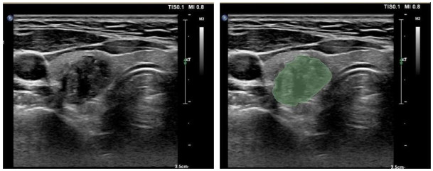
Thyroid nodules are now among the most frequent thyroid lesions, with a yearly rise in detection rates due to advancements in clinical diagnostic procedures and the widespread use of physical examinations. A non-invasive imaging assessment is crucial to the preoperative diagnosis of this condition. Dual-energy CT is a valuable addition to standard CT because it uses X-ray radiation from two distinct energy spectrums to offer a range of pictures with varied characteristics for the identification of thyroid nodules. This study provides an overview of dual-energy CT's multi-parameter imaging and basic principles. In order to generate ideas for the clinical application and future research of dual-energy CT in the diagnosis of thyroid nodules, the value of dual-energy CT in the diagnosis, differential diagnosis, and evaluation of lymph node metastasis in patients with thyroid nodules, as well as other potential application values, are reviewed concurrently.