


Value of quantitative 99mTc-MDP SPECT/CT in elderly osteoporotic spinal compression fractures
-
摘要:
目的 探讨定量单光子发射计算机断层扫描(SPECT)/CT获得的标准摄取值在老年骨质疏松性椎体压缩骨折的临床价值。 方法 回顾性分析2019年8月~2021年6月于我院就诊的65例骨质疏松性压缩骨折患者的临床资料。患者分别接受 99mTc-MDP 全身骨显像、局部SPECT/CT断层显像及磁共振成像。根据临床病史,将107个骨折椎体分为新鲜组(n=60)及陈旧组(n= 47),通过图像处理分析,分别计算两组椎体最大标准摄取值(SUVmax)以及平面图像和断层图像的靶区/非靶区比值(T/NT)并比较差异;绘制ROC曲线来获得SUVmax及T/NT的最佳临界值,并比较其诊断效能。 结果 新鲜组、陈旧组及正常组椎体SUVmax分别为43.20±17.17、10.71±3.17及9.78±2.95,新鲜组与陈旧组的差异有统计学意义(P < 0.01),而陈旧组与正常组的差异无统计学意义(P > 0.05);平面显像T/NT分别为2.22±0.70及1.41±0.45,差异有统计学意义(P < 0.01);断层显像T/NT分别为5.06±3.52及2.08±0.73,差异有统计学意义(P < 0.01)。通过绘制ROC曲线获得SUVmax、平面显像T/NT及断层显像T/NT的最佳临界值进行鉴别诊断,SUVmax的诊断敏感度、特异性、诊断一致性均高于平面显像T/NT及断层显像T/NT。 结论 定量SPECT/CT标准摄取值在老年骨质疏松性压缩骨折的诊断中具有重要的参考价值,为临床治疗提供客观依据。 -
关键词:
- 压缩骨折 /
- 单光子发射计算机断层摄影术 /
- SPECT/CT定量分析 /
- 骨质疏松 /
- 标准摄取值
Abstract:Objective To investigate the clinical value of standard uptake value obtained by quantitative single-photon-emission computed tomography (SPECT)/CT in elderly osteoporotic spinal compression fractures. Methods Sixty-five patients with osteoporotic compression fractures who underwent 99mTc-MDP whole body bone scan, SPECT/CT scan and magnetic resonance imaging(MRI) from August 2019 to June 2021 were enrolled. Based on the clinical history, 107 fractured vertebrae were divided into fresh group (n=60) and old group (n=47), the maximum standard uptake value (SUVmax) and the target/non-target ratio (T/NT) between the two groups was compared. Analyze the correlation between the SUVmax of the fractures and the injury time. ROC curves were plotted to obtain the cut-off values of SUVmax and T/NT and to compare their diagnostic value. Results The vertebral SUVmax was 43.20 ± 17.17, 10.71 ± 3.17 and 9.78 ± 2.95 in the fresh, old and normal groups, respectively, with significant differences between the fresh and old groups (P < 0.01) and no significant differences between the old and normal groups (P > 0.05). The planar T/NT was 2.22 ± 0.70 and 1.41 ± 0.45 in the fresh and old groups, the difference was statistically significant (P < 0.01). The tomo T/NT was 5.06±3.52 and 2.08±0.73 in the fresh and old groups, the difference was statistically significant (P < 0.01). The cut-off values of SUVmax, planar T/NT and tomo T/NT were obtained by plotting ROC curves for differential diagnosis, and the diagnostic sensitivity, specificity and diagnostic agreement of SUVmax were higher than planar T/ NT and tomo T/NT. Conclusion The standard uptake value of quantitative SPECT/CT has important reference value in the diagnosis of elderly osteoporotic spinal compression fractures, it can provide an objective basis for clinical treatment. -
Key words:
- compression fracture /
- SPECT/CT /
- quantitative analysis /
- osteoporosis /
- standard uptake value
-

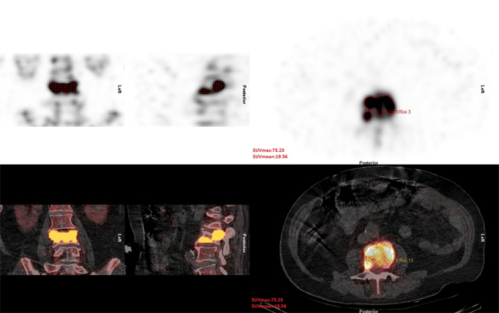
图 1 手动勾画靶区及非靶区并计算断层显像T/NT
Figure 1. Manually outline target and non-target areas and calculate tomo T/NT.

图 2 勾画受累椎体浓聚VOI并测量病灶SUVmax
Figure 2. Draw the concentrated VOI of the vertebrae and measure the SUVmax.

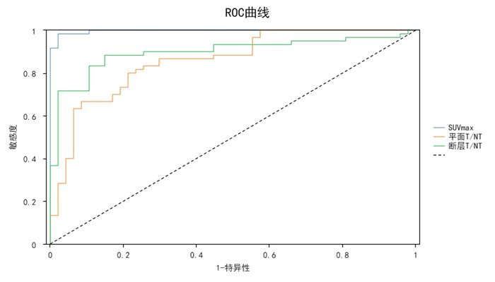
图 3 受累椎体SUVmax、平面显像T/NT及断层显像T/NT的ROC曲线
Figure 3. ROC curves of SUVmax, planar T/NT and tomo T/ NT of the vertebrae.
表 1 骨定量SUVmax、平面显像T/NT及断层显像T/NT的诊断效能对比
Table 1. Comparison of the diagnostic efficacy of bone quantification SUVmax, planar T/NT and tomo T/NT
方法 Cut-off AUC 敏感度 特异性 Kappa P SUVmax 18.500 0.997 0.983 0.979 0.943 < 0.01 平面T/NT 1.738 0.857 0.800 0.787 0.585 < 0.01 断层T/NT 2.573 0.897 0.883 0.851 0.715 < 0.01  下载: 导出CSV
-
[1] Hargunani R, Le Corroller T, Khashoggi K, et al. An overview of vertebroplasty: current status, controversies, and future directions[J]. Can Assoc Radiol J, 2012, 63(3): S11-7. [2] 蒋安清, 姜为民, 王根林, 等. 骨质疏松性椎体压缩骨折PKP术后再发椎体骨折的原因及相关危险因素分析[J]. 中国脊柱脊髓杂志, 2016, 26(10): 951-3. doi: 10.3969/j.issn.1004-406X.2016.10.15 [3] 边朝辉, 李国胜, 唐海. PKP治疗新鲜和陈旧骨质疏松性椎体压缩骨折[J]. 实用骨科杂志, 2015, 21(4): 366-70. https://www.cnki.com.cn/Article/CJFDTOTAL-SGKZ201504030.htm [4] 蔡金辉, 刘庆余, 曾玉蓉, 等. MRI预测经皮椎体强化术骨水泥椎间盘渗漏的价值[J]. 中国医学影像技术, 2017, 33(7): 1061-5. https://www.cnki.com.cn/Article/CJFDTOTAL-ZYXX201707033.htm [5] 闫伟, 杨莉. 骨质疏松性椎体压缩骨折的影像学诊断[J]. 中国CT和MRI杂志, 2017, 15(11): 135-7. doi: 10.3969/j.issn.1672-5131.2017.11.041 [6] Bailey DL, Willowson KP. An evidence- based review of quantitative SPECT imaging and potential clinical applications[J]. J Nucl Med, 2013, 54(1): 83-9. doi: 10.2967/jnumed.112.111476 [7] 冯飞, 邓介超, 唐海. 多发性骨质疏松性椎体压缩骨折中新鲜与陈旧骨折椎体的诊断与鉴别诊断[J]. 中华骨质疏松和骨矿盐疾病杂志, 2013, 6(2): 132-6. doi: 10.3969/j.issn.1674-2591.2013.02.007 [8] 胡正刚, 田俊松, 游玉峰. 高强度MRI与SPECT-CT在老年骨质疏松性椎体压缩骨折诊断中的价值研究[J]. 中国CT和MRI杂志, 2020, 18(10): 170-3. doi: 10.3969/j.issn.1672-5131.2020.10.051 [9] 康荘, 唐可, 肖艳, 等. 核素骨显像和MRI定位骨质疏松性椎体压缩骨折责任椎体的价值[J]. 中华创伤杂志, 2016(9): 789-93. doi: 10.3760/cma.j.issn.1001-8050.2016.09.005 [10] 周义, 赵御森, 张志敏, 等. SPECT/CT骨显像诊断新发骨质疏松性椎体压缩骨折的价值[J]. 中国医学影像技术, 2019, 35(4): 630-2. https://www.cnki.com.cn/Article/CJFDTOTAL-ZYXX201904055.htm [11] Armstrong IS, Hoffmann SA. Activity concentration measurements using a conjugate gradient (Siemens xSPECT) reconstruction algorithm in SPECT/CT[J]. Nucl Med Commun, 2016, 37(11): 1212-7. doi: 10.1097/MNM.0000000000000586 [12] Yamane T, Fukushima K, Shirotake S, et al. Test-retest repeatability of quantitative bone SPECT/CT[J]. Ann Nucl Med, 2021, 35(3): 338-46. doi: 10.1007/s12149-020-01568-2 [13] Hirschmann MT, Davda K, Rasch H, et al. Clinical value of combined single photon emission computerized tomography and conventional computer tomography (SPECT/CT) in sports medicine[J]. Sports Med Arthrosc Rev, 2011, 19(2): 174-81. doi: 10.1097/JSA.0b013e3181ec8707 [14] Okazaki T, Nakagawa H, Yagi K, et al. Bone scintigraphy for the diagnosis of the responsible level of osteoporotic vertebral compression fractures in percutaneous balloon kyphoplasty[J]. Clin Neurol Neurosurg, 2017, 152: 23-7. doi: 10.1016/j.clineuro.2016.11.007 [15] Moore AEB, Blake GM, Taylor KA, et al. Changes observed in radionuclide bone scans during and after teriparatide treatment for osteoporosis[J]. Eur J Nucl Med Mol Imaging, 2012, 39(2): 326-36. doi: 10.1007/s00259-011-1974-y [16] Matin P. The appearance of bone scans following fractures, including immediate and long-term studies[J]. J Nucl Med, 1979, 20 (12): 1227-31. [17] Spitz J, Lauer I, Tittel K. The age dependence of traumatically induced bone remodeling as studied in the bone scintigram[J]. Nuklearmedizin Nucl Med, 1991, 30(5): 155-60. doi: 10.1055/s-0038-1629568 [18] 石恩东, 张凯, 毛军胜, 等. 老年人骨质疏松椎体压缩性骨折不同治疗方法的临床分析[J]. 中华老年医学杂志, 2007, 26(9): 670-2. https://www.cnki.com.cn/Article/CJFDTOTAL-HLWK201602025.htm [19] 董智勇, 范学辉, 杨吉坤. 经皮椎体后凸成形术治疗后壁破裂的骨质疏松性椎体压缩骨折的安全性和有效性[J]. 中华骨与关节外科杂志, 2018, 11(4): 312-5. doi: 10.3969/j.issn.2095-9958.2018.04.007 [20] 杨辉, 张家立, 李奕军, 等. 骨质疏松骨折患者骨水泥分布类型对椎体强化术后再骨折的影响[J]. 实用医学杂志, 2019, 35(12): 1930-4. doi: 10.3969/j.issn.1006-5725.2019.12.015 [21] 杨静, 张斌青, 郭会利, 等. SPECT/CT对骨质疏松性椎体骨折的诊断价值及影像学特征[J]. 影像诊断与介入放射学, 2014, 23(5): 388-91. doi: 10.3969/j.issn.1005-8001.2014.05.006 -

 点击查看大图

计量
- 文章访问数: 198
- HTML全文浏览量: 106
- PDF下载量: 10
- 被引次数: 0