


Application of 99mTc-MIBI SPECT/CT and neck ultrasound in the preoperative diagnosis of primary hyperparathyroidism
-
摘要:
目的 探究99mTc-MIBI SPECT/CT及颈部超声在原发性甲状旁腺功能亢进症(PHPT)术前诊断中的应用。 方法 收集92例疑似PHPT患者资料,评价99mTc-MIBI SPECT/CT及颈部超声检查的诊断效能,分析99mTc-MIBI摄取情况与术前甲状旁腺素(PTH)及病灶最大直径的关系。 结果 颈部超声、99mTc-MIBI平面显像、SPECT/CT显像及联合检查敏感度分别为58.70%、71.74%、85.87%、93.48%,其中SPECT/CT显像、联合检查敏感度分别高于超声、99mTc-MIBI平面显像(P<0.05);99mTc-MIBI平面显像阳性组患者术前PTH和术后病灶最大直径均高于阴性组(P<0.05),且阳性组患者早期相、延迟相病灶与正常组织的摄取比值T/Ne、T/Nd分别与术前PTH、术后病灶最大直径呈正相关关系(P<0.05);ROC曲线显示,术前PTH预测99mTc-MIBI平面显像阳性的曲线下面积为0.780,95%CI为0.675~0.886,敏感度为80.30%,特异性为69.23%。 结论 患者术前PTH、病灶大小分别与99mTc-MIBI摄取呈正相关关系,99mTc-MIBI SPECT/CT及超声联合对PHPT术前诊断具有较高的应用价值。 -
关键词:
- 原发性甲状旁腺功能亢进症 /
- 99mTc-MIBI /
- SPECT/CT /
- 超声 /
- 术前诊断
Abstract:Objective To investigate the application of 99mTc-MIBI SPECT/CT and neck ultrasound in the preoperative diagnosis of primary hyperparathyroidism. Methods The data of 92 patients with suspected primary hyperparathyroidism were collected. The diagnostic efficiency of 99mTc-MIBI SPECT/CT and neck ultrasound was evaluated. The relationship between 99mTc-MIBI uptake and preoperative parathyroid hormone (PTH), the maximum diameter of lesions was analyzed. Results The sensitivity values of neck ultrasound, 99mTc-MIBI plane imaging, SPECT/CT imaging and combined detection were 58.70%, 71.74%, 85.87% and 93.48%, respectively. The sensitivity of SPECT/CT imaging and combined detection was higher than that of ultrasound and 99mTc-MIBI plane imaging (P<0.05). The preoperative PTH and postoperative maximum diameter of lesions in positive 99mTc-MIBI plane imaging group were higher than those in negative group (P<0.05). The uptake ratios T/Ne and T/Nd in early phase lesions, delayed phase lesions and normal tissues were positively correlated with preoperative PTH and postoperative maximum diameter of lesions (P<0.05). ROC curves showed that the AUC, 95%CI, sensitivity and specificity of preoperative PTH for predicting positive 99mTc-MIBI plane imaging were 0.780, 0.675-0.886, 80.30% and 69.23%, respectively. Conclusion The preoperative PTH and lesion size are positively correlated with 99mTc-MIBI uptake. The combination of 99mTc-MIBI SPECT/CT and ultrasound has high application value in the preoperative diagnosis of PHPT. -
Key words:
- primary hyperparathyroidism /
- 99mTc-MIBI /
- SPECT/CT /
- ultrasound /
- preoperative diagnosis
-

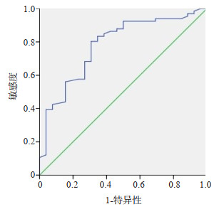
图 1 原发性甲状旁腺功能亢进症影像学表现
A:甲状腺彩超显示甲状腺左叶外侧缘弱回声结节,边界清晰,内可见点线状血流信号;B:甲状旁腺双时相显像中早期相和延迟相示甲状腺左叶下极区域显像剂异常浓聚;C:120 min时SPECT/CT断层融合显像示甲状腺左叶中份外侧缘低密度软组织结节影,边界清晰,显像剂异常浓聚.
Figure 1. Imaging features of primary hyperparathyroidism.

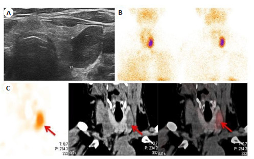
图 2 术前血清PTH预测99mTc-MIBI平面显像结果的ROC曲线
Figure 2. ROC curves of preoperative serum PTH for predicting the results of 99mTc- MIBI plane imaging.
表 1 影像学检查的诊断效能
Table 1. Diagnostic efficiency of imaging examinations (%)
检查方法 敏感度 敏感度 58.70(54/92) 99mTc-MIBI平面显像 71.74(66/92) SPECT/CT显像 85.87(79/92) 超声+99mTc-MIBI+SPECT/CT显像 93.48(86/92) χ2 37.508 P <0.001  下载: 导出CSV
表 2 99mTc-MIBI平面显像阳/阴性患者一般资料的比较
Table 2. Comparison of general data between patients with positive/negative 99mTc-MIBI plane imaging (Mean±SD)
组別 男/女(n) 年龄(岁) 术前PTH(ng/L) 术后病灶最大直径(mm) 阳性组(n=66) 21/45 55.13±13.27 265.48±32.96 1.98±0.52 阴性组(n=26) 7/19 53.50±10.45 105.67±21.37 1.49±0.34 t/χ2 0.211 0.561 22.861 4.438 P 0.646 0.576 <0.001 <0.001 PTH:甲状旁腺素.
下载: 导出CSV
-
[1] 陈健, 刘康, 张锡平, 等. 原发性甲状旁腺功能亢进合并胫腓骨骨折1例[J]. 中南医学科学杂志, 2021, 49(1): 106-9. https://www.cnki.com.cn/Article/CJFDTOTAL-HYYY202101021.htm [2] Kowalski GJ, Buła G, Żądło D, et al. Primary hyperparathyroidism [J]. Endokrynol Pol, 2020, 71(3): 260-70. doi: 10.5603/EP.a2020.0028 [3] 旷鹏昊, 吴国洋, 傅锦波, 等. 经口入路腔镜甲状旁腺切除术在原发性甲状旁腺功能亢进症治疗中的应用[J]. 中华普通外科杂志, 2021, 36 (12): 941-2. doi: 10.3760/cma.j.cn113855-20210315-00163-1 [4] 张晋, 韩建立, 赵浩亮, 等. 52例原发性甲状旁腺功能亢进症的外科诊疗分析[J]. 现代消化及介入诊疗, 2019, 24(S2): 1770. https://cdmd.cnki.com.cn/Article/CDMD-10114-1019612186.htm [5] 宋桉, 王鸥, 刘春晓, 等. 甲状旁腺四维CT在原发性甲状旁腺功能亢进症术前定位中的诊断价值[J]. 中华内科杂志, 2020, 59(10): 788-95. doi: 10.3760/cma.j.cn112138-20200413-00367 [6] Hofer T, Kronbichler J, Huber H, et al. 18F-choline PET/CT, MRI, and software-based image fusion analysis in patients with primary hyperparathyroidism[J]. Clin Nucl Med, 2021, 46(9): 710-6. [7] Mallick R, Malik J, Yip L, et al. Novel findings on SPECT-CT Tc-99 sestamibi imaging for primary hyperparathyroidism[J]. J Surg Res, 2020, 252: 216-21. doi: 10.1016/j.jss.2020.03.014 [8] Liu YM, Dang YH, Huo L, et al. Preoperative localization of adenomas in primary hyperparathyroidism: the value of 11 C-choline PET/CT in patients with negative or discordant findings on ultrasonography and 99m Tc-sestamibi SPECT/CT[J]. J Nucl Med, 2020, 61(4): 584-9. doi: 10.2967/jnumed.119.233213 [9] Newey PJ. Hereditary primary hyperparathyroidism[J]. Endocrinol Metab Clin North Am, 2021, 50(4): 663-81. doi: 10.1016/j.ecl.2021.08.003 [10] de Nova JLM, Sampedro-Nuñez M, Huguet-Moreno I, et al. A practical approach to normocalcemic primary hyperparathyroidism [J]. Endocrine, 2021, 74(2): 235-44. doi: 10.1007/s12020-021-02845-4 [11] 孔祥崇, 赵颖, 李霞. 超声引导下微波消融治疗继发性甲状旁腺功能亢进的疗效与价值[J]. 医学影像学杂志, 2020, 30(7): 1161-5. https://www.cnki.com.cn/Article/CJFDTOTAL-XYXZ202007009.htm [12] Pretet V, Rotania M, Helali M, et al. 18 F-fluorocholine PET and multiphase CT integrated in dual modality PET/4D-CT for preoperative evaluation of primary hyperparathyroidism[J]. J Clin Med, 2020, 9(6): 2005. doi: 10.3390/jcm9062005 [13] Ballester Vázquez E, Pérez García JI, López Mora DA, et al. Identification of occult adenomas in primary hyperparathyroidism with 18F-fluorocholine PET/CT[J]. Cir Esp (Engl Ed), 2020, 98(7): 395-402. doi: 10.1016/j.ciresp.2020.01.003 [14] Md SF, Md CC, Christophe Trésallet MD P, et al. Utility of a second 99mTc-MIBI scintigraphy before reoperation for patients with persistent sporadic primary hyperparathyroidism: results of a retrospective multicenter study[J]. Ann Surg Oncol, 2020, 27(10): 3831-9. doi: 10.1245/s10434-020-08428-3 [15] Gungor S, Dede F, Can B, et al. The value of parathyroid scintigraphy on lesion detection in patients with normocalcemic primary hyperparathyroidism[J]. Revista Española De Med Nucl E Imagen Mol Engl Ed, 2022, 41(2): 86-90. [16] 季艳会译, 孟召伟. 甲状旁腺核素显像在原发性甲状旁腺功能亢进症外科手术治疗中的作用[J]. 中华核医学与分子影像杂志, 2018, 38 (7): 508-14. [17] Agirre L, de la Quintana A, Martínez G, et al. Surgical results and the location of pathological glands in the treatment of primary sporadic hyperparathyroidism with negative preoperative 99mTc-sestamibi scintigraphy[J]. Cirugía Española Engl Ed, 2022, 100(1): 18-24. doi: 10.1016/j.ciresp.2020.11.011 [18] Lee SH, Shin E, Ha SJ, et al. Is dual-phase SPECT/CT with 99mTc-sestamibi better than single-phase SPECT/CT for lesion localization in patients with hyperparathyroidism?[J]. Medicine, 2020, 99(19): e19989. doi: 10.1097/MD.0000000000019989 [19] Wei WJ, Shen CT, Song HJ, et al. Comparison of SPET/CT, SPET and planar imaging using 99mTc-MIBI as independent techniques to support minimally invasive parathyroidectomy in primary hyperparathyroidism: a meta-analysis[J]. Hell J Nucl Med, 2015, 18 (2): 127-35. [20] Elif Ö, Mustafa G, Uğuray A, et al. Comparison of 99mTc-MIBI planar scintigraphy, SPET/CT and ultrasonography in detection of parathyroid adenoma in patients with primary hyperparathyroidism [J]. Hellenic J Nucl Med, 2020, 23(1): 21-6. [21] 张莹莹, 韩娜, 武凤玉, 等. 99Tc m-MIBI SPECT/CT显像在原发性甲状旁腺功能亢进症术前诊断中的价值及影响因素[J]. 中华核医学与分子影像杂志, 2021, 41(6): 345-9. doi: 10.3760/cma.j.cn321828-20200408-00142 [22] Asseeva P, Paladino NC, Guerin C, et al. Value of 123I/ 99mTc-sestamibi parathyroid scintigraphy with subtraction SPECT/ CT in primary hyperparathyroidism for directing minimally invasive parathyroidectomy[J]. Am J Surg, 2019, 217(1): 108-13. doi: 10.1016/j.amjsurg.2018.06.027 [23] 李永亮, 于亚萍, 雒瑾, 等. 原发性甲状旁腺功能亢进99mTc-MIBI SPECT/CT显像与血清PTH及钙磷水平相关性的研究[J]. 临床放射学杂志, 2021, 40(8): 1629-32. https://www.cnki.com.cn/Article/CJFDTOTAL-LCFS202108038.htm -

 点击查看大图
计量
- 文章访问数: 260
- HTML全文浏览量: 154
- PDF下载量: 5
- 被引次数: 0