


Investigation and analysis of pelvic tilt in MRI examination
-
摘要:
目的 探索MRI检查图像患者骨盆倾斜的情况。 方法 回顾性分析2013~2016年因各种妇科疾病行MRI检查的患者,构建骨盆三维模型,通过三维模型的正前面观判定患者骨盆位置是否有倾斜,在二维图像上测量倾斜的角度。 结果 在461例研究对象中,106例(23%)存在骨盆位置的倾斜,主要是左右倾斜,其中向右倾斜28例,偏转(6.28±2.14)°,向左倾斜有78例,偏转(5.88±1.67)°。 结论 常规MRI平卧位检查时存在骨盆倾斜的情况,导致患者正中矢状面选取存在偏差,利用正中矢状面进行的测量就存在偏差,引起骨盆左右倾斜的原因可能与骨盆旋转有关,而利用三维模型测量可以有效避免因骨盆偏斜引起的偏差。 Abstract:Objective To explore the pelvic tilt situation of patients who have done MRI examination. Methods Patients who have performed MRI tests for various gynecological diseases from 2013 to 2016 were included. After we reconstructed 3D model of patient's pelvis based on MRI images, frontal view of the three-dimensional model was used to determine whether the position of the pelvis was tilted or not. The angle of inclination was measured on the two-dimensional image. Results In the 461 subjects, 106 (23%) patients had a tilt pelvic position, mainly left-right tilt type. 28 ones were right tilt with average deflection of (6.28±2.14)°. 78 were leftward tilt with average deflection of (5.88±1.67)°. Conclusions The pelvic tilt in the cross-sectional view of the routine MRI supine test results in deviation of the median sagittal plane of the patient. The pelvic tilt may result from pelvic rotation. The use of three-dimensional model can effectively avoid bias due to pelvic tilt. -
Key words:
- magnetic resonance imaging /
- pelvic organ prolapse /
- pelvic /
- tilt /
- median sagittal plane /
- 3D models
-
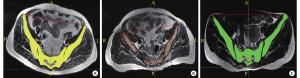
图 2 体位二维示意图
A: 体位左偏, L-R-P-A:左侧-右侧-后面-前面, 调整后坐标轴在原有坐标轴基础上左偏; B: 体位右偏, L-R-P-A:左侧-右侧-后面-前面, 调整后坐标轴在原有坐标轴基础上右偏; C: 体位正常, L-R-P-A: 左侧-右侧-后面-前面, 调整后坐标轴与原有坐标轴基本重合.
表 1 骨盆不同方向偏转的一般统计情况
患者体位 例数 最大值(°) 最小值(°) 均数±标准差(°) 偏斜 106 10.76 2.39 5.98±1.80 左偏 78 10.76 2.39 5.88±1.67 右偏 28 10.36 3.19 6.28±2.14  下载: 导出CSV
-
[1] 王 毅, 庹 勇, 何 敏, 等. 超声与磁共振对宫颈癌术前精准分期的价值及核磁弥散序列在随访过程中的意义分析[J]. 实用医院临床杂志, 2018, 36(1): 136-8 doi: 10.3969/j.issn.1672-6170.2018.01.043 [2] 史 珊, 罗 萍, 赵彦萍, 等. 直肠脱垂合并小肠疝的排粪造影及静态MRI征象初步分析[J]. 中国中西医结合影像学杂志, 2018, 15(2): 197-200 doi: 10.3969/j.issn.1672-0512.2018.02.031 [3] 张丹丹, 庄治国, 耿小川, 等. 肿瘤直径联合ADC值对不同亚型乳腺癌NAC疗效的评估价值[J]. 实用放射学杂志, 2018, 33(3): 366-9 doi: 10.3969/j.issn.1002-1671.2018.03.010 [4] 江 勇, 郭 华, 蔡新宇. MRI在评估孕妇分娩后早期盆底功能障碍中作用分析[J]. 中国CT和MRI杂志, 2018, 15(2): 51-3 doi: 10.3969/j.issn.1672-5131.2018.02.016 [5] Gousse AE, Barbaric ZL, Satlr MH, et al. Dynamic half Fourier acquisition single shot turbo spin-echo magnetic resonance imaging for evaluating the female pelvis[J]. J Urol, 2000, 164(6): 1606-13 [6] Colaiacomo MC, Masselli G, Polettini E, et al. Dynamic Mr imaging of the pelvic floor: a pictorial review[J]. Radiographics, 2009, 29(3): e35-9 doi: 10.1148/rg.e35 [7] Woodfield CA, Hampton BS, Sung V, et al. Magnetic resonance imaging of pelvic organ prolapse: comparing pubo-coccygeal and mid-pubic lines with clinical staging[J]. Int Urogynecol J Pelvic Floor Dysfunct, 2009, 20(7): 695-701 [8] Lienemann A, Sprenger D, Janssen U, et al. Assessment of pelvic organ descent by use of functional cine-MRI: which reference line should be used[J]. Neurourol Urodyn, 2004, 23(1): 33-7 doi: 10.1002/nau.v23:1 [9] Fauconnier A, Zareski E, Bader G, et al. Dynamic magnetic resonance imaging for grading pelvic organ prolapse by use of a new reference line[C]. Paris: ICS/IUGA annual meeting, 1994. [10] Ratnatunga K, Deen K, Prasad R. A protocol for dynamic magnetic resonance imaging of the pelvic floor[J]. Indian J Gastroenterol, 2013, 32(1): 43-8 doi: 10.1007/s12664-012-0223-z [11] Lakeman MM, Zijta FM, Peringa J, et al. Dynamic magnetic resonance imaging to quantify pelvic organ prolapse: reliability of assessment and correlation with clinical findings and pelvic floor symptoms[J]. Int Urogynecol J, 2012, 23(11): 1547-54 doi: 10.1007/s00192-012-1772-5 [12] Larson KA, Hsu YL. Magnetic resonance imaging based three-dimensional model of anterior vaginal wall position at rest and maximal strain in women with and without prolapse[J]. Int Urogynecol J Pelvic Floor Dysfunct, 2010, 21(1): 103-9 doi: 10.1007/s00192-009-0994-7 [13] Bump RC, Mattiasson A, Bø K, et al. The standardization of terminology of female pelvic organ prolapse and pelvic floor dysfunction[J]. Am J Obstet Gynecol, 1996, 175(1): 10-7 doi: 10.1016/S0002-9378(96)70243-0 [14] Yang A, Mostwin JL, Rosenshein NB, et al. Pelvic floor descent in women: dynamic evaluation with fast Mr imaging and cinematic display[J]. Radiology, 1991, 179(1): 25-33 doi: 10.1148/radiology.179.1.2006286 [15] Kruyt RH, Delemarre JB, Doornbos J, et al. Normal anorectum: dynamic Mr imaging anatomy[J]. Radiology, 1991, 179(1): 159-63 doi: 10.1148/radiology.179.1.2006269 [16] Fauconnier A, Zareski E, Abichedid J, et al. Dynamic magnetic resonance imaging for grading pelvic organ prolapse according to the International Continence Society classification: which line should be used[J]. Neurourol Urodyn, 2008, 27(3): 191-7 doi: 10.1002/(ISSN)1520-6777 [17] Pannu HK. Dynamic Mr imaging of female organ prolapse[J]. Radiol Clin North Am, 2003, 41(2): 409-23 doi: 10.1016/S0033-8389(02)00120-3 [18] Boyadzhyan L, Raman SS, Raz S. Role of static and dynamic Mr imaging in surgical pelvic floor dysfunction[J]. Radiographics, 2008, 28(4): 949-67 doi: 10.1148/rg.284075139 [19] 刘 萍, 郭传家, 陈春林, 等. 基于磁共振成像的在体女性骨盆数字化三维模型的构建[J]. 实用妇产科杂志, 2013, 29(6): 423-6 doi: 10.3969/j.issn.1003-6946.2013.06.009 [20] 黄 璐, 陈春林, 刘 萍, 等. 在体女性主韧带及宫骶韧带数字化三维模型构建及其意义[J]. 中国实用妇科与产科杂志, 2014, 30(6): 453-6 [21] 刘 萍, 陈若兰, 郭传家, 等. 基于磁共振成像的肛门外括约肌数字化三维模型构建[J]. 实用医学杂志, 2014, 30(2): 177-9 doi: 10.3969/j.issn.1006-5725.2014.02.004 [22] 刘 萍, 陈若兰, 陈春林, 等. MRI三维模型用于健康未育青年女性盆底肌群的解剖形态学特点分析[J]. 中华妇产科杂志, 2014, 49(5): 336-40 doi: 10.3760/cma.j.issn.0529-567x.2014.05.004 [23] 刘 萍, 唐连, 陈春林, 等. 盆腔器官脱垂盆腔器官及肛提肌动态三维模型构建方法研究[J]. 中国实用妇科与产科杂志, 2016, 14(4): 341-6 -


 点击查看大图

图(3) / 表(1)
计量
- 文章访问数: 1517
- HTML全文浏览量: 550
- PDF下载量: 22
- 被引次数: 0