


Advances in data processing and application of imaging transcriptomics
-
摘要: 随着全脑转录组数据,即全脑基因表达图谱(如艾伦人类脑图谱)的出现,影像转录组学为理解大脑分子尺度上的空间分布与宏观神经影像表型之间的关系开辟了新的机会。越来越多的研究揭示了基因表达的特定时空模式与大脑结构和功能的不同属性之间的联系。本文介绍了影像转录组学领域广泛使用的基因表达数据集,以及转录组数据处理的基本步骤和常用的工具箱,并概述了将基因表达数据与影像数据相关联的基本工作流程和三大类分析方法。近年来,影像转录组学已被广泛应用于理解脑神经发育和各类神经精神疾病中; 但该领域仍处于新兴阶段,必须克服一些方法学挑战以确保研究结果的稳健性。随着该领域的发展和现有方法的改进,未来的研究可以与越来越全面和精确的将转录组图谱数据相结合,这将会为识别体内观察到的与疾病相关的大脑变化的分子相关性提供一个强大可靠的框架。Abstract: With the advent of brain-wide transcriptomics data, i.e. brain-wide gene expression atlases such as the Allen human brain atlas, imaging transcriptomics has opened new opportunities for understanding the relationship between spatial variations on molecular scale of the brain and macroscopic neuroimaging phenotypes. A growing body of literature is demonstrating relationships between gene expression and different properties of brain structure and function. This article introduces the gene expression dataset widely used in the field of imaging transcriptomics, as well as the basic steps and the commonly used toolbox for transcriptomics data processing, and outlines the basic workflow and three kinds of analysis methods for associating gene expression data with image data. In recent years, imaging transcriptomics has been widely used to understand brain neurodevelopment and various neuropsychiatric disorders. However, the field is still nascent, and several methodological challenges must be overcome to ensure the robustness of the findings. As the filed develops and existing methodologies are refined, future studies can be combined with increasingly more comprehensive and precise transcriptional atlas data, which will offer a powerful and reliable framework for identifying the molecular correlates of disease-related brain changes observed in vivo.
-
Key words:
- imaging transcriptomics /
- Allen human brain atlas /
- gene expression /
- neuroimaging /
- brain disorders
-

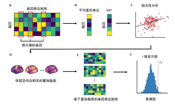
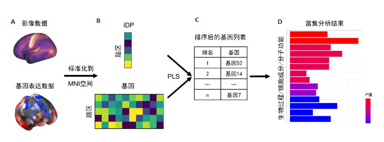
图 1 影像转录组学的一般分析流程
IDP: 影像衍生表型; MNI: 蒙特利尔神经科学研究所; PLS: 偏最小二乘法.
Figure 1. General analysis workflow of imaging transcriptomics.

图 3 考虑空间自相关的影像转录组学分析方法
Figure 3. Imaging transcriptomics analysis methods considering spatial autocorrelation.
表 1 转录组数据处理步骤和方法
Table 1. Transcriptomics data processing steps and methods
处理步骤 目标 可选择的方法 基因注释 使用可用的最新信息将探针重新分配给基因 可使用的软件和工具:Re-annotator[ 27 ]、BioMart (http://www.biomart.org/)、AnnotationDbi (https://bioconductor.org/packages/AnnotationDbi)数据过滤 采用适当的控制来区分表达信号和噪声,提高数据可靠性 在至少k%的样本中排除掉信号强度没有超过背景噪声的探针,阈值k%可自定义 探针选择 当有多个探针注释到同一个基因时,对探针进行选择 (1)计算一个基因的所有可用探针的平均值,(2)选择具有最大强度的探针,(3)选择跨大脑区域方差最大的探针,(4)选择6个供体大脑中差异稳定性最高的探针,(5) 选择与其他探针平均相关性最高的探针 样本分配 将组织样本映射到影像数据中的感兴趣区域 生成供体特异性的分割方案,分别分配左/右脑、皮层/皮下的样本,并设置2 mm的距离阈值,以确保不会分配距离分区超过给定阈值的样本 数据标准化 减少个体间差异 分别对每个供体的基因表达数据进行标准化,可采用z分数标准化、sigmoid标准化等方法 基因过滤 减少供体特异性差异,并根据研究问题选择感兴趣的基因 (1)选择差异稳定性值高的基因,(2)基于GWASs研究选择疾病特异性基因,(3)基于先验假设选择基因 GWASs: 全基因组关联研究.  下载: 导出CSV
-
[1] Brammer M. The role of neuroimaging in diagnosis and personalized medicine: current position and likely future directions[J]. Dialogues Clin Neurosci, 2009, 11(4): 389-96. doi: 10.31887/DCNS.2009.11.4/mbrammer [2] Opel N, Goltermann J, Hermesdorf M, et al. Cross-disorder analysis of brain structural abnormalities in six major psychiatric disorders: a secondary analysis of mega- and Meta- analytical findings from the ENIGMA consortium[J]. Biol Psychiatry, 2020, 88(9): 678-86. doi: 10.1016/j.biopsych.2020.04.027 [3] Patel Y, Parker N, Shin J, et al. Virtual histology of cortical thickness reveals shared neurobiology across six psychiatric disorders[J]. Biol Psychiatry, 2020, 87(9): S239-40. [4] Cassidy PJ, Radda GK. Molecular imaging perspectives[J]. J R Soc Interface, 2005, 2(3): 133-44. doi: 10.1098/rsif.2005.0040 [5] Arnatkevičiūtė A, Fulcher BD, Fornito A. A practical Guide to linking brain-wide gene expression and neuroimaging data[J]. NeuroImage, 2019, 189: 353-67. doi: 10.1016/j.neuroimage.2019.01.011 [6] Fornito A, Arnatkevičiūtė A, Fulcher BD. Bridging the gap between connectome and transcriptome[J]. Trends Cogn Sci, 2019, 23(1): 34- 50. doi: 10.1016/j.tics.2018.10.005 [7] Arnatkeviciute A, Fulcher BD, Bellgrove MA, et al. Where the genome meets the connectome: understanding how genes shape human brain connectivity[J]. NeuroImage, 2021, 244: 118570. doi: 10.1016/j.neuroimage.2021.118570 [8] Bigos KL, Weinberger DR. Imaging genetics-days of future past[J]. NeuroImage, 2010, 53(3): 804-9. doi: 10.1016/j.neuroimage.2010.01.035 [9] Baaré WFC, Hulshoff Pol HE, Boomsma DI, et al. Quantitative genetic modeling of variation in human brain morphology[J]. Cereb Cortex, 2001, 11(9): 816-24. doi: 10.1093/cercor/11.9.816 [10] Thompson PM, Ge T, Glahn DC, et al. Genetics of the connectome [J]. NeuroImage, 2013, 80: 475-88. doi: 10.1016/j.neuroimage.2013.05.013 [11] Arnatkeviciute A, Fulcher BD, Oldham S, et al. Genetic influences on hub connectivity of the human connectome[J]. Nat Commun, 2021, 12: 4237. doi: 10.1038/s41467-021-24306-2 [12] Elliott LT, Sharp K, Alfaro-Almagro F, et al. Genome-wide association studies of brain imaging phenotypes in UK Biobank[J]. Nature, 2018, 562(7726): 210-6. doi: 10.1038/s41586-018-0571-7 [13] Smith SM, Douaud G, Chen W, et al. An expanded set of genomewide association studies of brain imaging phenotypes in UK Biobank [J]. Nat Neurosci, 2021, 24(5): 737-45. doi: 10.1038/s41593-021-00826-4 [14] Choi JK, Kim SC. Environmental effects on gene expression phenotype have regional biases in the human genome[J]. Genetics, 2007, 175(4): 1607-13. doi: 10.1534/genetics.106.069047 [15] Cole SW. Social regulation of human gene expression[J]. Curr Dir Psychol Sci, 2009, 18(3): 132-7. doi: 10.1111/j.1467-8721.2009.01623.x [16] Hawrylycz MJ, Lein ES, Guillozet-Bongaarts AL, et al. An anatomically comprehensive atlas of the adult human brain transcriptome[J]. Nature, 2012, 489(7416): 391-9. doi: 10.1038/nature11405 [17] Hawrylycz M, Miller JA, Menon V, et al. Canonical genetic signatures of the adult human brain[J]. Nat Neurosci, 2015, 18(12): 1832-44. doi: 10.1038/nn.4171 [18] Shen EH, Overly CC, Jones AR. The Allen Human Brain Atlas: comprehensive gene expression mapping of the human brain[J]. Trends Neurosci, 2012, 35(12): 711-4. doi: 10.1016/j.tins.2012.09.005 [19] French L, Paus T. A FreeSurfer view of the cortical transcriptome generated from the Allen Human Brain Atlas[J]. Front Neurosci, 2015, 9: 323. [20] Gorgolewski KJ, Varoquaux G, Rivera G, et al. NeuroVault. org: a web-based repository for collecting and sharing unthresholded statistical maps of the human brain[J]. Front Neuroinform, 2015, 9: 8. [21] Rizzo G, Veronese M, Expert P, et al. MENGA: a new comprehensive tool for the integration of neuroimaging data and the Allen human brain transcriptome atlas[J]. PLoS One, 2016, 11(2): e0148744. doi: 10.1371/journal.pone.0148744 [22] Bhagwat N, Barry A, Dickie EW, et al. Understanding the impact of preprocessing pipelines on neuroimaging cortical surface analyses [J]. Gigascience, 2021, 10(1): 155. doi: 10.1093/gigascience/giaa155 [23] Oldham S, Iūtė AA, Smith RE, et al. The efficacy of different preprocessing steps in reducing motion-related confounds in diffusion MRI connectomics[J]. NeuroImage, 2020, 222: 117252. doi: 10.1016/j.neuroimage.2020.117252 [24] Carp J. On the plurality of (methodological) worlds: estimating the analytic flexibility of FMRI experiments[J]. Front Neurosci, 2012, 10(6): 149. [25] Parkes L, Fulcher B, Yücel M, et al. An evaluation of the efficacy, reliability, and sensitivity of motion correction strategies for restingstate functional MRI[J]. NeuroImage, 2018, 171: 415-36. doi: 10.1016/j.neuroimage.2017.12.073 [26] Markello RD, Arnatkeviciute A, Poline JB, et al. Standardizing workflows in imaging transcriptomics with the abagen toolbox[J]. eLife, 2021, 10: e72129. doi: 10.7554/eLife.72129 [27] Arloth J, Bader DM, Röh S, et al. re- annotator: annotation pipeline for microarray probe sequences[J]. PLoS One, 2015, 10(10): e0139516. doi: 10.1371/journal.pone.0139516 [28] Rizzo G, Veronese M, Heckemann RA, et al. The predictive power of brain mRNA mappings for in vivo protein density: a positron emission tomography correlation study[J]. J Cereb Blood Flow Metab, 2014, 34(5): 827-35. doi: 10.1038/jcbfm.2014.21 [29] Veronese M, Bodini B, García-Lorenzo D, et al. Quantification of[11C]PIB PET for imaging myelin in the human brain: a test-retest reproducibility study in high-resolution research tomography[J]. J Cereb Blood Flow Metab, 2015, 35(11): 1771-82. doi: 10.1038/jcbfm.2015.120 [30] Beliveau V, Ganz M, Feng L, et al. A high-resolution in vivo atlas of the human brain's serotonin system[J]. J Neurosci, 2017, 37(1): 120-8. doi: 10.1523/JNEUROSCI.2830-16.2016 [31] Komorowski A, James GM, Philippe C, et al. Association of protein distribution and gene expression revealed by PET and post-mortem quantification in the serotonergic system of the human brain[J]. Cereb Cortex, 2016, 27(1): 117-30. [32] Gryglewski G, Seiger R, James GM, et al. Spatial analysis and high resolution mapping of the human whole-brain transcriptome for integrative analysis in neuroimaging[J]. NeuroImage, 2018, 176: 259-67. doi: 10.1016/j.neuroimage.2018.04.068 [33] Ritchie J, Pantazatos SP, French L. Transcriptomic characterization of MRI contrast with focus on the T1-w/T2-w ratio in the cerebral cortex[J]. NeuroImage, 2018, 174: 504-17. doi: 10.1016/j.neuroimage.2018.03.027 [34] Shin J, French L, Xu T, et al. Cell-specific gene-expression profiles and cortical thickness in the human brain[J]. Cereb Cortex, 2017, 28 (9): 3267-77. [35] Liu F, Tian HJ, Li J, et al. Altered voxel-wise gray matter structural brain networks in schizophrenia: association with brain genetic expression pattern[J]. Brain Imaging Behav, 2019, 13(2): 493-502. doi: 10.1007/s11682-018-9880-6 [36] Patania A, Selvaggi P, Veronese M, et al. Topological gene expression networks recapitulate brain anatomy and function[J]. Netw Neurosci, 2019, 3(3): 744-62. doi: 10.1162/netn_a_00094 [37] Komorowski A, Weidenauer A, Murgaš M, et al. Association of dopamine D2/3 receptor binding potential measured using PET and [11C]-(+)-PHNO with post-mortem DRD2/3 gene expression in the human brain[J]. NeuroImage, 2020, 223: 117270. doi: 10.1016/j.neuroimage.2020.117270 [38] Patel Y, Shin J, Drakesmith M, et al. Virtual histology of multi-modal magnetic resonance imaging of cerebral cortex in young men[J]. NeuroImage, 2020, 218: 116968. doi: 10.1016/j.neuroimage.2020.116968 [39] Nørgaard M, Beliveau V, Ganz M, et al. A high-resolution in vivo atlas of the human brain's benzodiazepine binding site of GABAA receptors[J]. NeuroImage, 2021, 232: 117878. doi: 10.1016/j.neuroimage.2021.117878 [40] Richiardi J, Altmann A, Milazzo AC, et al. BRAIN NETWORKS. Correlated gene expression supports synchronous activity in brain networks[J]. Science, 2015, 348(6240): 1241-4. doi: 10.1126/science.1255905 [41] Fulcher BD, Fornito A. A transcriptional signature of hub connectivity in the mouse connectome[J]. Proc Natl Acad Sci USA, 2016, 113(5): 1435-40. doi: 10.1073/pnas.1513302113 [42] Arnatkevičiūtė A, Fulcher BD, Pocock R, et al. Hub connectivity, neuronal diversity, and gene expression in the Caenorhabditis elegans connectome[J]. PLoS Comput Biol, 2018, 14(2): e1005989. doi: 10.1371/journal.pcbi.1005989 [43] Oldham MC, Konopka G, Iwamoto K, et al. Functional organization of the transcriptome in human brain[J]. Nat Neurosci, 2008, 11(11): 1271-82. doi: 10.1038/nn.2207 [44] Forest M, Iturria-Medina Y, Goldman JS, et al. Gene networks show associations with seed region connectivity[J]. Hum Brain Mapp, 2017, 38(6): 3126-40. doi: 10.1002/hbm.23579 [45] Burt JB, Demirtaş M, Eckner WJ, et al. Hierarchy of transcriptomic specialization across human cortex captured by structural neuroimaging topography[J]. Nat Neurosci, 2018, 21(9): 1251-9. doi: 10.1038/s41593-018-0195-0 [46] Lau HYG, Fornito A, Fulcher BD. Scaling of gene transcriptional gradients with brain size across mouse development[J]. NeuroImage, 2021, 224: 117395. doi: 10.1016/j.neuroimage.2020.117395 [47] Markello RD, Misic B. Comparing spatial null models for brain maps [J]. NeuroImage, 2021, 236: 118052. doi: 10.1016/j.neuroimage.2021.118052 [48] Burt JB, Helmer M, Shinn M, et al[J]. NeuroImage, 2020, 220: 117038. [49] Alexander-Bloch AF, Shou HC, Liu SY, et al. On testing for spatial correspondence between maps of human brain structure and function [J]. NeuroImage, 2018, 178: 540-51. doi: 10.1016/j.neuroimage.2018.05.070 [50] Kirsch L, Chechik G. On expression patterns and developmental origin of human brain regions[J]. PLoS Comput Biol, 2016, 12(8): e1005064. doi: 10.1371/journal.pcbi.1005064 [51] Whitaker KJ, Vértes PE, Romero-Garcia R, et al. Adolescence is associated with genomically patterned consolidation of the hubs of the human brain connectome[J]. Proc Natl Acad Sci USA, 2016, 113 (32): 9105-10. doi: 10.1073/pnas.1601745113 [52] Romero-Garcia R, Whitaker KJ, Váša F, et al. Structural covariance networks are coupled to expression of genes enriched in supragranular layers of the human cortex[J]. NeuroImage, 2018, 171: 256-67. doi: 10.1016/j.neuroimage.2017.12.060 [53] Seidlitz J, VVVa F, Shinn M, et al. Morphometric similarity networks detect microscale cortical organisation and predict inter- individual cognitive variation[J]. SSRN Journal, 2018, 97(1): 231-47. [54] Arnatkeviciute A, Fulcher BD, Oldham S, et al. Genetic influences on hub connectivity of the human connectome[J]. Nat Commun, 2021, 12(1): 1-14. doi: 10.1038/s41467-020-20314-w [55] Krienen FM, Yeo BTT, Ge T, et al. Transcriptional profiles of supragranular-enriched genes associate with corticocortical network architecture in the human brain[J]. Proc Natl Acad Sci USA, 2016, 113(4): E469-78. [56] Vértes PE, Rittman T, Whitaker KJ, et al. Gene transcription profiles associated with inter-modular hubs and connection distance in human functional magnetic resonance imaging networks[J]. Philos Trans R Soc Lond B Biol Sci, 2016, 371(1705): 20150362. doi: 10.1098/rstb.2015.0362 [57] Fox AS, Chang LJ, Gorgolewski KJ, et al. Bridging psychology and genetics using large-scale spatial analysis of neuroimaging and neurogenetic data[J]. bioRxiv, 2014, DOI: 10.1101/012310. [58] Hansen JY, Markello RD, Vogel JW, et al. Mapping gene transcription and neurocognition across human neocortex[J]. Nat Hum Behav, 2021, 5(9): 1240-50. doi: 10.1038/s41562-021-01082-z [59] Rittman T, Rubinov M, Vértes PE, et al. Regional expression of the MAPT gene is associated with loss of hubs in brain networks and cognitive impairment in Parkinson disease and progressive supranuclear palsy[J]. NeurobiolAging, 2016, 48(1): 153-60. [60] Romme IAC, de Reus MA, Ophoff RA, et al. Connectome disconnectivity and cortical gene expression in patients with schizophrenia[J]. Biol Psychiatry, 2017, 81(6): 495-502. doi: 10.1016/j.biopsych.2016.07.012 [61] McColgan P, Gregory S, Seunarine KK, et al. Brain regions showing white matter loss in Huntington's disease are enriched for synaptic and metabolic genes[J]. Biol Psychiatry, 2018, 83(5): 456-65. doi: 10.1016/j.biopsych.2017.10.019 [62] Henderson MX, Cornblath EJ, Darwich A, et al. Spread of α- synuclein pathology through the brain connectome is modulated by selective vulnerability and predicted by network analysis[J]. Nat Neurosci, 2019, 22(8): 1248-57. doi: 10.1038/s41593-019-0457-5 [63] Morgan SE, Seidlitz J, Whitaker KJ, et al. Cortical patterning of abnormal morphometric similarity in psychosis is associated with brain expression of schizophrenia-related genes[J]. Proc Natl Acad Sci USA, 2019, 116(19): 9604-9. doi: 10.1073/pnas.1820754116 [64] Zheng YQ, Zhang Y, Yau Y, et al. Local vulnerability and global connectivity jointly shape neurodegenerative disease propagation [J]. PLoS Biol, 2019, 17(11): e3000495. doi: 10.1371/journal.pbio.3000495 [65] Anderson KM, Collins MA, Chin R, et al. Transcriptional and imaging-genetic association of cortical interneurons, brain function, and schizophrenia risk[J]. Nat Commun, 2020, 11: 2889. doi: 10.1038/s41467-020-16710-x [66] Vogel JW, Iturria-Medina Y, Strandberg OT, et al. Spread of pathological tau proteins through communicating neurons in human Alzheimer's disease[J]. Nat Commun, 2020, 11: 2612. doi: 10.1038/s41467-020-15701-2 [67] Shafiei G, Bazinet V, Dadar M, et al. Network structure and transcriptomic vulnerability shape atrophy in frontotemporal dementia[J]. Brain, 2022: 69. [68] Gilmore JH, Knickmeyer RC, Gao W. Imaging structural and functional brain development in early childhood[J]. Nat Rev Neurosci, 2018, 19(3): 123-37. doi: 10.1038/nrn.2018.1 [69] Amlien IK, Fjell AM, Tamnes CK, et al. Organizing principles of human cortical development-thickness and area from 4 to 30 years: insights from comparative primate neuroanatomy[J]. Cereb Cortex, 2014, 26(1): 257-67. [70] Patel Y, Shin J, Gowland PA, et al. Maturation of the human cerebral cortex during adolescence: myelin or dendritic Arbor?[J]. Cereb Cortex, 2018, 29(8): 3351-62. [71] Li J, Seidlitz J, Suckling J, et al. Cortical structural differences in major depressive disorder correlate with cell type-specific transcriptional signatures[J]. Nat Commun, 2021, 12: 1647. doi: 10.1038/s41467-021-21943-5 [72] Romero-Garcia R, Warrier V, Bullmore ET, et al. Synaptic and transcriptionally downregulated genes are associated with cortical thickness differences in autism[J]. Mol Psychiatry, 2019, 24(7): 1053-64. doi: 10.1038/s41380-018-0023-7 [73] Hess JL, Radonjić NV, Patak J, et al. Autophagy, apoptosis, and neurodevelopmental genes might underlie selective brain region vulnerability in attention-deficit/hyperactivity disorder[J]. Mol Psychiatry, 2021, 26(11): 6643-54. doi: 10.1038/s41380-020-00974-2 [74] Seidlitz J, Váša F, Shinn M, et al. Morphometric similarity networks detect microscale cortical organization and predict inter-individual cognitive variation[J]. Neuron, 2018, 97(1): 231-47. e7. doi: 10.1016/j.neuron.2017.11.039 [75] Grothe MJ, Sepulcre J, Gonzalez-Escamilla G, et al. Molecular properties underlying regional vulnerability to Alzheimer's disease pathology[J]. Brain, 2018, 141(9): 2755-71. [76] Vidal-Pineiro D, Parker N, Shin J, et al. Cellular correlates of cortical thinning throughout the lifespan[J]. Sci Rep, 2020, 10: 21803. doi: 10.1038/s41598-020-78471-3 [77] Wang DF, Liu S, Warrell J, et al. Comprehensive functional genomic resource and integrative model for the human brain[J]. Science, 2018, 362(6420): eaat8464. doi: 10.1126/science.aat8464 [78] Eze UC, Bhaduri A, Haeussler M, et al. Single-cell atlas of early human brain development highlights heterogeneity of human neuroepithelial cells and early radial glia[J]. Nat Neurosci, 2021, 24 (4): 584-94. doi: 10.1038/s41593-020-00794-1 [79] Bakken TE, van Velthoven CT, Menon V, et al. Single-cell and singlenucleus RNA-seq uncovers shared and distinct axes of variation in dorsal LGN neurons in mice, non-human Primates, and humans[J]. eLife, 2021, 10: e64875. doi: 10.7554/eLife.64875 -

 点击查看大图
图(3) / 表(1)
计量
- 文章访问数: 477
- HTML全文浏览量: 160
- PDF下载量: 95
- 被引次数: 0