


Quantitative study of valve structure and degree of regurgitation in mitral valve prolapse by RT-3D-TEE
-
摘要:
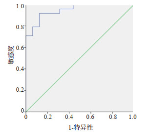
目的 应用实时三维经食管超声心动图(RT-3D-TEE)评估二尖瓣脱垂病变瓣器结构改变与反流程度之间的相关性。 方法 纳入于我院就诊行经胸超声心动图检查确诊为二尖瓣脱垂伴反流的患者40例,另选取10例作为对照组。50例患者均行RT-3D-TEE检查并采集二尖瓣三维图像,使用4D Auto MVQ脱机工作站进行图像后处理和定量分析。 结果 瓣环三维面积、瓣环二维面积、瓣环周长、前后直径、前外-后内侧直径、非平面角度、总瓣叶面积、后叶面积随反流程度增加而增大,瓣高联合比随反流程度增加而减小,组间差异均有统计学意义(P < 0.05)。瓣环三维面积、瓣环二维面积、瓣环周长、前后直径、总瓣叶面积与反流程度呈极强相关关系(r=0.847、0.843、0.845、0.854、0.854,P < 0.05)。A Total是导致重度二尖瓣反流发生的危险因素(P < 0.01,B=1.576,OR=4.834)。总瓣叶面积(截点值=8.9 cm2)预测重度二尖瓣反流的敏感度为91.7%,特异性为87.5%,曲线下面积为0.948(P < 0.01)。 结论 二尖瓣瓣环大小、扁平程度、瓣叶大小与反流程度呈正相关。总瓣叶面积是导致二尖瓣脱垂患者二尖瓣反流程度加重的危险因素。 Abstract:Objective To evaluate the correlation between valve structure changes and the degree of mitral regurgitation, and predict the risk factors for aggravating the degree of regurgitation in patients with mitral valve prolapse via real-time three-dimensional transesophageal echocardiography (RT-3D-TEE). Methods Forty patients who were diagnosed as mitral valve prolapse with mitral valve regurgitation by transthoracic echocardiography, and 10 volunteers in our hospital were involved. All the 50 enrolled patients underwent RT-3D-TEE and the three-dimensional images of the mitral valve were collected. Then the 4D Auto MVQ offline workstation was used for image post-processing and quantitative analysis. Results Annulus area 3D, annulus area 2D, annulus perimeter, anterior-to-posterior diameter, anterolateral to posteromedial diameter, non-planar angle, total leaflet area (A Total), posterior leaflet area increased and annular height to commissural width ratio decreased with the increase of the degree of regurgitation. The differences between two groups were statistically significant (P < 0.05). Annulus area 3D、annulus area 2D、annulus perimeter、anterior-to-posterior diameter、A Total were extremely strongly correlated with the degree of mitral regurgitation (r=0.847, 0.843, 0.845, 0.854, 0.854 respectively, P < 0.05). A Total was a risk factor for severe mitral regurgitation (P < 0.01, B=1.576, OR=4.834). The sensitivity of A Total (cut point value=8.9 cm2) in predicting severe mitral regurgitation was 91.7%, the specificity was 87.5%, and the area under the curve was 0.948 (P < 0.01). Conclusion The size of the mitral valve annulus, the degree of flattening and the size of the valve leaflet were positively correlated with the degree of regurgitation. Among them, A Total is a risk factor for the aggravation of mitral regurgitation in patients with mitral valve prolapse. -

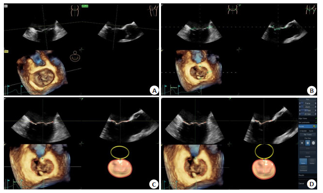
图 1 图像后处理
A: 调整轴线位置; B: 放置标记点; C: 预览图像; D: 点击result获得相关参数.
Figure 1. Image post-processing.

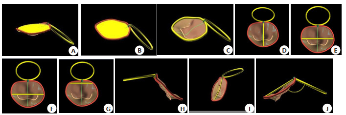
图 2 瓣环参数
A: 瓣环二维面积; B: 瓣环三维面积; C: 瓣环周长; D: 前-后直径; E: 前外-后内侧直径; F: 联合部直径; G: 三角间距离; H: 瓣环高度; I: 非平面角度; J: 二尖瓣环与主动脉瓣环平面夹角.
Figure 2. Annulus parameters.

图 3 瓣叶参数
A: 前叶面积; B: 后叶面积; C: 前叶长度; D: 后叶长度; E: 前叶角度; F: 后叶角度; G: 闭锁线长度3D;H: 幕状区高度; I: 幕状区容量自定义参数:总瓣叶面积.
Figure 3. Leaflet parameters.

图 4 二尖瓣外科观
A: 健康对照组的二尖瓣; B: 二尖瓣脱垂伴中度二尖瓣反流患者的二尖瓣; C: 二尖瓣脱垂伴重度二尖瓣反流患者的二尖瓣。随着反流的加重,二尖瓣环面积及各径向参数值逐渐增大,瓣环椭圆形逐渐消失.
Figure 4. Mitral valve surgical view.

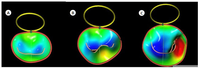
图 5 二尖瓣非平面性变化
A: 健康对照组的二尖瓣; B: 二尖瓣脱垂伴中度二尖瓣反流患者的二尖瓣; C: 二尖瓣脱垂伴重度二尖瓣反流患者的二尖瓣。随着反流程度的加重,二尖瓣环的非平面角度逐渐增大,瓣环变的扁平.
Figure 5. Mitral valve nonplanar changes.
表 1 两组一般临床资料比较
Table 1. Comparison of general clinical data between the two groups (Mean±SD)
指标 对照组(n=10) 非重度反流组(n=16) 重度反流组(n=24) 总计(n=50) 男/女(n) 6/4 11/5 15/9 32/18 年龄(岁) 36±14.66 48±13.13 45±12.73 44±13.64 身高(cm) 168±7.67 167±8.07 172±7.59 170±7.93 体质量(kg) 69±11.01 66±14.80 73±13.78 70±13.80 BM(kg/m2) 24±3.44 23±4.42 24±4.17 24±4.08 体表面积(m2) 1.79±0.17 1.75±0.23 1.87±0.20 1.82±0.21 LA(mm) 31±2.69 38±4.19* 44±6.17*# LV(mm) 46±3.16 54±5.77* 60±7.32*# EDV(mL) 107±21.51 152±32.95* 186±53.07*# 射血分数(%) 68(5.53) 64.71(6.67) 62(3.30)* *P < 0.05 vs对照组; #P < 0.05 vs非重度反流组. LA: 左房内径; LV: 左室内径; EDV: 舒张末期容量.  下载: 导出CSV
表 2 三组间二尖瓣瓣环参数比较
Table 2. Comparison of parameters of mitral annular among three groups (Mean±SD)
指标 对照组(n=10) 非重度反流组(n=16) 重度反流组(n=24) A3D(cm2) 5.26±0.70 6.83±1.19* 9.85±1.75*# A2D(cm2) 4.86±0.66 6.49±1.21* 9.51±1.73*# AP(cm) 8.24±0.53 9.34±0.82* 11.21±1.01*# DAP(cm) 2.09±0.21 2.54±0.30* 3.15±0.33*# DAL-PM(cm) 2.80±0.19 3.10±0.31* 3.70±0.36*# DC(cm) 2.79±0.23 3.09±0.29 3.65±0.34*# DI(cm) 2.02±0.27 2.24±0.21 2.34±0.36* SI(cm/cm) 0.75±0.07 0.82±0.08 0.86±0.08* NPA(°) 149±8.67 159±8.69* 166±6.97*# AHCWR(%) 16.23±2.82 13.75±3.01* 11.12±1.88*# *P < 0.05 vs对照组; #P < 0.05 vs非重度反流组. A3D:瓣环三维面积; A2D:瓣环二维面积; AP:瓣环周长; DAP:前后直径; DAL-PM:前外-后内侧直径; DC:联合部直径; DI:三角间距离; SI:球度指数; NPA:非平面角度; AHCWR:瓣高联合比.
下载: 导出CSV
表 3 三组间二尖瓣瓣叶参数比较
Table 3. Comparison of mitral valve parameters among three groups (Mean±SD)
指标 对照组U=10) 非重度反流组(》=16) 重度反流组(》=24) A Ant(cm2) 3.29±0.62 3.98±0.89 5.66±1.15*# A Post(cm2) 2.43±0.74 3.69±1.15* 5.66±1.62*# A Total(cm2) 5.72±0.85 7.67±1.39* 11.32±2.02*# L Ant(cm) 1.60±0.27 1.77±0.37 2.22±0.48*# L Post(cm) 0.91±0.23 1.18±0.54 1.44±0.58* LC Ant 3D(cm) 2.42±0.42 2.88±0.41 3.75±0.54*# LC Post 3D(cm) 2.56±0.59 2.94±0.37 3.75±0.57*# GAnt(°) 20±4.63 15±9.84 13±7.61* 0Post(°) 38±9.40 24±18.17 19±15.21* HTent(cm) 0.52±0.09 0.37±0.23 0.38±0.22* VTent(mL) 0.76±0.28 0.88±0.44 1.48±0.78*# *P < 0.05 vs对照组; #P < 0.05 vs非重度反流组. A Ant:前叶面积; A Post:后叶面积; A Total:总瓣叶面积; L Ant:前叶长度; L Post:后叶长度; LC Ant 3D:前闭锁线长度3D;LC Post 3D:后闭锁线长度3D;θAnt:前叶角度; θPost:后叶角度; H Tent:幕状区高度; V Tent:幕状区容量.
下载: 导出CSV
-
[1] Kukavica D, Guglielmo M, Baggiano A, et al. Arrhythmic mitral valve prolapse: introducing an era of multimodality imaging-based diagnosis and risk stratification[J]. Diagnostics (Basel), 2021, 11 (3): 467. doi: 10.3390/diagnostics11030467 [2] Dulgheru R, Bruls S, Lancellotti P. How I look at the regurgitant mitral valve—a stepwise echocardiographic assessment[J]. Eur Heart J Cardiovasc Imaging, 2020, 22(5): 491-3. [3] Avierinos JF, Gersh BJ, Melton LJ 3rd, et al. Natural history of asymptomatic mitral valve prolapse in the community[J]. Circulation, 2002, 106(11): 1355-61. doi: 10.1161/01.CIR.0000028933.34260.09 [4] Malkowski MJ, Boudoulas H, Wooley CF, et al. Spectrum of structural abnormalities in floppy mitral valve echocardiographic evaluation[J]. Am Heart J, 1996, 132(1): 145-51. doi: 10.1016/S0002-8703(96)90403-2 [5] Levine RA, Triulzi MO, Harrigan P, et al. The relationship of mitral annular shape to the diagnosis of mitral valve prolapse[J]. Circulation, 1987, 75(4): 756-67. doi: 10.1161/01.CIR.75.4.756 [6] Lee APW, Hsiung MC, Salgo IS, et al. Quantitative analysis of mitral valve morphology in mitral valve prolapse with real-time 3-dimensional echocardiography: importance of annular saddle shape in the pathogenesis of mitral regurgitation[J]. Circulation, 2013, 127 (7): 832-41. doi: 10.1161/CIRCULATIONAHA.112.118083 [7] 崔翼靖, 陈昕, 张慧慧, 等. 经食管实时三维超声研究不同反流程度二尖瓣脱垂患者瓣环动态变化[J]. 中国超声医学杂志, 2017, 33(4): 313-5. doi: 10.3969/j.issn.1002-0101.2017.04.010 [8] El-Tallawi KC, Zhang P, Azencott R, et al. Valve strain quantitation in normal mitral valves and mitral prolapse with variable degrees of regurgitation[J]. JACC Cardiovasc Imaging, 2021, 14(6): 1099-109. doi: 10.1016/j.jcmg.2021.01.006 [9] Lancellotti P, Tribouilloy C, Hagendorff A, et al. European Association of Echocardiography recommendations for the assessment of valvular regurgitation. Part 1: aortic and pulmonary regurgitation (native valve disease)[J]. Eur J Echocardiogr, 2010, 11 (3): 223-44. doi: 10.1093/ejechocard/jeq030 [10] Perk G, Lang RM, Garcia-Fernandez MA, et al. Use of real time three-dimensional transesophageal echocardiography in intracardiac catheter based interventions[J]. J Am Soc Echocardiogr, 2009, 22 (8): 865-82. doi: 10.1016/j.echo.2009.04.031 [11] Boltwood CM, Tei C, Wong M, et al. Quantitative echocardiography of the mitral complex in dilated cardiomyopathy: the mechanism of functional mitral regurgitation[J]. Circulation, 1983, 68(3): 498-508. doi: 10.1161/01.CIR.68.3.498 [12] Members WC, Otto CM, Nishimura RA, et al. 2020 ACC/AHA guideline for the management of patients with valvular heart disease: executive summary: a report of the American college of cardiology/American heart association joint committee on clinical practice guidelines[J]. J Am Coll Cardiol, 2021, 77(4): 450-500. doi: 10.1016/j.jacc.2020.11.035 [13] Bonow RO, O'Gara PT, Adams DH, et al. 2020 focused update of the 2017 ACC expert consensus decision pathway on the management of mitral regurgitation: a report of the American college of cardiology solution set oversight committee[J]. J Am Coll Cardiol, 2020, 75(17): 2236-70. doi: 10.1016/j.jacc.2020.02.005 [14] Grewal J, Suri R, Mankad S, et al. Mitral annular dynamics in myxomatous valve disease: new insights with real-time 3-dimensional echocardiography[J]. Circulation, 2010, 121(12): 1423-31. doi: 10.1161/CIRCULATIONAHA.109.901181 [15] Jensen MO, Jensen H, Levine RA, et al. Saddle-shaped mitral valve annuloplasty rings improve leaflet coaptation geometry[J]. J Thorac Cardiovasc Surg, 2011, 142(3): 697-703. doi: 10.1016/j.jtcvs.2011.01.022 [16] Connell PS, Azimuddin AF, Kim SE, et al. Regurgitation hemodynamics alone cause mitral valve remodeling characteristic of clinical disease states In vitro[J]. Ann Biomed Eng, 2016, 44(4): 954-67. doi: 10.1007/s10439-015-1398-0 [17] Reimink MS, Kunzelman KS, Verrier ED, et al. The effect of anterior chordal replacement on mitral valve function and stresses[J]. ASAIO J, 1995, 41(3): M754-62. doi: 10.1097/00002480-199507000-00114 [18] He ZM, Bhattacharya S. Mitral valve annulus tension and the mechanism of annular dilation: an in-vitro study[J]. J Heart Valve Dis, 2010, 19(6): 701-7. [19] Jiang LY, Owais K, Matyal R, et al. Dynamism of the mitral annulus: a spatial and temporal analysis[J]. J Cardiothorac Vasc Anesth, 2014, 28(5): 1191-7. doi: 10.1053/j.jvca.2014.03.020 [20] Faletra FF, Leo LA, Paiocchi VL, et al. Anatomy of mitral annulus insights from non-invasive imaging techniques[J]. Eur Heart J Cardiovasc Imaging, 2019, 20(8): 843-57. doi: 10.1093/ehjci/jez153 [21] Salgo IS, Gorman JH 3rd, Gorman RC, et al. Effect of annular shape on leaflet curvature in reducing mitral leaflet stress[J]. Circulation, 2002, 106(6): 711-7. doi: 10.1161/01.CIR.0000025426.39426.83 [22] Padala M, Hutchison RA, Croft LR, et al. Saddle shape of the mitral annulus reduces systolic strains on the P2 segment of the posterior mitral leaflet[J]. Ann Thorac Surg, 2009, 88(5): 1499-504. doi: 10.1016/j.athoracsur.2009.06.042 [23] Fernández-Ruiz I. Cilia defects linked to mitral valve prolapse[J]. Nat Rev Cardiol, 2019, 16(8): 456. [24] Calafiore AM, Totaro A, Paparella D, et al. Mimicking natural mitral adaptation to ischaemic regurgitation: a proposed change in the surgical paradigm[J]. Eur J Cardiothorac Surg, 2020, 58(1): 35-9. doi: 10.1093/ejcts/ezaa163 [25] Choi A, McPherson DD, Kim H. Biomechanical evaluation of the pathophysiologic developmental mechanisms of mitral valve prolapse: effect of valvular morphologic alteration[J]. Med Biol Eng Comput, 2016, 54(5): 799-809. doi: 10.1007/s11517-015-1371-y [26] Enriquez-Sarano M, Avierinos JF, Messika-Zeitoun D, et al. Quantitative determinants of the outcome of asymptomatic mitral regurgitation[J]. N Engl J Med, 2005, 352(9): 875-83. doi: 10.1056/NEJMoa041451 -


 点击查看大图
计量
- 文章访问数: 260
- HTML全文浏览量: 94
- PDF下载量: 10
- 被引次数: 0