


Diagnostic accuracy of nuchal translucency thickening combined with Tei index in fetal cardiac malformations in early pregnancy and impact factors of diagnostic accuracy
-
摘要:
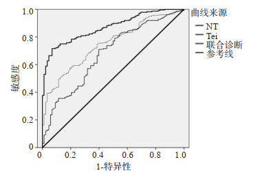
目的 研究超声检测颈部透明层(NT)增厚结合Tei指数对孕早期胎儿心脏畸形的诊断及诊断准确率影响因素。 方法 选择2015年1月~2020年2月在本院进行孕早期筛查的先天性疾病孕产妇108例作为研究对象,另选取同期进行孕检的正常产妇101例作为对照组,比较两组患者的NT以及Tei指数的差异,研究NT以及Tei指数联合诊断对于先天性心脏病的诊断效能,分析影响诊断准确率影响因素。 结果 观察组患者的NT(t=16.780,P < 0.001)和Tei指数(t=7.406,P < 0.001)高于对照组;NT以及Tei指数联合诊断对于先天性心脏病的诊断特异性显著高于单独检测,通过ROC曲线分析,NT以及Tei指数联合诊断的曲线下面积显著高于单独检测,同时通过临界值分析,对于先天性心脏病的诊断,NT的临界值为2.56 mm,Tei指数的临界值为0.51;准确诊断与假阳性以及假阴性患者的筛查时机、医生专业技能、胎儿体位之间的差异存在统计学意义(P < 0.05);多因素分析显示,筛查时机、医生专业技能、胎儿体位均是造成患者假阳性以及假阴性的影响因素。 结论 超声检测NT增厚结合Tei指数对孕早期胎儿心脏畸形的诊断具有积极的意义,在诊断中,筛查时机较小、医生专业技能较低、胎儿体位不配合均是影响诊断准确率的影响因素。 Abstract:Objective To study the diagnostic of fetal cardiac malformations in early pregnancy by ultrasound detection of nuchal translucency (NT) thickening combined with Tei index and the factors influencing the diagnostic accuracy. Methods A total of 108 cases of pregnant women who underwent early pregnancy screening for congenital diseases in our hospital from January 2015 to February 2020 were selected as the research object. 101 cases of normal pregnant women who underwent pregnancy examination at the same time were selected as the control group. The differences in NT and Tei index between the two groups were compared to study the diagnostic efficacy of combined diagnosis of NT and Tei index for congenital heart disease and to analyse the factors influencing diagnostic accuracy. Results NT (t=16.780, P < 0.001) and Tei index (t=7.406, P < 0.001) in the observation group were significantly higher than those in the control group. The diagnostic specificity of combined diagnosis of NT and Tei index for congenital heart disease was significantly higher than that of individual tests, and the area under the curve of combined diagnosis of NT and Tei index was significantly higher than that of individual tests by ROC curve analysis. At the same time, through critical value analysis, for the diagnosis of congenital heart disease, the critical value of NT was 2.56 mm and the critical value of Tei index was 0.51. There were significant differences between the timing of screening, doctor's expertise and fetal position for accurate diagnosis and false-positive and false-negative patients (P < 0.05). The multifactorial analysis showed that the timing of screening, doctor's expertise and fetal position were all influential factors in the false positives and false negatives. Conclusion Ultrasonic detection of NT thickening combined with Tei index has positive implications for the diagnosis of fetal cardiac malformation in early pregnancy. In the diagnosis, the smaller timing of screening, the low expertise of the physician and lack of cooperation of the fetal position are all factors affecting the diagnostic accuracy. -
Key words:
- congenital heart disease /
- nuchal translucency /
- ultrasound /
- Tei index /
- joint detection
-
表 1 两组患者的一般资料比较
Table 1. Comparison of general data between the two groups (Mean±SD)
组別 孕周(周) 年龄(岁) 产次(次) 观察组(n=108) 11.23±2.33 27.42±2.09 1.35±0.34 对照组(n=101) 11.44±2.43 27.71±2.43 1.42±0.32 t 0.637 0.922 1.533 P 0.525 0.358 0.127  下载: 导出CSV
表 2 两组的NT以及Tei指数比较
Table 2. Comparison of NT and Tei indexes between the two groups (Mean±SD)
组別 NT(mm) Tei 观察组(n=108) 2.74±0.33 0.54±0.22 对照组(n=101) 2.02±0.29 0.36±0.12 t 16.780 7.406 P < 0.001 < 0.001
下载: 导出CSV
表 3 NT以及Tei指数联合诊断对于先天性心脏病的诊断效能分析
Table 3. Analysis of diagnostic efficacy of combined diagnosis of NT and Tei index for congenital heart disease
诊断方法 真阳例数
(n)假阳例数
(n)真阴例数
(n)假阴例数
(n)准确率
(%)敏感度
(%)特异性
(%)阳性预测值
(%)阴性预测值
(%)NT 93 25 76 15 80.86 86.11 44.97 78.81 83.52 Tei 91 20 81 17 82.30 84.26 47.09 81.98 82.65 联合检测 90 5 96 18 89.00 83.33 51.61 94.74 84.21
下载: 导出CSV
表 4 ROC曲线分析
Table 4. ROC curve analysis [n(%)]
组别 准确诊断(n=186) 假阳、假阴(n=23) χ2 P 筛查时机(孕周) 4.662 0.031 < 10周 120(85.71) 20(14.29) >10周 66(95.65) 3(4.35) 医生专业技能 高级 91(94.79) 5(5.21) 6.092 0.014 非高级 95(84.07) 18(15.93) 胎儿体位 配合 80(95.24) 4(4.76) 5.592 0.018 非配合 106(84.80) 19(15.20)
下载: 导出CSV
表 5 假阳性以及假阴性患者的单因素分析
Table 5. Univariate analysis of false positive and false negative patients[n(%)]
诊断方法 标准误 AUC AUC(95%CI) P NT 11.231 0.775 0.520~0.872 0.017 Tei 9.252 0.603 0.600~0.746 < 0.001 联合检测 12.230 0.823 0.126~0.996 < 0.001
下载: 导出CSV
表 6 多因素分析
Table 6. Multi factor analysis
因索 β S.E. Wald P OR 95%CI 筛査时机(孕周) 1.018 2.361 1.322 0.001 1.019 1.009~1.926 医牛专业技能 1.062 3.269 1.333 0.002 1.632 1.331-2.320 胎儿休位 0.369 4.139 1.691 < 0.001 1.089 1.002~2.065
下载: 导出CSV
-
[1] 韩松岩, 王东辉, 张媛, 等. 四维超声联合二维超声检查对孕中期疑似心脏畸形胎儿的诊断价值[J]. 中国妇幼保健, 2021, 36(2): 407-10. https://www.cnki.com.cn/Article/CJFDTOTAL-ZFYB202102053.htm [2] 刘吉庆, 苏静, 孟秋霞. 四维超声心动图联合二维彩超在胎儿先天性心脏畸形筛查中的应用[J]. 中国妇幼保健, 2019, 34(19): 4574-6. https://www.cnki.com.cn/Article/CJFDTOTAL-ZFYB201919071.htm [3] 林健谊, 庞振华. 超声心动图在产前诊断胎儿先天性心脏畸形中的应用价值[J]. 江苏医药, 2018, 44(1): 106-7. https://www.cnki.com.cn/Article/CJFDTOTAL-YIYA201801035.htm [4] 贾赛玉, 王跃涛, 王冬艳, 等. 超声三步法快速筛查胎儿先天性心脏畸形的临床价值[J]. 江苏医药, 2018, 44(2): 162-5, 120. https://www.cnki.com.cn/Article/CJFDTOTAL-YIYA201802011.htm [5] 王媛, 赵旭, 杨娅, 等. 超声时间-空间关联成像技术及三维超声在胎儿先天性心脏畸形及心外畸形诊断中的应用价值[J]. 中国全科医学, 2019, 22(3): 346-50. https://www.cnki.com.cn/Article/CJFDTOTAL-QKYX201903028.htm [6] 李泞珊, 夏红梅, 邓曦, 等. 不合并心脏畸形的卵圆孔血流受限胎儿超声影像特征及预后[J]. 中华超声影像学杂志, 2019, 28(1): 36-41. doi: 10.3760/cma.j.issn.1004-4477.2019.01.008 [7] 刘世霞, 张加琪, 宋娟, 等. 产前超声诊断胎儿双上腔静脉伴复杂心脏畸形1例[J]. 中国现代医学杂志, 2019, 29(14): 125-6. doi: 10.3969/j.issn.1005-8982.2019.14.028 [8] Annabi MS, Zhang B, Bergler-Klein J, et al. Usefulness of the Btype natriuretic peptides in low ejection fraction, low-flow, lowgradient aortic Stenosis results from the TOPAS multicenter prospective cohort study[J]. Struct Heart, 2021, 5(3): 319-27. doi: 10.1080/24748706.2021.1900630 [9] Jin H, Wang J, Zhang GY, et al. A Chinese multicenter retrospective study of isolated increased nuchal translucency associated chromosome anomaly and prenatal diagnostic suggestions[J]. Sci Rep, 2021, 11(1): 5596. doi: 10.1038/s41598-021-85108-6 [10] Siegmund AS, Pieper PG, Bouma BJ, et al. Early N-terminal pro-Btype natriuretic peptide is associated with cardiac complications and function during pregnancy in congenital heart disease[J]. Neth Heart J, 2021, 29(5): 262-72. doi: 10.1007/s12471-021-01540-3 [11] Secchi F, Alì M, Monti CB, et al. Right and left ventricle native T1 mapping in systolic phase in patients with congenital heart disease [J]. Acta Radiol, 2021, 62(3): 334-40. doi: 10.1177/0284185120924563 [12] Reiner B. Myocardial protection with beta blocker treatment in infants with heart failure due to congenital heart defects and Duchenne muscular dystrophy[J]. Open J Thorac Surg, 2020, 10 (4): 81-8. doi: 10.4236/ojts.2020.104008 [13] Słodki M, Soroka M, Rizzo G, et al. Prenatal Atrioventricular Septal Defect (AVSD) as a planned congenital heart disease with different outcome depending on the presence of the coexisting extracardiac abnormalities (ECA) and/or malformations (ECM)[J]. J Matern Fetal Neonatal Med, 2020, 33(15): 2635-41. doi: 10.1080/14767058.2018.1556254 [14] Luo DL, Chen PY, Yang ZY, et al. High plasma adiponectin is associated with increased pulmonary blood flow and reduced right ventricular function in patients with pulmonary hypertension[J]. BMC Pulm Med, 2020, 20(1): 204. doi: 10.1186/s12890-020-01233-4 [15] Escribano-Subias P, López R, Almenar L, et al. Changes in REVEAL risk score in patients with pulmonary arterial hypertension treated with macitentan in clinical practice: results from the PRACMA study[J]. BMC Pulm Med, 2020, 20(1): 154. doi: 10.1186/s12890-020-01197-5 [16] 夏涛, 毛培明, 刘仕静, 等. 产前胎儿超声心动图在先天性心脏病产前诊断中的应用价值[J]. 影像科学与光化学, 2021, 39(2): 235-9. https://www.cnki.com.cn/Article/CJFDTOTAL-GKGH202102013.htm [17] 龚婷, 计晓娟, 郑敏, 等. 超声心动图与计算机断层扫描血管成像诊断儿童三房心及其预后分析[J]. 中华医学超声杂志: 电子版, 2021, 18(2): 150-4. doi: 10.3877/cma.j.issn.1672-6448.2021.02.005 [18] 王新霞, 马澜, 栗河舟, 等. 不同类型左无名静脉异常产前超声心动图特征及其临床意义[J]. 中国医学影像技术, 2021, 37(3): 328-32. https://www.cnki.com.cn/Article/CJFDTOTAL-ZYXX202103003.htm [19] 于佳慧, 谭雪莹, 张昕彤, 等. 双心室或右心室心肌致密化不全超声心动图表现[J]. 中国医学影像技术, 2021, 37(9): 1434-6. https://www.cnki.com.cn/Article/CJFDTOTAL-ZYXX202109045.htm [20] 郭玉霞, 赵博文, 戚夏近, 等. 胎儿超声心动图检查定量11~17周正常胎儿主动脉和肺动脉内径Z-评分的初步研究[J]. 中华医学超声杂志: 电子版, 2020, 17(1): 52-9. doi: 10.3877/cma.j.issn.1672-6448.2020.01.010 [21] 黄仪, 夏焙. 超声心动图Z值在先天性心脏病法洛四联症中的应用现状与展望[J]. 中华医学超声杂志: 电子版, 2021, 18(6): 529-33. doi: 10.3877/cma.j.issn.1672-6448.2021.06.001 [22] 郭雪丽, 钟柳丹. 超声心动图Tei指数及NT值与胎儿心功能异常的相关性分析[J]. 影像研究与医学应用, 2020, 4(18): 224-6. doi: 10.3969/j.issn.2096-3807.2020.18.122 [23] Schneider M, Moser M, Dannenberg V, et al. QRS Duration and Outcome Late after Repair of Tetralogy of Fallot: Neurohormonal Activation Differentiates between Mechanical and Electrical Dyssynchrony[J]. Congenit Heart Dis, 2020, 15(1): 51-8. doi: 10.32604/CHD.2020.011712 -

 点击查看大图
计量
- 文章访问数: 187
- HTML全文浏览量: 98
- PDF下载量: 7
- 被引次数: 0