


Value of CT and MRI multiple sequences in condition assessment of patients with traumatic brain injury
-
摘要:
目的分析CT和MRI多序列评估脑外伤患者病况的价值。 方法连续收集2018年2月~2020年4月本院收治的脑外伤患者70例的临床资料,所有入院后的患者均进行CT和磁共振T1WI、T2WI、液体衰减翻转翻转恢复序列(FLAIR)、弥散加权成像(DWI)、增强梯度回波T2*加权血管成像(ESWAN)序列扫描检查,记录病灶的位置、数目、形态等,并与手术病理学诊断结果进行对比,分析两种诊断方式对病情的评估价值;于伤后3月进行随访,记录患者有无神经症状,并进行格拉斯哥预后评分,采用Spearman相关进行比较分析。 结果60例脑外伤患者中手术诊断硬膜下血肿42例,蛛网膜下腔出血21例,硬膜外血肿17例,脑挫伤23例。MRI在硬膜下血肿、脑挫伤、蛛网膜下腔出血的诊断率高于CT检查(P < 0.05),两种检查方式在硬膜外血肿检查准确率差异无统计学意义(P>0.05);60例患者微出血病灶MRI多序列检查中ESWAN检查数目最多,其后依次是FLAIR序列、DWI序列、T2WI序列、T1WI序列,CT与T2WI序列检出数目差异无统计学意义(P>0.05)。微出血灶主要分布在额叶、颞枕顶叶、胼胝体、基底节、丘脑、脑干等区域,ESWAN序列检出出血病灶的总体积为288 557 mm3,以颞枕顶叶白质体积最大为63 153 mm3;60例脑外伤患者ESWAN序列发现出血性病灶数目、体积与患者入院时格拉斯哥昏迷评分之间经Spearman相关检验均存在明显负相关(r=-0.753, P < 0.01;r=-0.736,P < 0.01),伤后3月的格拉斯哥预后评分与ESWAN序列发现出血性病灶数目、体积负相关(r=-0.648, P < 0.01;r=-0.612,P < 0.01)。 结论与CT检查相比,MRI多序列联合检查在脑外伤患者硬膜下血肿、脑挫伤、蛛网膜下腔出血诊断准确率更高,且ESWAN序列在出血性病灶的数目、体积检出方面更有优势,对患者病情及远期预后有重要参考价值。 Abstract:ObjectiveTo analyze the value of CT and MRI multiple sequences in evaluating the condition of patients with traumatic brain injury. MethodsThe clinical data of 70 patients with traumatic brain injury admitted and treated in the hospital were collected from February 2018 to April 2020. All patients were undergone with CT, magnetic resonance T1WI, T2WI, fluid attenuated inversion recovery (FLAIR), diffusion weighted imaging (DWI) and enhanced gradient echo T2- star weighted angiography (ESWAN) sequence scanning. Location, number and shape of the lesion were recorded. Compared with the results of surgical pathology, the evaluation value of the two diagnostic methods was analyzed. The patients were followed up 3 months after injury, and the Glasgow Outcome Score (GOS) was recorded. Spearman correlation was used for comparative analysis. ResultsAmong the 60 patients with traumatic brain injury, 42 cases were diagnosed as subdural hematoma, 21 cases as subarachnoid hemorrhage, 17 cases as epidural hematoma and 23 cases as brain contusion. The diagnostic rates of subdural hematoma, brain contusion and subarachnoid hemorrhage by MRI were higher than those by CT (P < 0.05), but there was no significant difference in the accuracy between the two methods in diagnosis of epidural hematoma (P>0.05). For microbleeds in the 60 patients detected by MRI multiple sequences, the number detected by ESWAN was the largest, followed by FLAIR sequence, DWI sequence, T2WI sequence and T1WI sequence. There was no significant difference in the number between CT and T2WI sequence (P>0.05). Microbleeds were mainly distributed in the frontal lobe, temporooccipital parietal lobe, corpus callosum, basal ganglia, thalamus and brainstem. The total volume of bleeding detected by ESWAN sequence was 288 557 mm3 with the largest volume of 63153 mm3 in the white matter of temporal occipito parietal lobe. Spearman correlation test showed that there was a significant negative correlation between the number and volume of hemorrhagic foci found by ESWAN sequence and the GCS score at admission (r=-0.753, P < 0.01; r=-0.736, P < 0.01). The GOS score at 3 months after injury was negatively correlated with the number and volume of hemorrhagic foci found by ESWAN sequence (r=-0.648, P < 0.01; r=-0.612, P < 0.01). ConclusionCompared with CT, MRI multiple sequences combined examination is more accurate in the diagnosis of subdural hematoma, brain contusion and subarachnoid hemorrhage in patients with traumatic brain injury. Besides, ESWAN sequence has more advantages in detecting the number and volume of hemorrhagic foci, which has important reference value for patients' condition and long-term prognosis evaluation. -
Key words:
- CT /
- MRI /
- multiple sequences /
- traumatic brain injury /
- hematoma /
- hemorrhagic focus
-
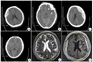
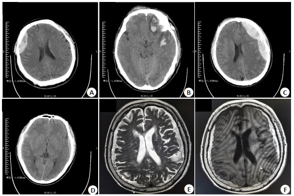
图 1 脑外伤患者的影像学表现
A:患者男, 46岁, 车祸伤, CT头颅常规含颅底平扫显示头部外伤, 创伤性硬膜外血肿; B:患者男, 45岁, 跌伤, 头颅CT显示硬膜下血肿、创伤性蛛网膜下腔血肿、颅骨骨折、脑挫伤; C:患者男, 32岁, 跌伤, 头颅CT显示硬膜外血肿、颅底骨折、创伤性蛛网膜下腔血肿; D:患者男, 29岁, 跌伤, 头颅CT显示硬膜下血肿、创伤性蛛网膜下腔血肿、脑挫伤; E、F:患者女, 34岁, 跌伤, MRI显示硬膜下血肿.
Figure 1. Images of patients with traumatic brain injury.
表 1 两种检查方式的诊断结果对比[n=60, n(%)]
Table 1. Comparison of diagnostic results of the two examination methods
检杳方式 硬膜下血肿 蛛网膜下腔出血 硬膜外血肿 脑挫伤 手术诊断 42 21 17 23 CT检杳 29 (69.05) 13 (61.90) 15 (88.24) 12(52.17) MRI检杳 41 (97.62) 21 (100.00) 13 (76.47) 22 (95.65) χ2/连续性χ2 12.343 7.216 0.202 11.275 P 0.001 0.007 0.653 0.001  下载: 导出CSV
表 2 CT和MRI多序列检出剪切灶和微出血病灶数目(n)
Table 2. Numbers of shear foci and microbleeds detected by CT and MRI multiple sequences
部位 T1WI T2WI FLAIR DWI ESWAN CT 胼胝体 54 89 126 138 534 93 颞枕顶叶灰质 48 67 98 79 421 68 颞枕顶叶白质 50 73 136 117 511 72 基底节丘脑 41 56 167 103 498 54 丘脑 32 60 112 82 191 63 额叶灰质 55 81 102 67 454 84 额叶白质 76 114 165 99 539 117 脑干 30 48 75 46 187 54 小脑 27 40 71 33 172 43
下载: 导出CSV
-
[1] Guilliams K, Wainwright MS. Pathophysiology and management of moderate and severe traumatic brain injury in children[J]. J Child Neurol, 2016, 31(1): 35-45. doi: 10.1177/0883073814562626 [2] 周静, 杨磊, 赵瑞, 等.双重任务在脑外伤康复中应用的研究进展[J].中国康复医学杂志, 2017, 32(11): 1314-6. doi: 10.3969/j.issn.1001-1242.2017.11.024 [3] 张志强, 刘丽娟, 张强, 等.移动CT和常规CT检查对颅脑损伤后脑继发性损害及治疗效果的影响[J].中华神经医学杂志, 2016, 15 (11): 1159-63. [4] 林炜, 陈建, 王化强, 等.创伤性颅脑损伤患者CT征象及预后分析[J].中国CT和MRI杂志, 2019, 17(1): 28-30, 91. [5] Yamazoe M, Maeda M, Umino M, et al. Cerebral microbleeds identified by susceptibility-weighted imaging in two cases of fabry disease without neurological symptoms[J]. Open J Med Imaging, 2015, 5(4): 194-8. doi: 10.4236/ojmi.2015.54024 [6] Zhang DM, Xu LP, Ma AD, et al. Value of conventional MRI and susceptibility Wei-ghted imaging in diagnosis of cerebral microbleeds[J]. Biomed Res, 2017, 28(20): 8905-8. [7] Brody DL, Benetatos J, Bennett RE, et al. The pathophysiology of repetitive concussive traumatic brain injury in experimental models; new developments and open questions[J]. Mol Cell Neurosci, 2015, 66(Pt B): 91-8. [8] 李文臣, 刘磊, 陈勃, 等.磁敏感加权成像对中度出血性弥漫轴索损伤的诊断价值和预后评价[J].中华神经外科创伤电子杂志, 2017, 3 (3): 136-41. [9] Abbas K, Shenk TE, Poole VN, et al. Alteration of default mode network in high school football Athletes due to repetitive subconcussive mild traumatic brain injury: a resting-state functional magnetic resonance imaging study[J]. Brain Connect, 2015, 5(2): 91-101. doi: 10.1089/brain.2014.0279 [10] Park MY. P4- 251: Distribution analysis of cerebral microbleeds in Alzheimer's disease and stroke with susceptibility weighted MR imaging[J].Alzheimer's Dement, 2015, 11(7S_Part_19): P877. [11] 曾旭东, 周旭峰, 温辉. MRI在评估2型糖尿病患者脑部小血管损伤的临床价值[J].中国CT和MRI杂志, 2019, 17(1): 44-6. [12] 蔡王莉, 汤光宇, 魏小二, 等. CT早期动态监测脑外伤手术治疗及其预后评估的价值[J].生物医学工程与临床, 2017, 21(1): 45-50. [13] 李譞婷, 袁俊亮, 范慧敏, 等.磁敏感加权成像诊断中枢神经系统疾病临床应用[J].中国实用内科杂志, 2017, 37(11): 972-6. [14] Ryan ME, Jaju A, Ciolino JD, et al. Rapid MRI evaluation of acute intracranial hemorrhage in pediatric head trauma[J]. Neuroradiology, 2016, 58(8): 793-9. doi: 10.1007/s00234-016-1686-x [15] 周丽景.颅脑CT对急性脑外伤患者手术疗效及预后的评估价值分析[J].中国CT和MRI杂志, 2017, 15(3): 19-21. [16] Roguski M, Morel B, Sweeney M, et al. Magnetic resonance imaging as an alternative to computed tomography in select patients with traumatic brain injury: a retrospective comparison[J]. J Neurosurg Pediatr, 2015, 15(5): 529-34. doi: 10.3171/2014.10.PEDS14128 [17] 南晓东, 张俊, 吴连强, 等.磁共振T2flair、DWI序列在各期脑出血诊断价值分析[J].中国CT和MRI杂志, 2017, 15(10): 10-3. [18] 罗伟, 姚景江, 张亚林.多序列MRI评估脑外伤微出血及弥漫性轴索损伤[J].医学影像学杂志, 2019, 29(9): 1446-9. [19] El-Serougy LG, El-Rakhawy MM, Ashamallah GA, et al. Reliability of magnetic susceptibility weighted imaging in detection of cerebral microbleeds in stroke patients[J]. Egypt J Radiol Nucl Med, 2017, 48 (1): 225-9. doi: 10.1016/j.ejrnm.2016.11.003 [20] 刘义. 3.0T MRI多序列联合应用在评估颅脑损伤严重程度中的应用[J].中国CT和MRI杂志, 2018, 16(2): 126-8. [21] Tu TW, Williams RA, Lescher JD, et al. Radiological-pathological correlation of diffusion tensor and magnetization transfer imaging in a closed head traumatic brain injury model[J]. Ann Neurol, 2016, 79 (6): 907-20. doi: 10.1002/ana.24641 -

 点击查看大图
计量
- 文章访问数: 686
- HTML全文浏览量: 306
- PDF下载量: 14
- 被引次数: 0