


Value of MSCT in diagnosis of common parotid benign tumor
-
摘要:
目的 探讨多层螺旋CT对腮腺常见良性肿瘤的诊断和鉴别诊断价值。 方法 回顾性分析30例经病理证实的腮腺良性肿瘤患者的CT资料,所有病例均行CT平扫和增强,对多形性腺瘤、腺淋巴瘤及基底细胞腺瘤的CT表现进行对照,分析评价其影像学表现特征。 结果 30例患者中,多形性腺瘤16例,腺淋巴瘤10例,基底细胞腺瘤4例。多形性腺瘤表现为特征性的渐进性强化;腺淋巴瘤表现为典型的“快进快出”;基底细胞腺瘤动脉期和静脉期均有较显著强化。3者增强方式不同,其强化差异有统计学意义(P<0.05)。 结论 腮腺良性肿瘤的多层螺旋CT表现有一定特征性,多层螺旋CT在腮腺良性肿瘤的诊断和鉴别诊断中有重要的作用。 Abstract:Objective To explore the value of MSCT in the diagnosis of common benign parotid tumors. Methods CT findings of 30 cases with parotid common benign tumor proved by pathology were analyzed retrospectively. All cases received plain and enhanced CT scanning.CT appearances between pleomorphic adenoma, gland lymphoma and BCA were compared. The image features were analyzed and evaluated. Results Sixteen cases were parotid pleomorphic adenoma, 10 cases were adenolymphoma and 4 cases were parotid basal cell tumor. A gradually increasing intensification of contrast was showed typically on pleomorphic adenomas. Adenolymphoma showed a typical intensification of contrast " quickly-in and quickly-out”, reached the peak time of enhancement at arterial phase. Basal cell tumor had a significant enhancement in the arterial and venous phase. The enhancement methods were significantly different among three tumors (P<0.05). The differences of enhancement in 3 groups were significant (P<0.05). Conclusion The parotid common benigns have characteristic CT signs. MSCT is important in the diagnosis and differential diagnosis. -
Key words:
- parotid gland /
- benign tumor /
- tomography /
- X-ray computed
-

图 2 腺淋巴瘤
双侧腮腺跨浅深叶结节, 右侧结节囊性为主, 边界清楚; 左侧结节实性为主, 内有小囊变, 平扫等密度; 增强后, 动脉期实性结节显著强化, 囊性结节无明显强化; 静脉期强化程度较动脉期明显减低; A: 平扫; B: 增强动脉期; C: 增强静脉期.
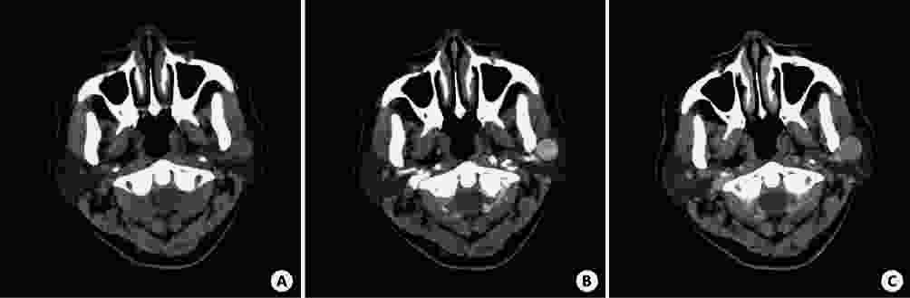
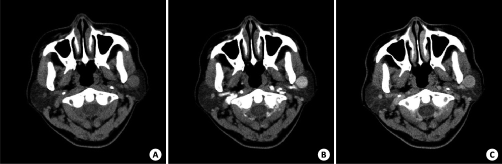
图 3 基底细胞腺瘤
左侧腮腺浅叶结节, 边界清楚; 平扫等密度; 增强后, 动脉期和静脉期结节显著强化, 两者无显著差别; A: 平扫; B: 增强动脉期; C: 增强静脉期.
表 1 3种腮腺肿瘤CT平扫、增强CT值比较(Mean±SD, HU)
肿瘤类型 位置(n, 浅/深) 平扫CT值 动脉期增加CT值 静脉期增加CT值 多形性腺瘤 12/4 35.3±8.5 13.4±8.8* 25.8±4.7 腺淋巴瘤 9/1 38.6±15.4 57.2±16.2 30.9±14.4 基底细胞腺瘤 4/0 40.8±16.3 61.3±14.5 58.6±10.5△ *P<0.05vs 腺淋巴瘤、基底细胞瘤; △P<0.05vs多形性腺瘤、腺淋巴瘤.  下载: 导出CSV
-
[1] Modlin IM, Shapiro MD, Kidd M. Primary carcinoid tumor of the parotid gland: a case report and review of the literature[J]. Ear Nose Throat J, 2006, 85(8): 533-9. https://www.researchgate.net/profile/Mark_Kidd/publication/6795678_Primary_carcinoid_tumor_of_the_parotid_gland_A_case_report_and_review_of_the_literature/links/0fcfd5101436a5f87a000000.pdf?inViewer=0&pdfJsDownload=0&origin=publication_detail [2] Perakis H, Heurle AD, Miller B, et al. Usefulness of CT and MRI in predicting arotid gland tumor histopathology[J]. Laryngoscope, 2011, 121(54): 44-7. doi: 10.1002/lary.22021/abstract [3] Dong Y, Lei GW, Wang SW, et al. Diagnostic value of CT perfusion imaging for parotid neoplasms[J]. Dentomaxillofac Radiol, 2014, 43(1): 20130237-42. doi: 10.1259/dmfr.20130237 [4] Wittekindt C, Streubel K, Arnold G, et al. Recurrent pleomorphic adenoma of the parotid gland: Analysis of 108 consecutive patients[J]. Head Neck, 2007, 29(9): 822-8. doi: 10.1002/(ISSN)1097-0347 [5] Singh S, 刘珍银. 腮腺良恶性肿瘤的CT与MRI诊断[J]. 临床医学工程, 2014, 21(8): 960-1. http://med.wanfangdata.com.cn/Paper/Detail?id=PeriodicalPaper_ylbjqj201408008 [6] 唐 坚, 夏建国, 魏仁华. 腮腺良恶性病变的MSCT 影像表现及鉴别诊断[J]. 中国医学工程, 2011, 19(10): 17-8. [7] Choi DS, Na DG, Byun HS, et al. Salivary gland tumors: evaluation with two-phase helical CT[J]. Radiology, 2000, 214(1): 231-6. doi: 10.1148/radiology.214.1.r00ja05231 [8] 陈 超, 刘红宇, 汪国余, 等. 腮腺基底细胞瘤的CT影像分析[J]. 医学影像学杂志, 2016, 26(2): 222-5. http://www.cqvip.com/QK/92832X/201211/43940963.html [9] 刘红刚. 头颈病理学[M]. 北京: 北京大学出版社, 2008: 300-5. [10] 董 越, 伍健林, 田 明. 多层螺旋CT在腮腺良性肿瘤中的诊断价值(附84例分析) [J]. 中国医学影像技术, 2007, 23(10): 1469-72. doi: 10.3321/j.issn:1003-3289.2007.10.012 [11] Yerli H, Aydin E, Coskun M, et al. Dynamic multislice computed tomography findings for parotid gland tumors[J]. J Comput Assist Tomogr, 2007, 31(2): 309-16. doi: 10.1097/01.rct.0000236418.82395.b3 [12] 许乙凯, 郑木明. 现代CT与MRI诊断技术[M]. 北京: 人民军医出版社, 2003. [13] Yerli H, Teksam M, Aydin E, et al. Basal cell adenoma of the parotid gland: dynamic CT and MRI findings[J]. British J Radiol, 2005, 78(931): 642-5. doi: 10.1259/bjr/32453517 [14] 刘春玲, 黄 飚, 周正根, 等. 腮腺基底细胞腺瘤的CT和MRI特点[J]. 中华放射学杂志, 2009, 43(6): 600-3. https://www.wenkuxiazai.com/doc/fde0aa0f0c22590103029da9-3.html [15] Miracle A, Rezaei A, Gandhi D, et al. CT perfusion of the Neek:intenal carotid artery versus external carotid artery as the referenee artery[J]. AJNR, 2009, 30(7): 1598-601. http://www.academia.edu/15908679/Noninvasive_Measurement_of_Regional_Cerebral_Blood_Flow_by_Near-Infrared_Spectroscopy_and_Indocyanine_Green [16] 邝平定, 张敏鸣, 邵国良, 等. 腮腺腺淋巴瘤的CT表现[J]. 中华放射学杂志, 2009, 43(12): 1324-6. doi: 10.3760/cma.j.issn.1005-1201.2009.12.022 [17] Bai M, Chen J, Raupach R, et al. Effect of nondinar three dimensional optimized Reconstruction algorithm filter on image quality and radiation dose validation on phantoms[J]. Med Phys, 2009, 36(1): 95-7. http://adsabs.harvard.edu/abs/2009MedPh..36...95B [18] 邝平定, 邵国良, 张敏鸣, 等. 腮腺基底细胞腺瘤的CT表现[J]. 实用放射学杂志, 2010, 26(10): 1420-2. doi: 10.3969/j.issn.1002-1671.2010.10.010 [19] 侯学文, 张利中. 腮腺肿瘤的多层螺旋CT表现[J]. 中国医疗设备, 2016, 31(5): 60-3. doi: 10.3321/j.issn:1000-7857.2009.18.021 [20] 元建鹏, 梁碧玲, 谢榜昆, 等. MRI征像在腮腺肿瘤定性诊断中的价值及其病理基础[J]. 癌症, 2003, 22(5): 514-9. [21] Chiu NC, Wu HM, Chou YH, et al. Basal cell adenoma versus pleomorphic adenoma of the parotid gland: CT findings[J]. Am J Roentgenol, 2007, 189(5): W254-61. doi: 10.2214/AJR.07.2292 [22] Jang M, Park D, Lee SR, et al. Basal cell adenoma in the parotid gland: CT and Mr findings[J]. AJNR Am J Neuroradiol, 2004, 25(4): 631-5. http://www.ajnr.org/content/ajnr/25/4/631.full.pdf -


 点击查看大图
图(3) / 表(1)
计量
- 文章访问数: 927
- HTML全文浏览量: 334
- PDF下载量: 7
- 被引次数: 0