


Diagnosis and localization of MRI negative temporal lobe focal cortical dysplasias by video electroencephalogram and diffusion trnsor imaging
-
摘要:
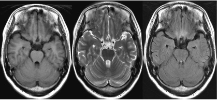
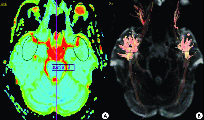
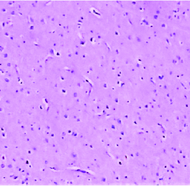
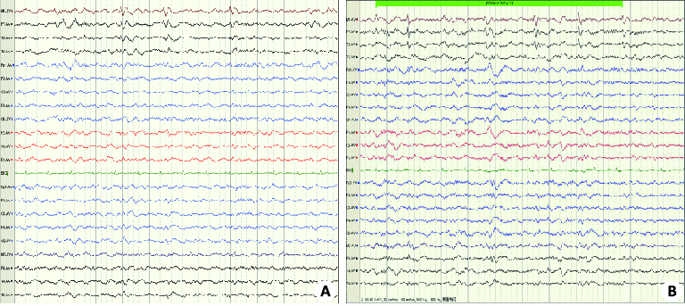
目的 探讨影像学表现阴性的颞叶局灶性皮质发育不良(FCD)的诊断和定位方法,为手术治疗提供依据。 方法 联合应用视频脑电图和弥散张量成像检查,通过视频脑电图特异表现,联合应用表观弥散系数值、部分各向异性值、弥散张量纤维束成像,诊断和定位影像学表现阴性的颞叶FCD。 结果 FCD区域的视频脑电图主要表现为棘波、尖波和阵发性或节律性的棘波活动,慢波出现较少或没有。FCD区域的表观弥散系数值较对侧显著升高,而部分各向异性值明显低于对侧,差异有统计学意义(P<0.05)。全部38例患者依据视频脑电图和弥散张量成像确定的颞叶FCD范围行手术切除。病理回报为FCDⅠA型11例,ⅠB型13例,ⅠC型10例,ⅡA型4例。术后随访12~26月,本组患者Engel'sⅠa级7例,Ⅰb级16例,Ⅱ 级10例,Ⅲ5级。 结论 联合应用视频脑电图和弥散张量成像技术可以准确定性、定位影像学检查阴性的颞叶FCD,为精确切除致痫灶,提高颞叶癫痫控制率提供保障。 Abstract:Objective To explore the diagnosis and localization of focal cortical dysplasia type I, and provide the basis for the surgical treatment. Methods Video EEG with combination of DTI were performed.VEEG specific performance, ADC value, fractional anisotropy, diffusion tensor imaging were used to diagnose and locate FCD I type with negative imaging . Results The apparent diffusion coefficient of FCD region was significantly higher than contralateral area.Fractional anisotropy was significantly lower than contralateral area (P<0.05). All patients were performed with resection based on FCD position and range, which determined by VEEG and DTI . The pathological return was type of FCD Ⅰ. The patients were followed up for 12-26 months,with 7 cases of Engel's grade Ⅰa, 28 cases of grade Ⅰb, 16 cases of gradeⅡ. Conclusion VEEG combined with DTI technology can accurately define and locate FCDⅠtype with negative imaging.It can provide protection for the precise excision of epileptogenic focus and improve seizure control. -
Key words:
- video electroencephalogram /
- diffusion trnsor imaging /
- focal cortical dysplasias /
- diagnosis /
- location
-
表 1 感兴趣区域与对侧颞叶对照区ADC和FA值(Mean±SD)
参数 感兴趣区 对侧颞叶对照区 ADC 1.55±0.14* 0.87±0.05 FA 0.107±0.034* 0.396±0.099 *P<0.05 vs对侧; ADC: 表观弥散系数值; FA: 部分各向异性.  下载: 导出CSV
-
[1] 梁晋川, 周晓平. 局部脑皮层发育不良与癫痫的关系[J]. 立体定向和功能性神经外科杂志, 2009, 22(1): 52-4. [2] 常 亮. 癫痫患者质量调整生命年的研究[J]. 中国全科医学, 2013, 16(13): 1506-9. [3] Blümcke I, Thom M, Aronica E, et al. The clinicopathologic spectrum of focal cortical dysplasias: a consensus classification proposed by an adhoc task force of the ILAE diagnostic methods commission[J]. Epilepsia, 2011, 52(1): 158-74. doi: 10.1111/epi.2011.52.issue-1 [4] 蔡立新, 李勇杰, 张国君, 等. 局灶性皮层发育不良癫痫患者头皮脑电图的特点及其定位意义[J]. 临床神经电生理学杂志, 2008, 17(1): 20-5. http://d.wanfangdata.com.cn/Thesis/Y1658812 [5] Reich DS, Smith SA, Jones CK, et al. Quantitative characterization of the corticospinal tract at 3T[J]. Am J Neurorad, 2006, 27(10): 2168-78. http://www.ajnr.org/content/27/10/2168 [6] Bello L, Cambini A, Castellano A, et al. Motor and language DTI fiber tracking combined with intraoperative subcortical mapping for surgical rmoval of glioma[J]. Neurolmage, 2008, 39(1): 369-82. doi: 10.1016/j.neuroimage.2007.08.031 [7] 李德军, 包尚联, 马 林, 等. 弥散张量成像在中枢神经系统中的应用[J]. 国外医学:生物医学工程分册, 2003, 26(5): 197-202. https://www.wenkuxiazai.com/doc/a22a4ad8ad51f01dc281f14b.html [8] Pavlisa G, Rados M, Pavlisa G. et aL the differences of water diffusion between brain tissue infiltrated by tumor and peritumoml vasogenic edema[J]. Clin Imaging, 2009, 33(2): 96-101. doi: 10.1016/j.clinimag.2008.06.035 [9] Rugg-Gunn FJ, Eriksson SH, Symms MR, et al. Diifusion tensor imaging in refractory epilepsy[J]. Lancet, 2002, 359(9319): 1748-51. doi: 10.1016/S0140-6736(02)08615-4 [10] 张 硕, 强金伟, 张康乐. 弥散张量成像对常规性临床应用的初步建立和实现[J]. 中国生物医学工程学报, 2007, 26(5): 796-800. [11] Keller SS, Richardson MP, Schoene-Bake 1, et al. Thalamotemporal alteration and postoperative seizure in temporal lobeepilepsy[J]. Ann Neural, 2015, 77(5): 760-74. doi: 10.1002/ana.24376 [12] Gascoigne M, Webster R, Barton B, et al. Accelerated long-term forgetting in children with temporal lobe epilepsy[J]. Neuropsychologia, 2013, 54(3, SI): 93-102. https://www.researchgate.net/publication/261954362_Accelerated_long-term_forgetting_in_children_with_temporal_lobe_epilepsy [13] Stewart CC, Swanson SJ, Sabsevitz DS, et al. Predictors of language lateralization in temporal lobe epilepsy[J]. Neuropsychologia, 2014, 60(7): 93-102. https://www.sciencedirect.com/science/article/pii/S0028393214001699 [14] Pope RA, Centeno M, Flügel D, et al. Neural correlates of de novo depression following left temporal lobe epilepsy surgery:A voxel based morphometry study of pre-surgical structural MRI[J]. Epilepsy Res, 2014, 108(3): 517-25. doi: 10.1016/j.eplepsyres.2013.12.003 [15] Shurtleff HA, Barry D, Firman T, et al. Impact of epilepsy surgery on development of preschool children: identification of a cohort likely to benefit from early intervention[J]. J Neurosurg Pediatr, 2015, 16(4): 383-92. doi: 10.3171/2015.3.PEDS14359 [16] 周 健, 鲍 民, 翟 锋, 等. MRI阴性的颞叶癫痫的外科治疗[J]. 中华神经外科杂志, 2011, 27(12): 1241-3. doi: 10.3760/cma.j.issn.1001-2346.2011.12.021 [17] Thom M. Review:hippocampal sclerosis in epilepsy:a neuropathology review[J]. Neuropathol Appl Neurobiol, 2014, 40(5): 520-43. doi: 10.1111/nan.12150 [18] Haneef Z,Chen DK.Functional neuro-imaging as a pre-surgical tool in epilepsy[J]. Ann Indian Acad Neural, 2014, 17(Suppl 1): S56-64. [19] 高觉民, 李 铭, 胡 静, 等. 颞叶癫痫的显微外科治疗[J]. 临床神经外科杂志, 2014, 11(4): 286-8. http://www.cqvip.com/QK/95268X/200402/10180777.html [20] Goellner E, Bianchin MM, Burneo JG, et al. Timing of early and late seizure recurrence after temporal lobe epilepsy surgery[J]. Epilepsia, 2013, 54(11): 1933-41. doi: 10.1111/epi.2013.54.issue-11 [21] Schaller K, Cabrilo I. Anterior temporal lobectomy[J]. Acta Neurochir, 2016, 158(1): 161-6. doi: 10.1007/s00701-015-2640-0 -

 点击查看大图
图(4) / 表(1)
计量
- 文章访问数: 706
- HTML全文浏览量: 294
- PDF下载量: 5
- 被引次数: 0