


The effect of myringotomy with ventilation tube insertion for otitis media in the formation of tympanosclerosis in guinea pigs
-
摘要:
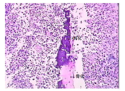
目的制作鼓室硬化豚鼠模型,验证分泌性中耳炎鼓膜置管是鼓室硬化的重要病因。 方法选择健康豚鼠50只,鼓室内注射肺炎链球菌液。注射7 d后选取有中耳积液的30只豚鼠,左耳置入鼓膜通气管,右耳行鼓膜切开作为对照。1个月、3个月、6个月后分别取A、B、C组豚鼠的中耳粘膜,观察其组织形态变化。 结果在制作模型3个月以及更长时间的实验耳中可出现鼓室硬化病理改变, 组织纤维化程度及钙质沉积与通气管放置时间成正比。 结论鼓膜置管是鼓室硬化重要病因。临床鼓膜置管手术需谨慎。 Abstract:Objective To evaluate the role of myringotomy with ventilation tube insertion for otitis media with effusion in thepathogenesis of experimentally induced tympanosclerosis. Methods Fitty guinea pigs were underwent bilaterallytympanocentesis and inoculated with a suspension of Streptococcus pneumoniae to establish the model of tympanosclerosis.After 7 days, 30 guinea pigs showed evidence of otitis media with effusion and were chosen to preform the followingexperiments. The right ears were treated with myringotomy and inserted with ventilation tube, whereas the left ears wereconducted myringotomy to serve as the control group. Middle ear membranes were obtained at 1, 3 and 6 months after theoperation to observe the morphology changes of tympanic membranes and middle ear mucosa. Results The histopathologicalchanges of tympanosclerosis were found in the right ears of the guinea pig models. There was a positive correlation betweenthe duration of ventilation tube insertion and the extent of calcium deposition and fibrosis. Conclusion Myringotomy withventilation tube insertion may be involved in the formation of tympanosclerosis. The surgical procedure should be consideredcarefully in clinical scenario. -
Key words:
- tympanosclerosis /
- guinea pig /
- myringotomy /
- ventilation tube insertion
-
表 1 鼓室硬化的实验结果
Group Research object Tympanosclerosis Percentage A(1 month) 9 0 0 B(3 months) 10 3 0.3 C(6 months) 10 4 0.4 N control 30 0 0  下载: 导出CSV
-
[1] Pereira MB, Pereira DR, Costa SS. Tympanostomy tube sequelae in children with otitis media with effusion: a three-year follow-up study[J]. Braz J Otorhinolaryngol, 2006, 71(4): 415-20. http://cn.bing.com/academic/profile?id=2140764601&encoded=0&v=paper_preview&mkt=zh-cn [2] Kay DJ, Nelson M, Rosenfeld RM. Meta-analysis of tympanostomy tube sequelae[J]. Otolaryngol Head Neck Surg, 2001, 124(4): 374-80. doi: 10.1067/mhn.2001.113941 [3] 李琰, 谢南屏. 鼓室硬化的病因学研究进展[J]. 听力学及言语疾病杂志, 2007, 15(5): 419-20. http://www.cnki.com.cn/Article/CJFDTOTAL-TLXJ200705034.htm [4] John D, Giles J. Tympanosclerosis in the rat tympanic membrane:an experimental study[J]. Laryngoscope, 2002, 112(6): 1663-6. http://cn.bing.com/academic/profile?id=2108847816&encoded=0&v=paper_preview&mkt=zh-cn [5] JJJassar P, Coatesworth A, Strachan DR. Long-term ventilation of the middle ear using a subannular tympanotomy technique: a follow-up study[J]. J Laryngol Otol, 2004, 118(12): 933-6. http://cn.bing.com/academic/profile?id=2078193174&encoded=0&v=paper_preview&mkt=zh-cn [6] Johnston LC, Feldman HM, Paradise JL, et al. Tympanic membrane abnormalities and hearing levels at the ages of 5 and 6 years in relation to persistent otitis media and tympanostomy tube insertion in the first 3 years of Life: a prospective study incorporating a randomized clinical trial [J]. Pediatrics, 2004, 114(1): e58-67. doi: 10.1542/peds.114.1.e58 [7] Tos M, Bonding P, Poulsen G. Tympanosclerosis of the drum in secretory otitis after insertion of grommets. A prospective, comparative study[J]. J Laryngol Otol, 1983, 97(6): 489-96. doi: 10.1017/S0022215100094445 [8] Karlidaga T. Comparison of free radicals and antioxidant enzymesin chronic otitis media with and without tympanosclerosis [J]. Laryngoscope, 2004, 114(1): 85-9. doi: 10.1097/00005537-200401000-00014 [9] Dingle AF, Flood LM, Kumar BU, et al. Tympanosclerosis and mini grommets: the relevance of grommet design[J]. J Laryngol Otol, 1995, 109(10): 922-5. http://cn.bing.com/academic/profile?id=2125454526&encoded=0&v=paper_preview&mkt=zh-cn [10] J KERR.Tympanosclerosis.Clin.Otolaryngol, 1993, 18(4): 341-9. http://journals.lww.com/thehearingjournal/Fulltext/2012/01000/Through_the_Otoscope__The_mysterious.5.aspx [11] Mcminn R. Electron microscopic observations on the repair of perforated tympanic membranes in the guinea-pig[J]. Anat , 1975, 120(9): 207-17. http://cn.bing.com/academic/profile?id=153838168&encoded=0&v=paper_preview&mkt=zh-cn [12] Wielinga E, Kuijpers W. The influence of re-aeration of the middle ear on tympanosclerotic lesions in otitis media with effusion.In:Tos [C]//M:The hague kugler publications, 1997: 629-30. [13] Uneri C, Sari M, Akboğa J, et al. Vitamin e-coated tympanostomy tube insertion decreases the quantity of free radicals in tympanic membrane[J]. Laryngoscope, 2006, 116(1): 140-3. doi: 10.1097/01.mlg.0000191460.32862.bf [14] Uneri C, Bağlam T, Yazici M. The effect of Vitamin E treatment on the development of myringosclerosis after ventilation tube insertion [J]. Int J Pediatr Otorhinolaryngol, 2006, 70(6): 1045-8. doi: 10.1016/j.ijporl.2005.10.019 [15] Aydogan F, Aydin E, Tastan E, et al. Is there any effect of coenzyme Q10 on prevention of myringosclerosis? Experimental study with rats [J]. Braz J Otorhinolaryngol, 2013, 79(3): 293-7. doi: 10.5935/1808-8694.20130053 -

 点击查看大图
图(1) / 表(1)
计量
- 文章访问数: 446
- HTML全文浏览量: 221
- PDF下载量: 0
- 被引次数: 0