


A Comparative observation of callus after surgery for fractures based on Digital Tomosythesis
-
摘要:
目的观察X线断层融合技术与数字摄影在骨折术后愈合进程中骨痂显示的差异。 方法54例骨折术后患者,分别于术后2周、1个月、2个月和3个月均行数字摄影和X线断层融合技术成像,对比两种检查方法获得的图像并作相应的统计学分析。 结果54 例患者中,X线断层融合技术和数字摄影两种方法所获得图像对骨痂的显示情况在术后2 周显示率分别为3.7%和14.81%,有显著差异(P<0.05),术后1 个月显示率分别为38.89%和77.78%,有显著差异(P<0.05),术后2 个月显示率分别为66.67%和87.04%,有显著差异(P<0.05),术后3个月显示率分别为88.89%和94.44%,无显著差异(P>0.05)。 结论X线断层融合技术对骨折术后早期骨痂显示具有重要的价值,较X线平片具有明显优势。 Abstract:Objective To observe the differences of the detection of callus in the early healing process after surgery of fractures between Digital Tomosynthesis (DTS) and Digital Rdiography (DR). Methods A total of 54 patients were performed on DR and DTS imaging after 2 weeks, 1 month, 2 months and 3 months of surgery for fractures and the obtained images were compared. Results In 54 patients, the detection rates of callus by DTS and DR were 3.7% and 14.81% respectively at the second week with significant difference (P<0.05), 38.89% and 77.78% after 1 month with significant difference (P<0.05), 66.67% and 87.04% at the 2 months with significant difference(P<0.05), and 88.89% and 94.44% at the 3 months, without significant difference(P>0.05). Conclusion DTS has an important value for the early detection of callus after surgery for fractures, which has more obvious advantages over the X-ray plain film. -
Key words:
- fracture /
- callus /
- tomography /
- X-ray
-
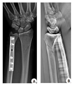
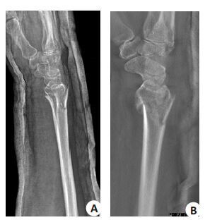
图 3 右侧尺桡骨骨折术后2 个月复查(图A,B为同一患者)
男, 48 岁, 常规X线显示骨折线锐利, 未见骨痂生长, 断层融合检查示骨折线部分消失, 提示少量骨痂生长.
表 1 两种方法对骨痂显示情况对照观察(n=45, %)
时间 DR检查 DTS检查 P 2 周 2(3.7) 8(14.81) <0.05 1 个月 21(38.89) 42(87.04) <0.05 2 个月 36(87.0) 47(87.04) <0.05 3 个月 48(88.89) 51(99.44) >0.05  下载: 导出CSV
-
[1] 尹东, 杨惠林. 影像学在监测和评估骨愈合中的研究现状[J]. 中国医学影像学杂志, 2003, 11(6): 463-4. http://www.cnki.com.cn/Article/CJFDTOTAL-ZYYZ200306034.htm [2] 王兴宇, 尹芸生, 苏晋生, 等. 基于X线断层融合技术对股骨颈骨折术后骨愈合的研究[J]. 中国临床医学影像杂志, 2011, 20(6): 447-8. http://www.cnki.com.cn/Article/CJFDTOTAL-LYYX201106025.htm [3] Anderson LD, Sisk TD, Tooms RE, et al. Compression plate fixation in acute diaphyseal fractures 0f the radiusand ulna[J]. J Bone Joint Surg, 1975, 57(4): 287-97. [4] 刘会玲, 张爱民, 王玮杰. CR钼靶X射线对应用抗骨增生胶囊治疗骨折模型大鼠骨痂密度的评价[J]. 中国组织工程研究与临床康复, 2008, 12(20): 3893-6. http://www.cnki.com.cn/Article/CJFDTOTAL-XDKF200820026.htm [5] 丁斌, 魏红. 骨痂在不同内固定骨折愈合过程中的差异[J]. 地方病通报, 2008, 23(2): 74-5, 77. http://www.cnki.com.cn/Article/CJFDTOTAL-DFBT200802036.htm [6] 赵怀志, 郝华, 卢明书, 等. X光片骨痂定量探讨与应用[J]. 中国中医骨伤科, 1995(4): 8-9, 3. http://www.cnki.com.cn/Article/CJFDTOTAL-ZGZG504.002.htm [7] 吕玉琦, 庞俊. 测量桡骨远端骨折骨痂生长的微机图像分析系统[J]. 中华放射学杂志, 1993, 7(5): 472-5. [8] 赵艳娥, 卢光明, 孙志远, 等. X线数字断层融合技术在骨折石膏固定摄影中的应用[J]. 中国临床医学影像杂志, 2009, 20(10): 797-8. http://www.cnki.com.cn/Article/CJFDTOTAL-LYYX200910026.htm [9] 高向东, 郝晓光. 数字化融合断层用于评价内固定术后骨折愈合情况分析[J]. 中国药物与临床, 2014, 12(7): 935-6. http://www.cnki.com.cn/Article/CJFDTOTAL-YWLC201407037.htm [10] 朱昭环, 耿敬标, 周轲, 等. X线数字断层融合技术在肋骨骨折诊断中的价值[J]. 重庆医学, 2012, 41(4): 370-1. http://www.cnki.com.cn/Article/CJFDTOTAL-WMIA201544042.htm [11] 丁昌懋, 卢振威, 王博, 等. X 线断层融合摄影在桡骨远端骨折复位石膏固定后随访中的应用价值[J]. 河南外科学杂志, 2015, 21(4): 40-1. http://www.cnki.com.cn/Article/CJFDTOTAL-HLWK201504024.htm [12] 陈仲平, 胡宴宾, 张文清, 等. X线数字断层融合技术在髌骨隐匿性骨折中的应用[J]. 河北医学, 2011, 17(9): 1145-7. http://www.cnki.com.cn/Article/CJFDTOTAL-HCYX201109005.htm [13] Clinical GH. Potential of digital linear tomosynthesis imaging of total joint arthroplasty[J]. J Digit Imaging, 2008, 21(3): 312-22. doi: 10.1007/s10278-007-9040-9 [14] 徐晶, 夏秀杰, 田宝方, 等. 数字断层融合技术在骨骼影像诊断中的临床应用价值[J]. 中国医疗设备, 2014(7): 166-8. http://www.cnki.com.cn/Article/CJFDTOTAL-YLSX201407070.htm -


 点击查看大图
图(4) / 表(1)
计量
- 文章访问数: 843
- HTML全文浏览量: 326
- PDF下载量: 4
- 被引次数: 0