


Diagnosis and differential diagnosis of magnetic resonance imaging for mass lesion in basal ganglia region
-
摘要:
目的探讨基底节区常见肿瘤的临床和MRI特点。 方法回顾分析2012年1月~2015年10月之间经病理证实的基底节区肿块性病变的临床、病理和影像资料,观察病变的影像特点及差异。 结果基底节区病变常见囊性变,以胶质瘤和生殖细胞肿瘤多见,脂肪信号仅见于生殖细胞肿瘤;生殖细胞肿瘤水肿程度较轻,低级别胶质瘤和胶质母细胞瘤更明显;出血多见于胶质母细胞瘤和生殖细胞肿瘤;低级别胶质瘤多表现为轻、中度强化,胶质母细胞瘤和淋巴瘤多表现为明显强化。 结论基底节区肿块样病变在影像表现各征象上有一定差别,可以为鉴别诊断提供帮助。 Abstract:Objective To explore the clinical and MRI characteristic of mass lesion in basal ganglia region. Methods A total of 52 patients of mass lesion in basal ganglia region received a pathologic diagnosis and MR scan during 2012.1~2015.10. The characteristic of clinical material and MRI were reviewed. Results Cystic degeneration was commonly seen in gliomas and germ cell tumors, fat signal was found in germ cell tumors, edema in gliomas were more serious than that in other lesion. Glioblastomas and lymphoma were significantly enhanced. Conclusion There is a difference of clinical maerial and MRI between mass lesions in basal ganglia region, which can help differential diagnosis. -
Key words:
- basal ganglia region /
- cystic degeneration /
- enhancement /
- Magnetic Resonance Imaging
-
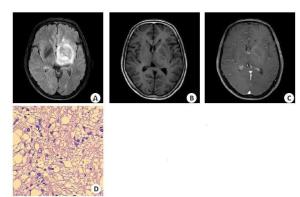
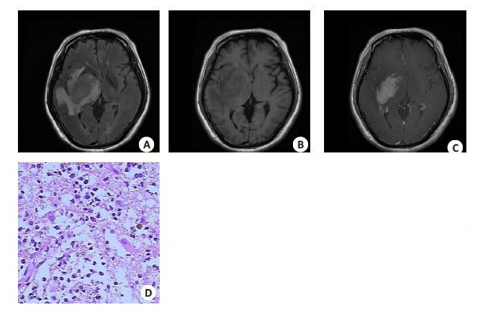
图 1 左侧基底节区低级别胶质瘤
A: FLAIR图像; B: T1WI图像; C:T1WI+C图像;D: 病理结果图像(HE染色, 20)。女, 58岁, 左侧基底节区肿块样病变, 呈混杂长T1长T2信号改变, 肿瘤周围明显水肿, 增强后轻度强化.
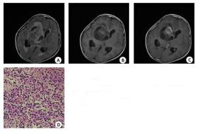
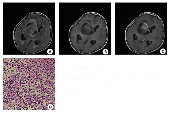
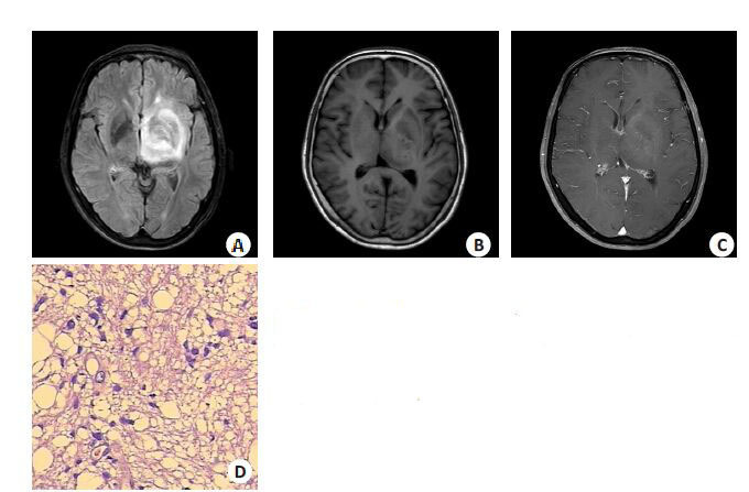
图 2 右侧基底节区混合性生殖细胞肿瘤
A: FLAIR图像; B: T1WI图像; C: T1WI+C图像; D: 病理结果图像(HE染色, 20). 女,10岁, 右侧基底节区肿块样病变, 呈混杂长T1长T2信号改变, 其内见短T1长T2脂肪信号影, 肿瘤周围轻度水肿, 增强后中度强化.
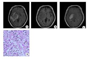
图 3 右侧基底节区大B细胞淋巴瘤
A: FLAIR图像; B: T1WI图像; C: T1WI+C图像; D: 病理结果图像(HE染色, 20). 男, 53岁,右侧基底节区肿块样病变, 呈混杂长T1略长T2信号改变, 肿瘤周围轻度水肿, 增强后明显强化.
表 1 MRI扫描参数
Sequence TR(ms) TE(ms) IR(ms) FOV(cm) Thickness(mm) Spacing(mm) NSA(NEX) T2WI(Brivo) 5100 130 - 24 6 1 1 T1WI(Brivo) 2705 24 860 24 6 1 1  下载: 导出CSV
-
[1] Phi JH, Cho BK, Kim SK, et al. Germinomas in the basal ganglia: magnetic resonance imaging classification and the prognosis[J]. J Neurooncol, 2010, 99(2): 227-36. doi: 10.1007/s11060-010-0119-7 [2] Nagata K, Nikaido Y, Yuasa T, et al. Germinoma causing wallerian degeneration-Case report and review of the literature [J]. J Neurosurg, 1998, 88(1): 126-8. doi: 10.3171/jns.1998.88.1.0126 [3] 刘永辉, 张水兴, 罗剑云, 等. 神经节细胞胶质瘤的影像学表现[J]. 实用放射学杂志, 2012, 28(10): 1647-9. [4] 陆兆丰, 邱永明, 程小兵, 等. 12例基底节区巨大生殖细胞瘤的治疗分析[J]. 上海交通大学学报:医学版, 2012, 35(07): 935-9. http://www.cnki.com.cn/Article/CJFDTOTAL-SHEY201207028.htm [5] 黄翔, 张荣, 黄峰平. 儿童丘脑基底节肿瘤的治疗策略[J]. 中华神经外科杂志, 2012, 28(10): 982-6. [6] 张荣, 沈文倩, 周良辅. 儿童原发性中枢神经系统肿瘤763例临床分析[J]. 中华医学杂志, 2007, 27(07): 442-7. http://www.cnki.com.cn/Article/CJFDTOTAL-ZHYX200707004.htm [7] 黄芳, 张家春, 黄燕. 右侧基底节区非霍奇金淋巴瘤1例[J]. 四川医学, 2012, 35(09): 1702-3. http://www.cnki.com.cn/Article/CJFDTOTAL-SCYX201209094.htm [8] 杨丰忠, 杨春红, 刘春荣, 等. 左额颞叶基底节区原始神经外胚瘤1例并文献复习[J]. 临床神经外科杂志, 2011, 18(02): 106-8. http://www.cnki.com.cn/Article/CJFDTOTAL-LCSW201102034.htm [9] 雷军, 姚丽青, 池彬, 等. 基底节区混合性生殖细胞肿瘤的临床病理分析[J]. 实用癌症杂志, 2014, 21(01): 97-9. http://www.cnki.com.cn/Article/CJFDTOTAL-SYAZ201401034.htm [10] 王志群, 李坤成, 王亮, 等. 基底节区生殖细胞瘤的MRI和MRS研究 [J]. 放射学实践, 2007, 26(05): 448-51. http://www.cnki.com.cn/Article/CJFDTOTAL-FSXS200705005.htm [11] 邱晓光, 罗世祺, 马振宇, 等. 28例基底节区生殖细胞瘤诊断性放疗的评价[J]. 中华神经外科杂志, 2006, 43(05): 290-2. http://www.cnki.com.cn/Article/CJFDTOTAL-ZHSW200605017.htm [12] 金晶, 周义成. 脑多发胶质瘤影像与病理对照研究[J]. 中国医学影像学杂志, 2012, 20(2): 84-7. http://www.cnki.com.cn/Article/CJFDTOTAL-ZYYZ201202005.htm [13] 徐晨阳, 丁炳谦, 李振江, 等. 基底节区胶质瘤的手术治疗分析[J]. 中国当代医药, 2013(25): 32-3. http://www.cnki.com.cn/Article/CJFDTOTAL-ZGUD201325015.htm [14] 段崇锋, 张丕宁, 高耸, 等. 基底节区生殖细胞瘤的C T、MR I诊断价值[J]. 实用放射学杂志, 2014(4): 565-7. [15] 胡炜, 张士中, 王献文. 立体定向置管引流术治疗丘脑、基底节区脑出血60例[J]. 郑州大学学报(医学版), 2006, 41(5): 1000-1. http://www.cnki.com.cn/Article/CJFDTOTAL-HNYK200605090.htm [16] 李海龙, 张剑宁, 米良, 等. 基底节区生殖细胞瘤的临床及影像学特点分析[J]. 临床与病理杂志, 2015, 44(05): 762-6. http://www.cnki.com.cn/Article/CJFDTOTAL-WYSB201505014.htm -

 点击查看大图
图(3) / 表(1)
计量
- 文章访问数: 670
- HTML全文浏览量: 265
- PDF下载量: 2
- 被引次数: 0