


Comparison study of three-dimensional and two-dimensional transesophageal echocardiography in measurement of atrial septal defect
-
摘要:
目的比较实时三维、多平面二维经食管超声心动图在房间隔缺损(atrial septal defect, ASD)测量的应用价值。 方法对67例继发孔型房间隔缺损患者分别应用经食管二维超声常规切面法、经食管二维超声十二切面法、实时三维经食管超声法进行测量,并将各测量结果与封堵器大小行相关性分析。 结果经食管二维超声常规切面法在整体(P < 0.001)、大ASD组(P < 0.001)、椭圆形ASD组(P < 0.01)测量ASD最大径小于实时三维经食管超声法,差异有统计学意义;经食管二维超声十二切面法在小ASD组(P < 0.05)测量ASD最大径大于实时三维经食管超声法,差异有统计学意义;实时三维经食管超声法测量房间隔缺损最大径与封堵器大小的相关性高于经食管二维超声常规切面法及十二切面法。 结论实时三维经食管超声可准确测量房间隔缺损,对封堵器选择有重要指导作用。经食管二维超声十二切面法可与实时三维经食管超声法互为补充。 -
关键词:
- 实时三维经食管超声心动图 /
- 多平面二维经食管超声心动图 /
- 房间隔缺损
Abstract:Objective To campare the application value of three-dimensional (3D) and two-dimensional (2D) transesophageal echocardiography (TEE) in measuring atrial septal defect (ASD). Methods Sixty-seven patients with ASD who undergoing percutaneous trans catheter closure treatment were enrolled. The diameter of ASDs were measured by conventional 2DTEE, 12-plane 2DTEE and 3 DTEE respectively, and correlation with the maximal diameter of ASD and the size of occluder was analyzed. Results The maximal diameter of ASD measured by conventional 2DTTE was less 3DTEE in integral group (P < 0.001), large group (P < 0.001) and Oval group (P < 0.01); the maximal diameter of ASD measured by 12-plane 2DTEE was greater than 3DTTE in small group (P < 0.05). The correlation between the maximal diameter of ASD measured by 3DTEE and the size of the occluder was greater than conventional 2DTEE and 12-plane 2DTEE in all groups. Conclusion 3DTEE can accurately measure the diameter of ASD, it plays an important role in occluder selection. 12-plane 2DTEE and 3DTEE can complement each other in ASD measurement. -
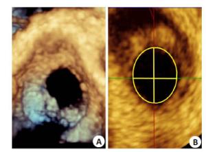
图 2 实时三维经食管超声多平面重建测量法示意图
(A:ASD三维成像; B:MPR功能测量ASD.)
Figure 2. Measurement of ASD by MPR
表 1 3DTEE法与2DTEE常规切面法、十二切面法测量结果对比
Table 1. Comparison of 3DTTE,conventional 2DTEE and 12-plane 2DTEE in ASD measurement(Mean±SD, mm)
组别 整体 小 大 圆形 椭圆形 3DTEE法 19.04±5.63 14.73±4.16 23.24±3.13 20.63±6.12 17.98±5.08 2DTEE常规切面法 18.15±5.52*** 14.30±4.43 21.88±3.57*** 19.96±5.90 16.93±4.95** 2DTEE十二切面法 19.31±5.64 15.21±4.55* 23.29±3.21 21.07±6.08 18.13±5.05 与3DTEE相比*P<0.05, **P<0.01,***P<0.001.  下载: 导出CSV
表 2 3DTEE法、2DTEE常规切面法、2DTEE十二切面法测量结果与封堵器大小的相关性
Table 2. Correlation between the size of the occluder and the maximal diameter of ASD measured by 3DTEE, conventional 2DTEE and 12-plane 2DTEE
组别 2DTEE常规切面法 2DTEE十二切面法 3DTEE法 r P r P r P 整体 0.949 0 0.964 0.000 0.979 0 小 0.921 0 0.958 0.000 0.968 0 大 0.865 0 0.86 0.000 0.931 0 圆形 0.919 0 0.948 0 0.963 0 椭圆形 0.945 0 0.962 0.000 0.973 0
下载: 导出CSV
-
[1] Sampaio F, Ribeiro J, Marcos A, et al. Real-time three-dimensional transesophageal echocardiography-an initial experience[J]. Rev Port Cardiol, 2009, 28(6): 671-82. http://www.academia.edu/12457305/Real-time_three-dimensional_transesophageal_echocardiography--an_initial_experience [2] Johri AM, Witzke C, Solis J, et al. Real-Time Three-Dimensional transesophageal echocardiography in patients with secundum atrial septal defects: outcomes following transcatheter closure[J]. J Am Soc Echocardiogr, 2011, 24(4): 431-7. doi: 10.1016/j.echo.2010.12.011 [3] Taniguchi M, Akagi T, Watanabe N, et al. Application of Real-Time Three-Dimensional transesophageal echocardiography using a matrix array probe for transcatheter closure of atrial septal defect[J]. J Am Soc Echocardiogr, 2009, 22(10): 1114-20. doi: 10.1016/j.echo.2009.06.008 [4] Lodato JA, Cao QL, Weinert L, et al. Feasibility of real-time three-dimensional transoesophageal echocardiography for guidance of percutaneous atrial septal defect closure[J]. Eur J Echocardiogr, 2009, 10(4): 543-8. doi: 10.1093/ejechocard/jen337 [5] Butera G, Romagnoli E, Carminati M, et al. Treatment of isolated secundum atrial septal defects:impact of age and defect morphology in 1,013 consecutive patients[J]. Am Heart J, 2008, 156(4): 706-12. doi: 10.1016/j.ahj.2008.06.008 [6] Bhaya M, Mutluer FO, Mahan E, et al. Live/real time Three-Dimensional transesophageal echocardiography in percutaneous closure of atrial septal defects[J]. Echocardiography, 2013, 30(3): 345-53. doi: 10.1111/echo.2013.30.issue-3 [7] Garcia-Fuertes D, Mesa-Rubio D, Ruiz-Ortiz M, et al. Monitoring complex secundum atrial septal defects percutaneous closure with real time Three-Dimensional echocardiography[J]. Echocardiography, 2012, 29(6): 729-34. doi: 10.1111/j.1540-8175.2012.01679.x [8] Roberson DA, Cui W, Patel D, et al. Three-Dimensional transesophageal echocardiography of atrial septal defect: a qualitative and quantitative anatomic study[J]. J Am Soc Echocardiogr, 2011, 24 (6): 600-10. doi: 10.1016/j.echo.2011.02.008 -


 点击查看大图
计量
- 文章访问数: 549
- HTML全文浏览量: 193
- PDF下载量: 2
- 被引次数: 0