


Application value analysis of BI-RADS grading standard in breast lesions
-
摘要:
目的通过分析乳腺病变的声像图特征,并与病理诊断结果比较,探讨BI-RADS-US分级标准在乳腺病变检查中的应用价值。 方法对2014年1月~2015年6月在本院进行乳腺超声检查的412例BI-RADS分级结果及随访做一回顾性分析。 结果412例乳腺病变BI-RADS分级病理结果中,良性病变367例,恶性病变45例。BI-RADS-US分级对乳腺病变诊断的敏感性为88.89%,特异度为85.83%,准确度为86.17%,其中,Ⅲ~Ⅴ中,PPV逐步增高。 结论BI-RADS分级对于乳腺病变的诊断具有重要的临床指导价值,实施BI-RADS分级可为乳腺病变的规范化及标准化诊断提供参考。 Abstract:Objective To investigate the value of BI-RADS-US classification in the diagnosis of breast lesions by analyzing the ultrasonographic characteristics of breast lesions and to compare with the pathological diagnosis. Methods A retrospective analysis was made on 412 cases of grade BI-RADS grade of breast ultrasound in our hospital from January 2014 to June 2015. Results In 412 cases of breast lesions, 367 cases of benign lesions, 45 cases of malignant lesions, cases of malignant lesions, cases of malignant lesions. The sensitivity of BI-RADS-US classification to the diagnosis of breast lesions was 88.89%, specificity was 85.83%, accuracy was 86.17%, and the PPV was gradually increased by III~V. Conclusion BI-RADS classification has important clinical value in the diagnosis of breast lesions. The implementation of BI-RADS grading can provide a reference for the standardization and standardization of breast lesions. -
Key words:
- breast lesion /
- color Doppler /
- pathology /
- biopsy
-

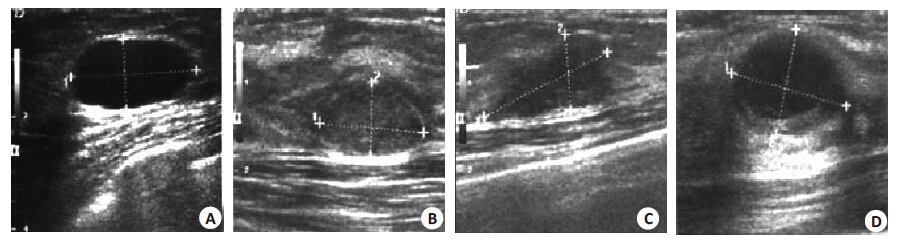
图 1 典型声像图
A: BI-RADS-US 分级为Ⅱ 级,术后病理证实为乳腺单纯囊肿; B: BI-RADS-US 分级为Ⅲ 级,术后病理证实为乳腺纤维瘤; C:BI-RADS-US分级为V级,术后病理证实为乳腺浸润导管癌; D: BI-RADS-US分级为Ⅱ 级,术后病理证实为乳腺浸润导管癌.
表 1 412例乳腺病变BI-RADS-US分级病理结果(n,%)
组别 Ⅱ级 Ⅲ级 Ⅳ级 Ⅴ级 ⅣA ⅣB ⅣC 良性(n=367) 65 232 18 38 12 2 恶性(n=45) 1 2 2 12 13 15 PPV 1.15 0.96 9.09 24.14 51.52 86.96 BI-RADS-US分级为II、III及IVA为良性病变;ⅣB~Ⅴ分级为恶性病变.  下载: 导出CSV
-
[1] 王小燕, 康利克, 蓝春勇, 等. 乳腺恶性肿块超声造影特征表现及诊断[J]. 中国超声医学杂志, 2012, 28(8): 705-8. http://cpfd.cnki.com.cn/Article/CPFDTOTAL-CSYX201207001285.htm [2] 肖晓云, 智慧, 杨海云, 等. 超声造影5分法诊断乳腺肿物的价值初探[J]. 中华超声影像学杂志, 2012, 21(4): 328-31. [3] Stöblen F, Landt S, Ishaq R, et al. High-frequency breast ultrasoundfor the detection of microcalcifications and associated masses inBI-RADS 4a patients[J]. Anticancer Res, 2011, 31(8): 2575-81. [4] 刘赫, 姜玉新, 戴晴, 等. 超声造影对乳腺导管内乳头状瘤的诊断价值[J]. 协和医学杂志, 2014, 5(1): 31-4. http://www.cnki.com.cn/Article/CJFDTOTAL-XHYX201401013.htm [5] 张渊, 江泉, 赵玉华, 等. 超声造影诊断乳腺肿瘤的价值[J]. 中国超声医学杂志, 2011, 27(5): 413-5. http://www.cnki.com.cn/Article/CJFDTOTAL-ZGCY201105018.htm [6] 曾锦树, 陈世良, 许翔, 等. 超声造影在乳腺良恶性病灶鉴别诊断中的应用[J]. 中国超声医学杂志, 2013, 29(6): 500-3. http://www.cnki.com.cn/Article/CJFDTOTAL-ZGCY201306010.htm [7] Yoon JH, Kim MJ, Moon HJ, et al. Subcategorization ofultrasonographic BI-RADS category 4: positive predictive valueand clinical factors affecting it[J]. Ultrasound Med Biol, 2011, 37(5): 693-9. doi: 10.1016/j.ultrasmedbio.2011.02.009 [8] 唐黎之, 王志刚, 冉海涛, 等. BI-RADS3级乳腺病变的回顾分析[J]. 中华超声影像学杂志, 2011, 20(3): 230-3. http://cdmd.cnki.com.cn/Article/CDMD-10631-1011173956.htm [9] Gweon HM, Son EJ, Youk JH, et al. Value of the US BI-RADS finalassessment following mastectomy: BI-RADS 4 and 5 lesions[J].Acta Radiol, 2012, 53(3): 255-60. doi: 10.1258/ar.2011.110597 [10] Raza S, Goldkamp AL, Chikarmane SA, et al. US of breast massescategorized as BI-RADS 3, 4, and 5: pictorial review of factorsinfluencing clinical management[J]. Radiographics, 2010, 30(5):1199-213. doi: 10.1148/rg.305095144 [11] Hsu HH, Yu JC, Lee HS, et al. Complex cystic lesions of the breaston ultrasonography: feature analysis and BI-RADS assessment[J].Eur J Radiol, 2011, 79(1): 73-9. doi: 10.1016/j.ejrad.2009.12.037 [12] Moon HJ, Kim MJ, Kwak JY, et al. Probably benign breast lesionson ultrasonography: a retrospective review of ultrasonographicfeatures and clinical factors affecting the BI-RADS categorization[J]. Acta Radiol, 2010, 51(4): 375-82. doi: 10.3109/02841851003662780 [13] 马慧娟, 冷振鹏, 王萍, 等. 乳腺超声影像报告数据系统在乳腺病灶诊治中的应用价值[J]. 中国超声医学杂志, 2013, 29(11): 971-4. http://www.cnki.com.cn/Article/CJFDTOTAL-ZGCY201311004.htm [14] 陈燕, 张江宇, 马小燕, 等. 超声BI-RADS 分级在乳腺肿块诊断中的应用[J]. 中华临床医师杂志: 电子版, 2013(6): 2421-4. -

 点击查看大图
图(1) / 表(1)
计量
- 文章访问数: 632
- HTML全文浏览量: 305
- PDF下载量: 7
- 被引次数: 0