


The clinical significance of Diffusion Tensor Imaging for resection of basal ganglia region Gliomas
-
摘要:
目的探讨弥散张量成像(DTI)对指导手术切除基底节区胶质瘤及降低致残率的临床意义。 方法收集2009~2014年间我科收治的48例基底节区胶质瘤患者,术前行MRI+DTI,用纤维束示踪方法重建出锥体束,明确肿瘤和锥体束的三维空间结构关系,以避开锥体束设计手术入路,按纤维束受肿瘤侵袭程度指导手术切除肿瘤范围,术后评估神经功能状况。同时选取同期30例基底节区胶质瘤未行DTI的手术患者作为对照组,了解两者两组全切率及术后4周KPS评分的差异。 结果对照组镜下全切19例,次全切7例,部分切除4例,全切率63.3%;DTI组镜下全切29例,次全切13例,部分切除6例,镜下全切率60.4%,两组镜下全切率比较,P>0.05,差别无统计学差异;对照组KPS评分为77.67±19.09分;DTI组KPS评分为87.29±14.84分,两组KPS评分比较,P < 0.05,差别有统计学意义。 结论术前DTI检查对指导手术切除基底节区胶质瘤有重要临床意义,起到降低患者致残率和提高术后生存质量的作用,但无提高肿瘤全切率。 Abstract:Objective To explore the clinical significance of diffusion tensor imaging (DTI) of instructing to remove basal ganglia region Gliomas and decreasing the disability after operation. Methods Fourty-eight cases of basal ganglia region Gliomas which were carried MRI and DTI scanning before operation from 2009 to 2014 were as the DTI Group. While 30 cases in controlled group were only took MRI scanning before operation in the same term. In the DTI group, pyramidal tract was reconstructed by the technique of Fiber Tracking and the 3D structure relationship between the fiber tracts and the tumor was known well before operation. Then, the surgical approach was chosen according to its away from the pyramidal tract. Otherwise, resection range of the tumor tissue was decided according to invasive severity of the pyramidal tract by the tumor. The difference of the rate of resection and the KPS scores were compared between two groups at four weeks after operation. Results In the controlled group, the rate of total resection, subtotal resection and biopsy was respectively 63.3% (19/30), 23.3%(7/30) and 13.3% (4/30); While in the DTI group, they were respectively 60.4% (29/48), 27.1% (13/48) and 12.5% (6/48). There was no significant difference in total resection between these two groups (P>0.05). There was a significant difference in KPS scores between two groups (P < 0.05) due to compare 87.29±14.84 in the DTI group with 77.67±19.09 in the controlled group. Conclusion DTI presurgically is good for instructing to remove basal ganglia region Gliomas due to its decreasing the disability rate and increasing living quality of patients after operation. But it did not contribute to total resection of tumors. -
Key words:
- diffusion tensor imaging /
- glioma /
- basal ganglia region /
- surgery
-

图 1 1例基底节区前部胶质瘤的手术入路设计及肿瘤切除程度
A: MRI 增强扫描示左基底节区前方肿瘤,累及豆状核及内囊前肢;B: DTI 示左侧锥体束被肿瘤推向后方且受肿侵犯不完整,采用前外侧入路(白色箭头所示); C: MRI 增强扫描示肿瘤切除较彻底; D:DTI示左侧锥体束位于术区后方,与术前相比无新增受损.
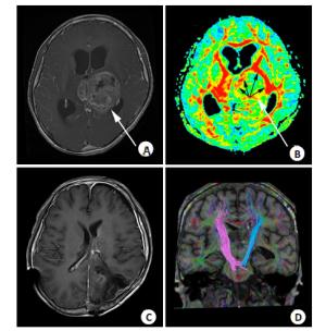
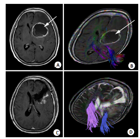
图 2 1例基底节区后部胶质瘤的手术入路选择及手术切除范围
A: MRI增强扫描示左侧丘脑肿瘤,累及内囊后肢及对侧丘脑; B: 彩图示左侧椎体束被肿瘤推向前方及内外侧,呈“C”形开口向后(黑色箭头所示),锥体束较完整,选择后外侧入路(白色箭头所示); C: MRI增强扫描示肿瘤切除较彻底; D: DTI 示肿瘤双侧锥体束较完整,肿瘤侧锥体束无明显受损.
表 1 两组镜下全切率比较
组别 例数 手术入路 肿瘤切除范围 内侧 前方 外侧 前外侧 后外侧 后方 全切除 次全切除 部分切除 全切率(%) 对照组 30 18 0 2 3 17 0 19 7 4 63.3 DTI组 48 26 2 4 7 7 2 29 13 6 60.4* *为P>0.05 两组肿瘤全切率比较无统计学差异.  下载: 导出CSV
-
[1] Lévy S, Chapet S, Mazeron JJ. Management of gliomas.CancerRadiother [J]. Cancer Radiothérapie Journal De La SociétéFrançaise De Radiothérapie Oncologique, 2014, 18(5): 461-7. [2] Albright AL. Feasibility and advisability of resections of thalamictumors in pediatric patients[J]. J Neurosurg, 2004, 100(5 SupplPediatrics): 468-72. [3] 李琳, 何琦玮, 孙夕林, 等. DTI多参数值及DTT技术在颅内肿瘤中的应用价值研究[J]. 哈尔滨医科大学学报, 2010, 44(3): 291-5. http://www.cnki.com.cn/Article/CJFDTOTAL-HYDX201003033.htm [4] 刘敏, 张璐, 孙丽华, 等. 辽宁省肿瘤化疗患者KPS评分情况分析[J]. 中国肿瘤, 2013, 22(8): 635-7. http://www.cnki.com.cn/Article/CJFDTOTAL-ZHLU201308010.htm [5] Franzini A, Leocata F, Cajola L, et al. Low-grade glial tumors inbasal ganglia and thalamus: natural history and biologicalreappraisal [J]. Neurosurgery, 1994, 35(5): 817-20; discussion 820-1. doi: 10.1227/00006123-199411000-00003 [6] Baroncini M, Vinchon M, Minéo JF, et al. Surgical resection ofthalamic tumors in children: approaches and clinical results[J].Childs Nerv Syst, 2007, 23(7): 753-60. doi: 10.1007/s00381-007-0299-4 [7] Sanai N, Berger MS. Glioma extent of resection and its impact onpatient outcome[J]. Neurosurgery, 2008, 62(4): 753-64; discussion264-6. doi: 10.1227/01.neu.0000318159.21731.cf [8] Berman J. Diffusion Mr tractography as a tool for surgical planning[J]. Magn Reson Imaging Clin N Am, 2009, 17(2): 205-14. doi: 10.1016/j.mric.2009.02.002 [9] Moshel YA, Elliott RE, Monoky DJ, et al. Role of diffusion tensorimaging in resection of thalamic juvenile pilocytic astrocytoma[J].J Neurosurg Pediatr, 2009, 4(6): 495-505. doi: 10.3171/2009.7.PEDS09128 [10] Li YO, Yang FG, Nguyen CT, et al. Independent componentanalysis of DTI reveals multivariate microstructural correlations ofwhite matter in the human brain[J]. Hum Brain Mapp, 2012, 33(6):1431-51. doi: 10.1002/hbm.v33.6 [11] Ferda J, Kastner J, Mukensnabl P, et al. Diffusion tensor magneticresonance imaging of glial brain tumors[J]. Eur J Radiol, 2010, 74(3): 428-36. doi: 10.1016/j.ejrad.2009.03.030 [12] Stadlbauer A, Hammen T, Grummich P, et al. Classification ofperitumoral fiber tract alterations in gliomas using metabolic andstructural neuroimaging[J]. J Nucl Med, 2011, 52(8): 1227-34. doi: 10.2967/jnumed.111.090597 -


 点击查看大图
图(2) / 表(1)
计量
- 文章访问数: 580
- HTML全文浏览量: 256
- PDF下载量: 6
- 被引次数: 0