


Analysis of the CT imaging of abdominal necrotizing fasciitis caused by Escherichia-co-li-infected appendicitis
-
摘要:
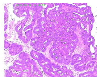
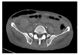
目的对大肠埃希菌引起阑尾炎所致腹壁坏死性筋膜炎案例进行探讨。 方法采用回顾性研究,总结1例由盲肠癌侵犯阑尾,由大肠埃希菌感染引起腹壁坏死性筋膜炎的影像学、组织细胞学、及病理学综合分析。 结果该病例CT平扫表现为全腹壁弥漫性水肿,皮下广泛性积气,腰大肌脓肿,增强后呈环形强化,盲肠管壁不规则增厚,阑尾包绕其中,阑尾远端增粗、短缩,增强后明显强化,阑尾周围炎性渗出,取腹壁脓液细菌培养为大肠埃希菌,经肠镜及病理检查,证实盲肠腺癌,阑尾开口闭塞,经临床有效治疗后复查,腹壁脓肿、皮下积气明显减少。 结论对于大肠埃希菌继发引起阑尾炎所致腹壁坏死性筋膜炎,应根据腹壁坏死性筋膜炎症状、体征及特点早期做出诊断,采取正确的检查方法,了解病变的范围及病因,为临床治疗提供及时、准备的信息。 -
关键词:
- 大肠埃希菌 /
- 阑尾炎 /
- 腹壁坏死性筋膜炎CT影像学表现 /
- CT影像学表现
Abstract:ObjectiveTo probe into the case of abdominal necrotizing fasciitis caused by Escherichia-coli-infected appendicitis. MethodsRetrospectively study and comprehensively sum up the analysis of medical imaging, histology, cytology and pathology of appendix abdominal necrotizing fasciitis, which is a case caused by colon cancer invasion and Escherichia coli infection. ResultsCT scan of the case shows abdominal diffuse edema, extensive subcutaneous pneumatosis, psoas abscess, ring-shaped enhancement after strengthening; irregular cecum wall thickening, appendix wrapped around the cecum, thickening and shortening of appendix distal, obvious enhancement after strengthening, periappendiceal inflammatory infiltration. Bacteria cultured from abdominal wall fester are Escherichia coli, which is confirmed to be cecum adenocarcinoma by endoscopy and pathological examination. CT scan also shows appendiceal orifice occlusion. After effective clinical treatment and recheck, there is significant reduction of abdominal wall abscess and subcutaneous pneumatosis. ConclusionFor abdominal necrotizing fasciitis caused by Escherichia-coli-infected appendicitis, early diagnosis should be made according to abdominal necrotizing fasciitis symptoms, signs and characteristics. Proper examination methods should be adopted to understand the extent of lesion and the pathogeny so as to provide timely and prepared information for clinical treatment. -
Key words:
- Escherichia coli /
- appendicitis /
- abdominal necrotizing fasciitis /
- CT imaging
-
[1] 陈咏君, 高波, 张延, 等.大肠埃希菌性阑尾炎的耐药性分析[J].沈阳医学院学报, 2012, 14(01): 19-5. http://www.cnki.com.cn/Article/CJFDTOTAL-SYYX201201010.htm [2] 肖丽华, 管有理. 40例阑尾炎并发腹壁坏死性筋膜炎的护理体会[J].贵阳中医学院学报, 2012, 34(05): 158-60. http://www.cnki.com.cn/Article/CJFDTOTAL-GYZX201205085.htm [3] 李林强, 孟庆辉, 梁德森, 等.肛周脓肿致腹壁及腹膜后坏死性筋膜炎1例[J].中国实用外科杂志, 2010, 30(07): 619. http://www.cnki.com.cn/Article/CJFDTOTAL-ZGWK201007038.htm [4] 张诚, 刘毅, 孙晓晨, 等.腹壁坏死性筋膜炎合并广泛皮肤软组织缺损的修复[J].解放军医药杂志, 2013, 25(12): 6-10. http://www.cnki.com.cn/Article/CJFDTOTAL-HBGF201312002.htm [5] 刘娜, 王江滨, 赵立梅. 1例肛周脓肿并发腹壁及腹膜后坏死性筋膜炎的护理体会[J].中国伤残医学, 2010, 18(6): 195-6. http://www.cnki.com.cn/Article/CJFDTOTAL-XCYZ201006173.htm [6] 苗强.肛周脓肿致腹壁急性坏死性筋膜炎2例分析[J].中国社区医师(医学专业), 2011, 13(20): 237. http://www.cnki.com.cn/Article/CJFDTOTAL-ZGSQ201120247.htm [7] 沈均良, 樊长姝.结节性筋膜炎的影像学表现与病理所见[J].实用医学影像杂志, 2005, 21(2). http://www.cnki.com.cn/Article/CJFDTOTAL-SYXY200501011.htm [8] 肖恩华.坏死性筋膜炎临床和影像学表现[J].临床放射学杂志, 2002.5. http://www.cnki.com.cn/Article/CJFDTOTAL-LCFS200205030.htm [9] 戚乐, 向军益, 戴平丰等.阑尾石多排螺旋CT征象诊断阑尾炎的价值[J].实用放射学杂志, 2012, 28(7): 1045-7. http://d.wanfangdata.com.cn/Periodical/syfsxzz201207015 [10] 王浩, 魏冉, 王兰云等.穿孔性阑尾炎与非穿孔性阑尾炎的CT鉴别诊断[J].医学影像学杂志, 2012, 22(1): 106-10. http://www.cnki.com.cn/Article/CJFDTOTAL-XYXZ201201037.htm -

 下载:
下载:
 点击查看大图
图(2)
计量
- 文章访问数: 690
- HTML全文浏览量: 158
- PDF下载量: 0
- 被引次数: 0