


Comparison of CT scanning and X ray in measuring the volume of the effusion for the cavity caused by pulmonary tuberculosis or lung carcinoma
-
摘要:
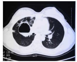
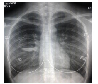
目的对比胸部CT扫描与X线平片对肺结核及肺癌空洞积液量的测量差异。 方法收集同时进行X线胸片及胸部增强CT扫描确诊空洞并积液的肺结核及肺癌患者患者各36及32例,均在CT引导下行积液穿刺,对比3种测量方法下的积液量差异。 结果结核及癌性空洞患者均表现为胸部增强CT所测得的积液量明显较X线平片多(P < 0.05),且穿刺实际所抽出积液量同样较X线平片多(P < 0.05),但胸部CT所测积液量与穿刺所得无统计学差异。 结论对于同一患者在同一时间点,胸部CT增强扫描所测得的空洞积液量更加接近实际体积,提示CT的测量精度更高。 Abstract:ObjectiveTo compare the CT scanning and χ ray in measuring the volume of the effusion for the cavity caused by pulmonary tuberculosis or lung carcinoma. MethodsWe collected simultaneous χ-ray sternum and breast enhancement CT scan confirmed empty and effusion of the 36 and 32 patients with tuberculosis and lung cancer patients, both in CT guiding effusion puncture, compare the three methods of measuring the amount of effusion. ResultsThe tuberculosis hole and cancer patients were measured by enhanced CT chest effusion quantity more obvious than χ-ray plain film (P < 0.05), and piercing actual amount drawn effusion more equally than χ-ray plain film (P < 0.05), but the chest CT and the quantity measured effusion puncture income no statistical difference. ConclusionFor patients with the same point at the same time, the chest CT enhanced scan cavity effusion quantity closer to actual measured by volume, prompt CT measurement precision was higher. -
Key words:
- tuberculosis /
- lung cancer /
- empty /
- Chest CT enhanced scan /
- χ-ray plain film
-
表 1 不同类型空洞积液体积比较
病变类型 结核空洞积液体积
(ml, n=36)癌性空洞积液体积
(ml, n=32)X线平片 63.57±10.84# 46.27±8.91# 胸部CT增强扫描 84.72±13.69* 62.34±11.07* 穿刺 81.14±11.98* 59.19±10.52* F -5.991 -4.126 P 0.013 0.021 与X线平片比较,*P < 0.05;与穿刺抽液比较,#P < 0.05.  下载: 导出CSV
-
[1] 陈勤, 姚亮平, 许家亮.不同年龄组继发性肺结核空洞与肺内其他病变的CT评价[J].实用医学影像杂志, 2010, 11(6): 351-3. http://www.cnki.com.cn/Article/CJFDTOTAL-SYXY201006005.htm [2] 闫瑞冬. CT引导下经皮肺穿刺介入治疗空洞型肺结核36例临床观察[J].临床肺科杂志, 2013, 18(4): 760. http://www.cnki.com.cn/Article/CJFDTOTAL-LCFK201304095.htm [3] 王海彦, 庄一平, 张晋, 等. CT引导下细针穿刺活检对肺部空洞性病变的诊断价值[J].临床放射学杂志, 2013, 32(7): 1021-5. http://www.cnki.com.cn/Article/CJFDTOTAL-LCFS201307034.htm [4] 邓祥俊.肺结核空洞与肺癌空洞的CT鉴别诊断[J].现代医药卫生, 2014, 30(21): 3286-7. http://www.cnki.com.cn/Article/CJFDTOTAL-XYWS201421042.htm [5] 杨欣, 杨州, 李唯, 等.肺部多发空洞性病变的CT诊断[J].实用医学影像杂志, 2011, 12(3): 152-5. http://www.cnki.com.cn/Article/CJFDTOTAL-SYXY201103005.htm [6] 田葵, 余辉山, 沙晋璐.低剂量CT定位下的介入治疗在耐药空洞型肺结核中的应用[J].放射学实践, 2013, 28(10): 1069-72. http://www.cnki.com.cn/Article/CJFDTOTAL-FSXS201310026.htm [7] 陈根铭, 赵双全, 朱少乾, 等.初治痰涂片阳性肺结核单发空洞治疗前后的影像学观察[J].医学影像学杂志, 2013, 23(5): 710-4. http://www.cnki.com.cn/Article/CJFDTOTAL-XYXZ201305016.htm [8] 莫国友, 姚亮平, 陈勤.中青年继发性肺结核空洞与空洞外肺结核病变的CT表现分析[J].实用放射学杂志, 2010, 7: 953-6. http://d.wanfangdata.com.cn/Periodical/syfsxzz201007010 [9] 阿孜肯, 古丽巴合提.大空洞周围型肺癌CT误诊结核分析[J].现代医用影像学, 2010, 19(3): 174-5. http://www.cnki.com.cn/Article/CJFDTOTAL-XDYY201003017.htm [10] 郭少贤, 廖星明. CT在空洞型肺结核诊断中的应用[J].临床医学工程, 2010, 17(2): 73-4. http://www.cnki.com.cn/Article/CJFDTOTAL-YBQJ201002038.htm -

 点击查看大图
图(2) / 表(1)
计量
- 文章访问数: 638
- HTML全文浏览量: 190
- PDF下载量: 3
- 被引次数: 0