


The value of transvaginal ultrasound elasticity imaging in the diagnosis of cervical carcin oma
-
摘要:
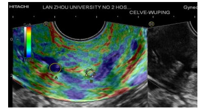
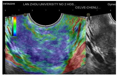
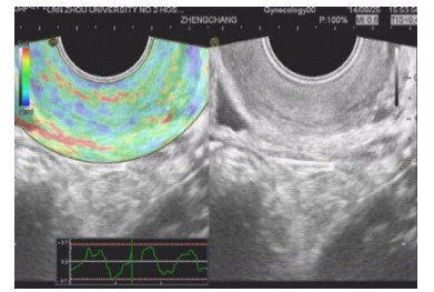
目的探讨探讨超声弹性成像技术在宫颈癌诊断的应用价值。 方法应用超声弹性成像技术检测2013年6月~2014年9月就诊于我院的32例宫颈癌患者病灶和宫颈上皮内瘤变(CIN)患者5例及20例正常宫颈,记录图像及测量相关数值进行分析统计。 结果32例宫颈癌患者,有5例诊断为良性,5例CIN患者中,有2例诊断为恶性,对于20正常宫颈中有3例诊断为恶性,超声弹性成像诊断宫颈癌的敏感度84.37%、特异度80%、准确度82.45%。 结论超声弹性成像在宫颈癌的诊断中有较高的应用价值,具有临床推广意义,但是超声弹性成像在癌前病变与宫颈癌的评分中存在交叉,由于样本量的限制,需更大的样本量进行验证。同时超声弹性成像有望用于宫颈癌化疗效果的评价以及在临床分期中提供客观的依据。 Abstract:ObjectiveTo discuss the application of ultrasound elastography imagine in the diagnosis of cervical cancer. MethodsApplication of ultrasound elastography imaging detection on June 6, 2013 -September 2013 clinic in our hospital of 32 cases of cervical cancer patients with lesions and cervical intraepithelial neoplasia (CIN) patients in 5 cases and 20 cases of normal cervix, numerical analysis of statistical record images and measurement. Results32 cases of cervical cancer patients, 5 cases were diagnosed as benign, 5 cases of CIN patients, 2 cases were diagnosed as malignant, there are 3 in 20 normal cervix cases diagnosed as malignant, the sensitivity of ultrasound elastography in the diagnosis of cervical cancer by 84.37%, specific degree 80%, 82.45% accuracy. ConclusionUltrasound elastography imagine in the diagnosis of cervical cancer has higher application value and clinical significance, but the ultrasound elastography imagine in the score of precancerous lesions and cervical cancer crossed, because of the limitation of sample size should be larger sample size for validation. Ultrasound elasticity imaging at the same time is expected to be used for cerical cancer chemotherapy effect evaluation and provide objective basis in clinical stage. -
Key words:
- ultrasound elastography imagin /
- cervical cancer /
- accuracy
-
[1] 曹泽毅.中国妇科肿瘤学, 7[M].北京:人民军医出版社, 2011: 797-811. [2] Thomas A, Kümmel S, Gemeinhardt O, et al. Real-Time sonoelastography of the cervix: tissue elasticity of the normal and abnormal cervix[J]. Acad Radiol, 2007, 14(2): 193-200. doi: 10.1016/j.acra.2006.11.010 [3] Shepherd JH, Milliken D. Conservative surger for carnoma of thecervix[J]. Clin Oncol, 2008, 20(6): 395-400. doi: 10.1016/j.clon.2008.05.002 [4] Pickerell DM. Elastography:imaging of tomorrow[J]. J Diagn Med Sonogr, 2010, 26(3): 109-13. doi: 10.1177/8756479310370482 [5] Pecorelli S. Revised FIGO staging for carcinoma of the vulva, cervix, and endometrium[J]. International Journal of Gynecology & Obstetrics, 2009, 105(2): 103-4. https://www.ncbi.nlm.nih.gov/pubmed/19367689 [6] Ophir J, Céspedes I, Ponnekanti H, et al. Elastography: a quantitative method for imaging the elasticity of biological tissues [J]. Ultrason Imaging, 1991, 13(2): 111-34. doi: 10.1177/016173469101300201 -

 下载:
下载:
 点击查看大图
图(3)
计量
- 文章访问数: 508
- HTML全文浏览量: 279
- PDF下载量: 5
- 被引次数: 0