


Correlation between the ultrasonic BI-RADS features and the expression of the Ki-67 in breast cancer
-
摘要:
目的探讨应用乳腺影像学报告及数据系统(BI-RADS)描述乳腺癌超声指标与Ki-67表达的相关性及其临床意义。 方法对174名在南方医科大学附属深圳妇幼保健院乳腺外科住院治疗并经病理证实的乳腺癌患者共计194个乳腺癌病灶的超声BI-RADS指标与免疫组织化学方法测定的肿瘤Ki-67表达结果进行对比分析。 结果194个乳腺癌病灶中,超声诊断乳腺癌的敏感度为86.1%,Ki-67阳性表达123例,阳性表达率63.4%(123/194)。单因素分析显示Ki-67阳性表达的乳腺癌肿块中边缘成角、边缘毛刺、内部微钙化、血流丰富指标出现比率高于阴性肿块(P < 0.05)。多因素分析显示边缘毛刺和血流丰富进入回归模型(P < 0.05)。 结论乳腺癌超声BI-RADS指标与Ki-67表达具有一定的相关性,可根据乳腺癌的超声表现初步预测其预后,为临床治疗方案的选择提供一定的帮助。 -
关键词:
- 乳腺癌 /
- 超声 /
- 乳腺影像学报告及数据系统 /
- Ki-67
Abstract:ObjectiveTo investigate the clinical prognosis of the correlation between ultrasonic features described by breast imaging reporting and data system (BI-RADS) and expression of the Ki-67 of breast cancers. MethodsThere were 174 patients hospitalized in breast surgery in Shenzhen Maternity & Child Healthcare Hospital Affiliated to Southern Medical University and 194 lesions of these patients were confirmed by pathology. Their ultrasonic features and expression of the Ki-67 examined by immunohistochemistry were correlation analyzed. ResultsThe sensitivity of ultrasound in diagnosis of breast cancer was 86.1%.The expression rate of the Ki-67 were 63.4% (123/194). Univariate analysis showed angled edge, spiculesign, microcalcifications and rich bloodstream were higher in Ki-67-positive tumors than Ki-67-negative tumors (P < 0.05). Multivariate analysis showed spiculesign and rich bloodstream entered into the regression model (P < 0.05). ConclusionsThere is significant correlation between ultrasound BI-RADS features and the expression of the Ki-67 in breast cancers.According to the ultrasound features to predict prognosis of breast cancers preliminarily will provid some help for clinical treatment. -
Key words:
- breast cancer /
- ultrasound /
- breast imaging reporting and data system /
- Ki-67
-
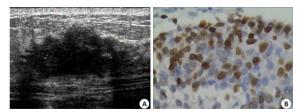
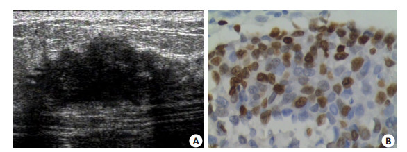
图 1 右乳浸润性导管癌
A:二维超声声像图示右乳11点距离乳头4.0 cm处一个大小约2.5 cm×1.3 cm肿块, 肿块边缘呈粗细、长短不一的发射状低回声, 即“边缘毛刺”征; B:Ki-67免疫组织化学染色阳性, 指数约90%(HE×40).
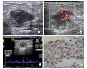
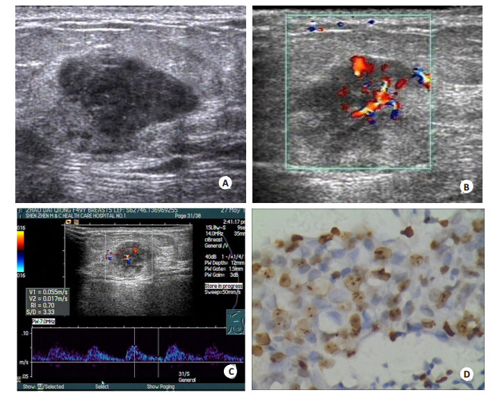
图 2 左乳浸润性导管癌
A:二维超声声像图示左乳12点距乳头6.0 cm处一个大小约1.6 cm×1.0 cm肿块;B:彩色多普勒血流成像显示肿块内可探及3条以上血管相互连通, 交织成网状, 为Ⅲ级血流丰富; C:脉冲多普勒血流成像检测肿块内最大流速5.5 cm/s, 阻力指数0.7; D:Ki-67免疫组织化学染色阳性, 指数约60%(HE×40).
表 1 乳腺癌超声BI-RADS与Ki-67表达的单因素分析
超声BI-RADS Ki-67阴性n(%) Ki-67阳性n(%) OR P 最大直径 < 2.0 45(63.4) 66(53.7) 1 ≥2.0 26(36.6) 57(46.3) 1.495 0.188 形态 卵圆形 28(39.4) 34(27.6) 1 圆形 10(14.1) 15(12.2) 1.235 0.661 不规则形 33(46.5) 74(60.2) 1.847 0.063 边缘 边缘清楚 31(43.7) 3(25.2) 1 边缘模糊 15(21.1) 14(11.4) 0.933 0.878 边缘微分叶 15(21.1) 22(17.9) 1.467 0.362 边缘成角 9(12.7) 23(18.7) 2.556 0.045 边缘毛刺 (1.4) 33(26.8) 33.000 0.001 边界 清楚锐利 62(87.3) 95(77.2) 1 强回声晕 9(12.7) 28(22.8) 2.030 0.089 包膜 包膜完整 6(8.5) 8(6.5) 1 包膜不完整 5(7.0) 5(4.1) 0.750 0.729 无包膜 60(84.5) 110(89.4) 1.375 0.572 方向 方向平行 58(81.7) 94(76.4) 1 方向垂直 13(18.3) 29(23.6) 1.376 0.392 内部回声 等回声 3(4.2) 0(0) 1 低回声 67(94.4) 118(95.9) 2.845 0.999 混合回声 (1.4) 5(4.1) 8.077 0.999 后方回声 增强 6(8.5) 3(2.4) 1 不变 53(74.6) 98(79.7) 3.698 0.072 衰减 12(16.9) 22(17.9) 3.667 0.101 微钙化 无 49(69.0) 53(43.1) 1 有 22(31.0) 70(56.9) 2.942 0.001 粗钙化 无 67(94.4) 118(95.9) 1 有 4(5.6) 5(4.1) 0.710 0.618 血流 无 49(69.0) 7(57.7) 1 少量 16(22.5) 18(14.6) 0.776 0.517 宁-富 6(8.5) 34(27.6) 3.911 0.005 淋巴结 无 7(100) 11(90.2) 1 有 0(0) 12(9.8) 1.033 0.999 OR:比数比, OR为1代表该亚变量为参照类, 其余每一类与参照类比较  下载: 导出CSV
表 2 乳腺癌超声BI-RADS与Ki-67表达的多因素Lo-gistic回归分析
超声BI-RADS OR (95%CI) P 边缘毛刺 22.087(2.719~179.421) 0.004 血流丰富 4.022(1.371~11.796) 0.011
下载: 导出CSV
-
[1] 陈万青, 张思维, 曾红梅, 等.中国2010年恶性肿瘤发病与死亡[J].中国肿瘤, 2014, 23(1): 1-10. http://www.cnki.com.cn/Article/CJFDTOTAL-ZHLU201401001.htm [2] Gerdes J, Schwab U, Lemke H, et al. Production of a mouse monoclonal antibody reactive with a human nuclear antigen associated with cell proliferation[J]. Int J Cancer, 1983, 31(1): 13-20. doi: 10.1002/(ISSN)1097-0215 [3] Schlüter C, Duchrow M, Wohlenberg C, et al. The cell proliferation-associated antigen of antibody Ki-67: a very large, ubiquitous nuclear protein with numerous repeated elements, representing a new kind of cell cycle-maintaining proteins[J]. J Cell Biol, 1993, 123(3): 513-22. doi: 10.1083/jcb.123.3.513 [4] Yerushalmi R, Woods R, Ravdin PM, et al. Ki67 in breast cancer: prognostic and predictive potential[J]. Lancet Oncol, 2010, 11(2): 174-83. doi: 10.1016/S1470-2045(09)70262-1 [5] Sedgwick E. The breast ultrasound lexicon:breast imaging reporting and data system (BI-RADS)[J].Semin Roentgenol, 2011, 46(4): 245-51. doi: 10.1053/j.ro.2011.04.001 [6] Adler DD, Carson PL, Rubin JM, et al. Doppler ultrasound color flow imaging in the study of breast cancer: preliminary findings[J]. Ultrasound Med Biol, 1990, 16(6): 553-9. doi: 10.1016/0301-5629(90)90020-D [7] 王宝娜, 王翔, 王靖, 等. Ki67在乳腺癌中的表达及临床意义[J].中华肿瘤杂志, 2014, 36(4): 273-5. http://d.wanfangdata.com.cn/Periodical/zhzl201404007 [8] Sparano JA, Fazzari M, Kenny PA. Clinical application of gene expression profiling in breast cancer[J]. Surg Oncol Clin N Am, 2010, 19(3): 581. doi: 10.1016/j.soc.2010.03.008 [9] 王景宇, 冬冬, 戴春来, 等.胃癌CT征象与组织分化及p53、Ki67表达的相关性[J].中国医学科学院学报, 2011, 33(5): 555-9. http://www.cnki.com.cn/Article/CJFDTOTAL-ZYKX201105016.htm [10] Liu M, Lawson G, Delos M, et al. Predictive value of the fraction of Cancer cells immunolabeled for proliferating cell nuclear antigen or Ki67 in biopsies of head and neck carcinomas to identify lymph node metastasis: comparison with clinical and radiologic examinations[J]. Head Neck, 2003, 25(4): 280-8. doi: 10.1002/(ISSN)1097-0347 [11] Cheang MC, Chia SK, Voduc D, et al. Ki67 index, HER2 status, and prognosis of patients with luminal B breast cancer[J]. J Natl Cancer Inst, 2009, 101(10): 736-50. doi: 10.1093/jnci/djp082 [12] Dowsett M, Nielsen TO, A'hern R, et al. Assessment of Ki67 in breast cancer: recommendations from the International Ki67 in breast cancer working group[J]. J Natl Cancer Inst, 2011, 103(22): 1656-64. doi: 10.1093/jnci/djr393 [13] Viale G, Giobbie-Hurder A, Regan MM, et al. Prognostic and predictive value of centrally reviewed Ki-67 labeling index in postmpausal womenwith endocrine-responsive breast can sults from Breast International Group Trial 1-98 comparingadjuvant tamoxifen with letrozole[J]. J Clin Oncol, 2008, 26(34): 5569-75. doi: 10.1200/JCO.2008.17.0829 [14] Viale G, Regan MM, Mastropasqua MG, et al. Predicive value of tumor Ki-67 expression in two randomized trials of adjuvant chemedocrine therapyfor node-negative breast cancer[J]. J Natl Cancer Inst, 2008, 100(3): 207-12. doi: 10.1093/jnci/djm289 [15] 雷双根, 余小芬, 谢春伟, 等.乳腺癌Ki67和CK5/6的表达及与临床病理特征的关系[J].广东医学, 2013, 34(19): 2971-4. http://www.cnki.com.cn/Article/CJFDTOTAL-GAYX201319028.htm [16] 闫山英, 李海平, 马力, 等.乳腺癌组织中Ki67的表达及临床意义[J].河北医药, 2013, 35(12): 1769-71. http://www.cnki.com.cn/Article/CJFDTOTAL-HBYZ201312004.htm [17] 王华毅, 张兆祥, 胡余昌, 等.乳腺癌和非癌组织中Syk、survivin和Ki-67的表达及其相关性[J].临床与实验病理学杂志, 2008, 24(2): 158-61. http://www.cnki.com.cn/Article/CJFDTOTAL-LSBL201001012.htm [18] Korpraphong P, Tritanon O, Tangcharoensathien W, et al. Ultrasonographic characteristics of mammographically occult small breast cancer[J]. J Breast Cancer, 2012, 15(3): 344-9. doi: 10.4048/jbc.2012.15.3.344 [19] Folkman J. Fighting cancer by attacking its blood supply[J]. Sci Am, 1996, 275(3): 150-4. doi: 10.1038/scientificamerican0996-150 [20] Nakopoulou L, Stefanaki K, Panayotopoulou E, et al. Expression of the vascular endothelial growth factor receptor-2/Flk-1 in breast carcinomas: correlation with proliferation[J]. Hum Pathol, 2002, 33 (9): 863-70. doi: 10.1053/hupa.2002.126879 [21] Cox RF, Morgan MP. Microcalcifications in breast cancer: Lessons from physiological mineralization[J]. Bone, 2013, 53(2): 437-50. doi: 10.1016/j.bone.2013.01.013 [22] Stavros AT, Thickman D, Rapp CL, et al. Solid breast nodules: use of sonography to distinguish between benign and malignant lesion [J]. Radiology, 1995, 196(1): 123-34. doi: 10.1148/radiology.196.1.7784555 -

 点击查看大图
图(2) / 表(2)
计量
- 文章访问数: 616
- HTML全文浏览量: 216
- PDF下载量: 7
- 被引次数: 0