


Prenatal ultrasound imaging characteristics and clinical significance of abnormal development of the inferior vena cava
-
摘要:
目的 总结胎儿下腔静脉(IVC)发育异常超声图像特征,探讨产前超声检查价值及临床意义。 方法 回顾性分析我院2018年1月~2023年2月产前诊断为胎儿IVC发育异常的88例孕妇的超声图像特征及合并心内外异常情况;不同类型IVC发育异常合并心内外异常构成比行多列表卡方检验,采用Bonferroni方法行组间多重比较。所有结果与产后MRA及超声心动图或尸检结果进行对比。 结果 88例IVC发育异常胎儿中,下腔静脉异常连接(ECIVC)18例,肾后段左下腔静脉(LIVC)42例,双下腔静脉(DIVC)28例。18例ECIVC胎儿胸腹斜冠状切面及四腔心切面均表现异常,其中腹部横切面异常17例,腹部横切面异常征象不典型1例;2例右心房纵切面表现异常;42例LIVC胎儿超声IVC冠状切面均呈“S”征;28例DIVC胎儿超声IVC冠状切面均呈“h”征。88例IVC发育异常胎儿分别合并心血管系统、骨骼系统、泌尿系统、消化系统、呼吸系统及感觉系统异常;不同类型IVC发育异常孤立性及合并心内外异常构成比总体差异有统计学意义(P < 0.001);孤立性ECIVC与孤立性DIVC及孤立性LIVC构成比、ECIVC合并心内异常与LIVC及DIVC合并心内异常构成比的差异有统计学意义(P < 0.05)。88例IVC发育异常胎儿,5例经引产证实;83例随访至出生后6月,均行MRI及超声心动图检查,与产前诊断一致。 结论 胎儿IVC发育异常在超声图像上具有特异性,超声能准确识别其有无合并心内外结构异常,分析不同类型IVC发育异常合并心内外结构异常构成比可以为临床评估预后及处理提供理论依据,提高认识、及早诊断对临床具有重要指导价值。 Abstract:Objective To explore the value and clinical significance of prenatal ultrasound examination by summarizing the ultrasound image features of fetal inferior vena cava (IVC) development abnormalities. Methods A retrospective analysis was conducted on the ultrasound image characteristics and combined intracardiac and extracardiac abnormalities of 88 cases diagnosed with fetal IVC developmental abnormalities in our hospital from January 2018 to February 2023. Multiple tabular chi square tests were performed on the composition ratio of IVC developmental abnormalities with intracardiac and extracardiac abnormalities of different types. The Bonferroni method was used for multiple comparisons between groups. All the results with postpartum MRA, echocardiography, or autopsy results were compared. Results There were 88 fetuses with abnormal IVC development, including 18 ectopic connection of inferior vena cava (ECIVC) patients, 42 left inferior vena cava (LIVC) patients and 28 double inferior vena cava (DIVC) patients. 18 ECIVC fetuses showed abnormalities in both the oblique coronal and four chamber views of the chest and abdomen, with 17 cases showing abnormalities in the transverse section of the abdomen and 1 case showing atypical abnormalities in the transverse section of the abdomen; Two cases showed abnormal longitudinal section of the right atrium; 42 LIVC fetuses showed an "S" sign on the coronal section of the inferior vena cava on ultrasound; 28 cases of DIVC fetuses showed an "h" sign on the coronal section of the inferior vena cava on ultrasound. 88 fetuses with IVC developmental abnormalities were associated with abnormalities in the cardiovascular system, skeletal system, urinary system, digestive system, respiratory system, and sensory system, respectively. There was a statistically significant overall difference in the proportion of isolated and combined intracardiac and extracardiac abnormalities in different types of IVC development (P < 0.001); The ratio of isolated ECIVC to isolated DIVC and isolated LIVC, as well as the ratio of ECIVC with intracardiac abnormalities to LIVC and DIVC with intracardiac abnormalities, and it showed statistically significant differences (P < 0.05). 88 fetuses with abnormal IVC development, of which 5 were confirmed by induced labor; 83 cases were followed up until 6 months after birth, and all underwent MRI and echocardiography examinations, which were consistent with prenatal diagnosis. Conclusion Fetal IVC developmental abnormalities have specificity in ultrasound images. Ultrasound can accurately identify whether there are concomitant abnormalities in the endocardial and endocardial structures. Analyzing the composition ratio of different types of IVC developmental abnormalities combined with abnormalities in the endocardial and endocardial structures can provide theoretical basis for clinical evaluation, prognosis, and management. Improving understanding and early diagnosis have important guiding value for clinical practice. -
Key words:
- fetal /
- prenatal /
- ultrasound /
- inferior vena cava abnormality /
- dysplasia
-

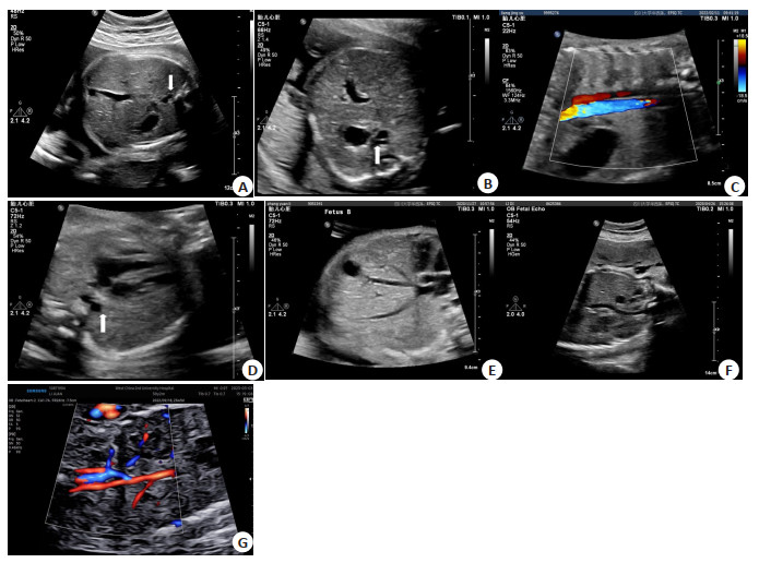
图 1 不同类型IVC发育异常的胎儿超声图像特征
Figure 1. Fetal ultrasound image characteristics of different types of IVC developmental abnormalities. A: Cross section of fetal abdomen: Azygos vein dilated at the right rear of the abdominal aorta, azygos vein (white arrow); B: Cross section of fetal abdomen: left posterior dilated semi azygos vein of abdominal aorta, semi azygos vein (white arrow); C: Oblique coronal section of fetal chest and abdomen: dilated azygos vein and thoracic aorta entering the chest cavity; D: Fetal four chamber view: right dilated azygos vein of thoracic aorta, azygos vein (white arrow); E: Longitudinal section of fetal right atrium: Three hepatic veins converge into the right atrium; F: The coronal section of the fetal inferior vena cava shows an "S" sign; G: The coronal section of the fetal inferior vena cava presents an "h" sign.
表 1 胎儿IVC发育异常合并系统异常明细
Table 1. Details of fetal IVC developmental abnormalities combined with systemic abnormalities
Merge exception Detailed classification Cardiovascular system (n=13) 4 cases of left atrial isomerism, 2 cases of left atrial isomerism combined with persistent left superior vena cava, 1 case of left atrial isomerism combined with dextrocardia, 2 cases of persistent left superior vena cava, 2 cases of ventricular septal defect, 1 case of right subclavian artery deviation, and 1 case of aortic arch narrowing. Skeletal system (n=3) 1 case of right foot fourth toe short and dorsally curved, 1 case of double foot varus, and 1 case of small mandible. Urinary system (n=3) 1 case of right renal accessory renal artery, 1 case of right renal polycystic renal dysplasia, and 1 case of pelvic ectopic kidney. Digestive system (n=2) Middle position of gastric acinus in 1 case, duodenal atresia in 1 case. Respiratory system (n=2) Two cases of pulmonary cystadenoma. Sensory system (n=1) A case of left cerebellar malformation. Other (n=1) One case of visceral inversion.  下载: 导出CSV
表 2 不同类型IVC发育异常孤立性及合并心内外异常结果
Table 2. Isolation of different types of IVC developmental abnormalities and results of combined extracardiac and extracardiac abnormalities[n(%)]
Group Isolation Combined intracardiac abnormalities Concomitant extracardiac abnormalities Simultaneously merging with intracardiac and extracardiac abnormalities Total (n) ECIVC 7(38.9) 7(38.9) 2(11.1) 2(11.1) 18 LIVC 34(81.0) 1(2.4) 5(11.9) 2(4.80) 42 DIVC 25(89.3) 1(3.6) 2(7.1) 0(0.0) 28 ECIVC: Ectopic connection of inferior vena cava; LIVC: Left inferior vena cava; DIVC: Double inferior vena cava.
下载: 导出CSV
表 3 不同类型IVC发育异常合并心内外异常构成比组间两两比较
Table 3. Pairwise comparison of the composition of different types of IVC developmental abnormalities combined with intracardiac and extracardiac abnormalities between groups
Group Isolation Intracardiac abnormalities Extracardiac abnormalities Simultaneously merging with intracardiac and extracardiac abnormalities n P n P n P n P ECIVC 7 < 0.05 7 < 0.05 2 > 0.05 2 > 0.05 LIVC 34 1 5 2 ECIVC 7 < 0.05 7 < 0.05 2 > 0.05 2 > 0.05 DIVC 25 1 2 0 LIVC 34 > 0.05 1 > 0.05 5 > 0.05 2 > 0.05 DIVC 25 1 2 0
下载: 导出CSV
-
[1] Oliveira JD, Martins I. Congenital systemic venous return anomalies to the right atrium review[J]. Insights Imag, 2019, 10(1): 115. doi: 10.1186/s13244-019-0802-y [2] 李胜利, 罗国阳. 胎儿畸形产前超声诊断学[M]. 2版. 北京: 科学出版社, 2017: 333-41. [3] 高熠洲, 吾米提·吾夏尔, 詹发亮, 等. 8例无症状性下腔静脉肝下段缺如患者的临床影像特征分析[J]. 中国心血管杂志, 2023, 28(2): 136-9. [4] Johnson AJ, Srour H. Extracorporeal membrane oxygenation complicated by an interrupted inferior vena Cava[J]. ASAIO J, 2021, 67(9): e160-2. [5] Castro VA, Díaz-Peromingo JA. Vena Cava atresia and deep vein thrombosis: a case report and systematic review[J]. Int J Angiol, 2022, 31(2): 88-91. doi: 10.1055/s-0041-1732434 [6] 刘慧冰, 李战飞, 裘毓雯, 等. 胎儿肾后段左下腔静脉畸形的产前超声诊断及预后分析[J]. 实用妇科内分泌电子杂志, 2023, 10(6): 97-9. [7] Bass JE, Redwine MD, Kramer LA, et al. Spectrum of congenital anomalies of the inferior vena Cava: cross-sectional imaging findings[J]. RadioGraphics, 2000, 20(3): 639-52. doi: 10.1148/radiographics.20.3.g00ma09639 [8] 程松玲, 栾泽东. 产前超声诊断下腔静脉离断伴半奇静脉异位连接1例[J]. 医学影像学杂志, 2021, 31(5): 877, 899. [9] 顾彬, 侯莉, 张冬梅, 等. 二维超声心动图结合STIC-HD Live Flow技术诊断胎儿下腔静脉离断中的价值探讨[J]. 中国产前诊断杂志: 电子版, 2021, 13(3): 29-33. [10] 颜华英, 何丽红, 张春国, 等. 产前超声诊断胎儿下腔静脉异常连接[J]. 中国医学影像学杂志, 2023, 31(7): 756-60. [11] Shin DS, Sandstrom CK, Ingraham CR, et al. The inferior vena cava: a pictorial review of embryology, anatomy, pathology, and interventions[J]. Abdom Radiol (NY), 2019, 44(7): 2511-27. doi: 10.1007/s00261-019-01988-3 [12] Kim SS, Shin HC, Hwang JA, et al. Various congenital anomalies of the inferior vena cava: review of cross-sectional imaging findings and report of a new variant[J]. Abdom Radiol, 2018, 43(8): 2130-49. doi: 10.1007/s00261-017-1430-y [13] Klinkhachorn P, Ritz BK, Umstot S, et al. Duplication of the inferior vena cava: evidence of a novel type Ⅳ[J]. Folia Med Cracov, 2020, 60(2): 5-13. [14] Lin LL, Zhang K, Yang X, et al. Orthostatic proteinuria due to inferior vena cava interruption without nutcracker phenomenon in an old obese female: a case report and literature review[J]. BMC Nephrol, 2023, 24(1): 225. doi: 10.1186/s12882-023-03279-y [15] Bækgaard N, Broholm R, Just S, et al. Long-term results using catheter-directed thrombolysis in 103 lower limbs with acute iliofemoral venous thrombosis[J]. Eur J Vasc Endovasc Surg, 2010, 39(1): 112-7. doi: 10.1016/j.ejvs.2009.09.015 [16] Kardum D. Left-sided inferior vena Cava associated with proximal deep venous thrombosis of the left lower extremity[J]. Acta Clin Croat, 2022, 61(1): 145-8. [17] Chen SJ, Wu MH, Wang JK. Clinical implications of congenital interruption of inferior vena cava[J]. J Formos Med Assoc, 2022, 121(10): 1938-44. doi: 10.1016/j.jfma.2022.01.021 [18] 安鹏, 王瑜, 冯伟, 等. 胎儿左侧异构综合征产前超声与病理解剖及血管铸型的对照研究[J]. 中国超声医学杂志, 2019, 35(5): 472-5. [19] Shaikh S, Awad H, Kelly A, et al. Azygos- lung neoplasm[J]. Eur J Case Rep Intern Med, 2021, 8(3): 002385. [20] 焦北鱼, 韩玉娜, 丁媛. 产前超声诊断胎儿下腔静脉变异[J]. 第二军医大学学报, 2017, 38(1): 124-7. [21] 赵映, 张烨, 刘晓伟, 等. 孤立性下腔静脉回流异常的产前超声诊断[J]. 心肺血管病杂志, 2016, 35(8): 628-31. [22] 吕国荣. 胎儿颅脑和心脏畸形超声诊断[M]. 北京: 北京大学医学出版社, 2010. [23] 沈祥君, 陈琳, 周柳英, 等. STIC-HD live flow技术在胎儿下腔静脉畸形诊断中的价值探讨[J]. 中国超声医学杂志, 2020, 36(8): 763-6. [24] 潘婷, 燕飞雷, 王艺璇, 等. 产前超声诊断胎儿双下腔静脉并马蹄肾1例[J]. 中国医学影像技术, 2022, 38(10): 1600. [25] 鲁琳, 张姣, 苏婧. 四维彩超在胎儿下腔静脉走行变异中的诊断价值[J]. 海南医学, 2022, 33(23): 3096-8. [26] 田艾军, 谌立军, 骆迎春, 等. 胎儿下腔静脉离断的超声诊断分析[C]. //昆明: 中华医学会第十三次全国超声医学学术会议论文汇编, 2013: 195. [27] 张紫薇, 王军梅. 胎儿期超声诊断左位下腔静脉及双下腔静脉[J]. 浙江大学学报: 医学版, 2019, 48(4): 446-52. [28] 李陶, 铁红霞, 马斌. 产前二维超声联合STIC技术诊断胎儿复杂双下腔静脉畸形1例[J]. 中国临床医学影像杂志, 2023, 34(5): 378-9. -

 点击查看大图
计量
- 文章访问数: 58
- HTML全文浏览量: 25
- PDF下载量: 5
- 被引次数: 0