


CT angiography and magnetic resonance angiography visually showing spinal vessels and spinal involvement in diagnosing spinal vascular malformations
-
摘要:
目的 研究CT血管造影(CTA)与磁共振血管成像(MRA)诊断脊髓血管畸形的临床应用。 方法 本研究为前瞻性研究,以2020年2月~2022年12月在我院诊断并进行治疗的脊髓血管畸形患者30例作为试验组,另选取30例同期进行椎体MR平扫加增强的无脊髓血管畸形患者作为对照组,对所有入组对象行磁共振对比增强血管造影(CE-MRA)序列以及重建脊髓血管CTA检查。比较CE-MRA序列以及重建脊髓血管CTA检查的诊断一致性,分析CE-MRA序列以及重建脊髓血管CTA检查联合诊断效能。 结果 CE-MRA以及CTA检查中硬脊膜动静脉瘘、脊髓动静脉畸形、髓周动静瘘之间的差异无统计学意义(P > 0.05),在对瘘口显示情况的分析中,CE-MRA以及CTA检查中对于供血动脉以及瘘口显示情况的差异无统计学意义(P > 0.05);CE-MRA与金标准的检查一致性较好,Kappa值为0.758;CTA与金标准的检查一致性较好,Kappa值为0.881,CE-MRA以及CTA检查联合诊断的敏感度高于单独检测;ROC曲线分析显示,CE-MRA以及CTA检查联合诊断的曲线下面积高于单独检测(P < 0.001)。 结论 在对脊髓血管畸形患者的诊断中,CTA以及CE-MRA的联合诊断可显著提升患者的诊断效能,可作为临床诊断的重要依据。 -
关键词:
- 脊髓血管畸形 /
- CT血管造影 /
- 磁共振对比增强血管造影 /
- 联合诊断
Abstract:Objective To explore the clinical application of CT angiography (CTA) and magnetic resonance angiography (MRA) in the diagnosis of spinal cord vascular malformations. Methods This prospective study included 30 patients with spinal cord vascular malformation diagnosed and treated in our hospital from February 2020 to December 2022 as the experimental group. In addition, 30 patients without spinal vascular malformation who underwent vertebra MR Scan plus enhancement were selected as the control group. Contrast enhanced magnetic resonance angiography (CE-MRA) sequence and reconstructed spinal vascular CTA were performed on all enrolled subjects. The diagnostic consistency of CE-MRA sequence and reconstructed spinal vascular CTA was compared, and the combined diagnostic efficacy of CE-MRA sequence and reconstructed spinal vascular CTA was analyzed. Results There was no statistical significance in dural arteriovenous fistula, spinal arteriovenous malformation, and perimedullary arteriovenous fistula in CE-MRA and CTA examination (P > 0.05). In the analysis of fistula, there was no statistical significance in the display of supplying artery and fistula in CE-MRA and CTA examination (P > 0.05). CE-MRA had a good consistency by compare with the gold standard, with a kappa value of 0.758, and CTA had a good consistency by compare with the gold standard, with a kappa value of 0.881. The sensitivity of CE-MRA and CTA combined diagnosis were significantly higher than that of the single detection. ROC curve analysis showed that the area under the curve of CE-MRA and CTA combined diagnosis was significantly higher than that of single detection(P < 0.001). Conclusion In the diagnosis of spinal cord vascular malformations, the combined diagnosis of CTA and CE-MRA can significantly improve the diagnostic efficiency of patients, and can be used as an important basis for clinical diagnosis. -

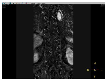
图 1 脊髓血管畸形的3D-CE-MRA图像分析
Figure 1. 3D-CE-MRA image analysis of spinal vascular malformations. Obviously thickened and tortuous drainage vein in thoracic vertebral canal, fistula and blood supplying artery could be seen on the right side at T11-12 level.

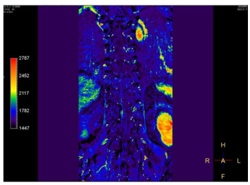
图 2 脊髓血管畸形的伪彩图
Figure 2. False-color images of vascular malformations in the spinal cord. Orifice fistula and blood supplying artery can be seen on the right side at T11-12 level.
表 1 试验组以及对照组的一般资料以及慢性病情况比较
Table 1. Comparison of general data and chronic diseases between the experimental group and the control group (n=30)
Group Gender(n, Male/Female) Age (years, Mean±SD) BMI (kg/m2, Mean±SD) Hypertension [n(%)] Diabetes [n(%)] Dyslipemia [n(%)] Experimental group 12/18 45.26±1.02 24.27±2.65 15(50.00) 9(30.00) 11(36.67) Control group 14/16 45.30±1.51 24.30±1.96 12(40.00) 10(33.33) 12(40.00) χ2/t 0.271 0.120 0.050 0.606 0.077 0.071 P 0.602 0.905 0.960 0.436 0.781 0.791  下载: 导出CSV
表 2 CE-MRA以及CTA检查的一致性分析
Table 2. Consistency analysis of CE-MRA and CTA tests (n=30)
Examination method Classification of disease Display of fistula Dural arteriovenous fistula Spinal cord arteriovenous malformation Perimedullary static fistula Supplying artery display Fistula display CE-MRA 12(40.00) 13(43.33) 5(16.67) 28(93.33) 29(96.67) CTA 15(50.00) 11(36.67) 4(13.33) 29(96.67) 27(90.00) χ2 0.611 0.350 1.071 P 0.737 0.554 0.301 CE-MRA: Contrast enhanced magnetic resonance angiography; CTA: CT angiography.
下载: 导出CSV
表 3 CE-MRA与金标准的检查一致性分析
Table 3. Consistency analysis of CE-MRA and gold standard (n)
Index Gold standard + - CE-MRA + 25 4 - 5 26 Kappa 0.758
下载: 导出CSV
表 4 CTA与金标准的检查一致性分析
Table 4. Analysis of consistency between CTA and gold standard (n)
Index Gold standard + - CTA + 24 3 - 6 27 Kappa 0.881
下载: 导出CSV
表 5 CE-MRA以及CTA检查单独检测与联合检测的诊断效能比较
Table 5. Comparison of the diagnostic efficacy of CE-MRA and CTA tests by single detection and combined detection
Diagnostic method Number of true positive (n) Number of false positive (n) Number of true negative (n) Number of false negative (n) Accuracy rate (%) Sensitivity (%) Specificity (%) Positive predictive value (%) Negative predictive value (%) CE-MRA 25 21 9 5 56.67 83.33 30.00 54.35 64.29 CTA 24 20 10 6 56.67 80.00 62.50 54.55 62.50 Combined diagnosis 27 5 25 3 86.67 90.00 89.29 84.38 89.29
下载: 导出CSV
表 6 ROC曲线分析
Table 6. ROC curve analysis
Diagnostic method Standard error AUC AUC(95% CI) P CE-MRA 0.027 0.778 0.762-0.869 < 0.001 CTA 1.027 0.792 0.762-0.870 < 0.001 Combined diagnosis 2.027 0.832 0.760-0.871 < 0.001
下载: 导出CSV
-
[1] 戴嵬, 孙园园, 王娟, 等. 复合手术在脊髓血管病治疗中的应用[J]. 中国脑血管病杂志, 2021, 18(10): 704-12. https://www.cnki.com.cn/Article/CJFDTOTAL-NXGB202110008.htm [2] 李晓宇, 张鸿祺, 凌锋, 等. 脊髓海绵状血管畸形术中神经电生理监测: 预警标准敏感性和特异性分析[J]. 中国现代神经疾病杂志, 2020, 20(11): 970-4. https://www.cnki.com.cn/Article/CJFDTOTAL-XDJB202011013.htm [3] 陈功, 秦宣锋, 安庆祝, 等. 进展型脊髓髓内海绵状血管畸形的手术时机探讨[J]. 中华神经外科杂志, 2021, 37(2): 158-63. [4] 杨城斌, 马永杰, 陈思畅, 等. 手术治疗外伤十年后硬脊膜动静脉瘘一例并文献复习[J]. 中国脑血管病杂志, 2020, 17(9): 543-5, 549. https://www.cnki.com.cn/Article/CJFDTOTAL-NXGB202009008.htm [5] 樊欣鑫, 纪祯龙. 脊髓动静脉畸形的治疗研究进展[J]. 东南大学学报: 医学版, 2021, 40(4): 560-4. https://www.cnki.com.cn/Article/CJFDTOTAL-NJTD202104025.htm [6] Kouskouras C, Charitanti A, Giavroglou C, et al. Intracranial aneurysms: evaluation using CTA and MRA. Correlation with DSA and intraoperative findings[J]. Neuroradiology, 2004, 46(10): 842-50. doi: 10.1007/s00234-004-1259-2 [7] 李忠, 商金元. CTA联合CE-MRA在诊断下肢动脉病变中的应用价值[J]. 中国医疗器械信息, 2022, 28(1): 85-7. https://www.cnki.com.cn/Article/CJFDTOTAL-ZGQX202201021.htm [8] Camargo A, Zacharia T, Kanekar S, et al. Value of CTA/MRA in the setting of intraparenchymal hemorrhage in the emergency department[J]. Neuroradiology, 2023, 65(1): 97-103. doi: 10.1007/s00234-022-03080-y [9] 袁小入, 吴钦, 左政, 等. 影像学研究硬脊膜动静脉瘘进展[J]. 中国介入影像与治疗学, 2022, 19(6)374-377 https://www.cnki.com.cn/Article/CJFDTOTAL-JRYX202206013.htm [10] Vatit P, Schneider JR. Progressive myelopathy due to apinal srteriovenous malformation[J]. J Neuroimaging, 2021, 31(2): 416. [11] Shetty Gurucharan S, Vivek S, Nandan PS, et al. Spinal epidural fistulas-a separate entity to dural fistulas with different angioarchitecture and treatment approach[J]. World Neurosurg, 2021, 149: e600-11. doi: 10.1016/j.wneu.2021.01.126 [12] Jung SH, Jung S, Moon KS, et al. Tailored surgical approaches for benign craniovertebral junction tumors[J]. J Korean Neurosurg Soc, 2010, 48(2): 139-44. doi: 10.3340/jkns.2010.48.2.139 [13] Jacqueline M, Miranda Ileana C, Miller Andrew D, et al. A review of proliferative vascular disorders of the central nervous system of animals[J]. Vet Pathol, 2020, 58(5). [14] Kwasnicki A, Calandriello A, Nikas D. Spontaneous spinal epidural hematoma in an infant presenting with Horner syndrome[J]. Child's Nerv Syst, 2021, 38: 827-830. [15] 方永康, 薛峥, 闵喆. 髓外硬膜下脊髓海绵状血管瘤导致的蛛网膜下腔出血的临床分析(附1例报告)[J]. 临床神经病学杂志, 2022, 35 (4): 285-9. https://www.cnki.com.cn/Article/CJFDTOTAL-LCSJ202204008.htm [16] 康永生, 梅伟, 王庆德, 等. 基于CT二维图像的分类复位手术治疗下颈椎单节段关节突脱位[J]. 中华创伤杂志, 2023, 39(4): 331-40. [17] 杨晓燕, 刘盛秀. 抑癌基因P53在血管瘤发生及靶向治疗中的作用[J]. 皮肤性病诊疗学杂志, 2009, 26(5): 319-22. https://www.cnki.com.cn/Article/CJFDTOTAL-LPFZ201905019.htm [18] 胡勤勤, 张德川, 曾国飞, 等. 基于对比剂分段注射的颈髓双期CT血管成像[J]. 实用放射学杂志, 2021, 37(5): 716-9, 723. [19] 李大胜, 黄河, 王娜娜, 等. CT血管造影和MRI在脊髓血管畸形术后随诊中的价值[J]. 中国医疗设备, 2021, 36(2): 69-72. https://www.cnki.com.cn/Article/CJFDTOTAL-YLSX202102027.htm -

 点击查看大图
计量
- 文章访问数: 50
- HTML全文浏览量: 23
- PDF下载量: 3
- 被引次数: 0