


Value and application of BI-RADS classification in breast invasive non-special type carcinoma
-
摘要:
目的 分析BI-RADS标准化超声分级4C类病灶征象运用于乳腺浸润性癌非特殊型诊断时与病理征象的关联性及此类型癌的年龄分布,探讨BI-RADS分级标准应用于乳腺浸润性癌非特殊型诊断中的价值。 方法 随机收集福建省立医院超声科于2020年5月~2023年5月所收治的88例超声BI-RADS 4C类的乳腺癌患者作为研究对象,针对患者年龄及病理检查结果进行分类,并对不同年龄段患不同类型乳腺疾病的患者的病灶超声特征与病理特征相关性进行讨论。 结果 超声检查及病理追踪分析结果发现,在BI-RADS 4C类88例患者中,≥40岁的患者人数占90%, < 40岁的患者人数占10%;在乳腺浸润性非特殊癌中,≥40岁的患者人数占比95%, < 40岁的患者人数占比5%。单纯浸润性非特殊癌在病理检查结果中占总数的64.77%,其余非单纯浸润性非特殊癌占35.23%。 结论 综合数据分析超声检查与BI-RADS分级结合在单纯浸润性非特殊癌中的检出率明显较高,有利于制定对应的治疗措施,更加突出BI-RADS分级在乳腺单纯浸润性非特殊癌中的检出率和运用。 -
关键词:
- BI-RADS分级 /
- 乳腺单纯浸润性非特殊癌 /
- 超声诊断
Abstract:Objective To analyze the association of BI-RADS standardized ultrasound grade 4C lesion signs in the diagnosis of breast invasive carcinoma and the age distribution of this type of cancer, and explore the value of BI-RADS classification criteria in the diagnosis of non-specific type of breast invasive carcinoma. Methods We randomly collected 88 cases of breast cancer patients with ultrasound BI-RADS grade 4C from May 2020 to May 2023 as study data, and classified the patients' age and pathological examination results. The correlation between lesion ultrasound characteristics and pathological characteristics of patients with different age groups with different types of breast diseases were analyzed. Results The ultrasound examination and pathological tracking analysis showed that among 88 patients in BI-RADS 4C, 90% were patients younger than 40 years; more than 95% were 40 years and 5% were patients younger than 40 years. Simple invasive non-special carcinoma accounted for 64.77% of the total pathological examination results, while the remaining non-simple invasive non-special carcinoma accounted for 35.23. Conclusion Comprehensive data analysis the detection rate of ultrasound examination and BI-RADS grade in simple invasive non-special cancer is obviously high, which is conducive to the formulation of corresponding treatment measures, and highlights the detection rate and application of BI-RADS grade in simple breast invasive non-special cancer. -

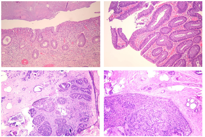
图 2 乳腺浸润性导管癌,组织学分级Ⅱ级(HE染色,×200)
Figure 2. Invasive ductal carcinoma of the breast, histological grade level 2 (HE staining, ×200).
表 1 BI-RADS分级4C类病理结果频数分析
Table 1. Frequency analysis of class 4C pathology results in BI-RADS grades
Pathology type Caes(n) % Middle-high-grade apocrine-type ductal carcinoma in situ of the breast 1 1.14 Medium-high-grade ductal carcinoma in situ of the breast 1 1.14 Mid-grade ductal in situ carcinoma of the breast 4 4.55 Metaplastic lesions of the breast 1 1.14 In-situ solid papillary carcinoma of the breast 1 1.14 Breast complex sclerotic lesions with breast sclerotic adenopathy 1 1.14 Complex sclerotic lesions of the breast 1 1.14 Solid papillary carcinoma of the breast was associated with invasion 1 1.14 Invasive micropapillary carcinoma of the breast 1 1.14 Invasive breast cancer 3 3.41 Invasive carcinoma of the breast (mainly mucinous carcinoma) 1 1.14 Invasive non-specific carcinoma of the breast 57 64.77 Mixed-type mucinous carcinoma of the breast 1 1.14 Malignant tumor of breast 1 1.14 Fibroepithelial tumor of the breast 1 1.14 Fibroadenoma of breast 1 1.14 High-grade ductal carcinoma in situ of the breast 5 5.68 Malignant cell 1 1.14 Malignancy, prioritizing poorly differentiated invasive carcinomas 1 1.14 Infiltrative, poorly differentiated carcinoma 1 1.14 Invasive carcinoma, metaplastic breast cancer is to be excluded 1 1.14 At least for the papillary intraductal carcinoma changes 1 1.14 Microscopic perilobular granulomatous inflammation was observed 1 1.14  下载: 导出CSV
-
[1] Reeder-Hayes K, Anderson B. Breast cancer disparities at home and abroad: a review of the challenges and opportunities for system-level change[J]. Clin Cancer Res, 2017, 23: 2655-2664. doi: 10.1158/1078-0432.CCR-16-2630 [2] 艾勇彪, 张丹峰, 李文仿, 等. 青年和老年乳腺癌病理特征及预后差异的分析[J]. 现代肿瘤医学, 2018, 26(23): 3764-7. doi: 10.3969/j.issn.1672-4992.2018.23.015 [3] Colzani E, Liljegren A, Johansson ALV, et al. Prognosis of patients with breast cancer: causes of death and effects of time since diagnosis, age, and tumor characteristics[J]. J Clin Oncol, 2011, 29(30): 4014-21. doi: 10.1200/JCO.2010.32.6462 [4] 陈光玉, 金永红, 项金凤. 超声对乳腺结节BI-RADS分类的声像图表现与病理结果的对比分析[J]. 蚌埠医学院学报, 2019, 44(8): 1097-9, 1103. https://www.cnki.com.cn/Article/CJFDTOTAL-BANG201908035.htm [5] Tang LC, Jin X, Yang HY, et al. Luminal B subtype: a key factor for the worse prognosis of young breast cancer patients in China [J]. BMC Cancer, 2015, 15: 201. doi: 10.1186/s12885-015-1207-z [6] 关海燕, 彭秋香, 曹艳红. 乳腺彩超BI-RADS分类在乳腺结节中的临床应用[J]. 医疗装备, 2023, 36(17): 60-2. https://www.cnki.com.cn/Article/CJFDTOTAL-YLZB202317017.htm [7] 郑蔚朗, 王丹丹. 超声诊断乳腺BI-RADS Ⅳa类结节在外科诊疗中的价值及病理结果对比[J]. 中国卫生标准管理, 2022, 13(18): 96-100. https://www.cnki.com.cn/Article/CJFDTOTAL-WSBZ202218021.htm [8] 刘迎春, 陈林, 庞芸, 等. 基于自动乳腺容积扫描、乳腺X线摄影及MRI的BI-RADS分类鉴别乳腺良恶性肿块[J]. 中国医学影像技术, 2021, 37(3): 386-90. https://www.cnki.com.cn/Article/CJFDTOTAL-ZYXX202103021.htm [9] Hao SY, Ou B, Li LJ, et al. Could ultrasonic elastography help the diagnosis of breast cancer with the usage of sonographic BI-RADS classification?[J]. Eur J Radiol, 2015, 84(12): 2492-500. doi: 10.1016/j.ejrad.2015.08.015 [10] 李树玲. 乳腺肿瘤学[M]. 北京: 科学技术文献出版社, 2000: 9-10. [11] 杨媛, 孔祥海, 杨迎青. 67例BI-RADS 3和4级乳腺病变患者超声诊断与病理结果相关性分析[J]. 皖南医学院学报, 2018, 37(4): 371-4. https://www.cnki.com.cn/Article/CJFDTOTAL-WNYX201804020.htm [12] 杨尊敬, 杜先玲, 杜先艳. 阿帕替尼联合放疗治疗晚期胃癌的临床疗效及安全性分析[J]. 实用癌症杂志, 2019, 34(1): 122-4. https://www.cnki.com.cn/Article/CJFDTOTAL-SYAZ201901035.htm [13] 卢建明, 赵淑红, 田锦, 等. 乳腺超声检查中BI-RADS分级诊断标准对乳腺肿瘤的诊断价值[J]. 宁夏医学杂志, 2015, 37(2): 140-2. https://www.cnki.com.cn/Article/CJFDTOTAL-NXYX201502022.htm [14] 张亘华, 柯林申. 超声造影在常规超声BI-RADS 4类乳腺肿块的诊断价值[J]. 医学影像学杂志, 2017, 27(7): 1403-5. https://www.cnki.com.cn/Article/CJFDTOTAL-XYXZ201707059.htm [15] 梁永超, 贾春梅, 薛影, 等. 超声造影对乳腺BI-RADS4类肿块的诊断价值[J]. 中华医学杂志, 2018, 98(19): 1498-502. doi: 10.3760/cma.j.issn.0376-2491.2018.19.009 [16] 朱桂新, 武爽, 董立阳, 等. 超声造影调整乳腺BI-RADS 3、4类肿块的临床价值[J]. 临床超声医学杂志, 2020, 22(11): 862-5. https://www.cnki.com.cn/Article/CJFDTOTAL-LCCY202011024.htm [17] 孙卫平. 高频彩超及X线钼靶检查对乳腺原位癌早期诊断作用分析[J]. 影像研究与医学应用, 2022, 6(15): 134-6. https://www.cnki.com.cn/Article/CJFDTOTAL-YXYY202215044.htm [18] 洪玺玺. 高频彩超与核磁共振在早期乳腺癌中的诊断效果[J]. 中国妇幼保健, 2021, 36(13): 3149-51. https://www.cnki.com.cn/Article/CJFDTOTAL-ZFYB202113071.htm [19] 刘跃辉, 郑春红, 李倩, 等. 二甲双胍联合阿帕替尼治疗中晚期胃癌患者的疗效及对肠道菌群的影响[J]. 国际消化病杂志, 2020, 40(1): 36-40. https://www.cnki.com.cn/Article/CJFDTOTAL-GWXH202001010.htm [20] 林梅清, 杨燕霞, 古艳, 等. 超声诊断乳腺浸润性导管癌14例[J]. 智慧健康, 2021, 7(9): 13-5. https://www.cnki.com.cn/Article/CJFDTOTAL-ZHJK202109005.htm [21] 林锦娜, 刘强. 第4版《年轻女性乳腺癌国际共识指南》更新要点解读[J]. 中华乳腺病杂志: 电子版, 2021, 15(4): 195-200. https://www.cnki.com.cn/Article/CJFDTOTAL-ZHRD202104001.htm -

 点击查看大图
图(2) / 表(1)
计量
- 文章访问数: 59
- HTML全文浏览量: 36
- PDF下载量: 2
- 被引次数: 0