


Predictive value of enhanced CT radiomics nomogram in muscular invasion of bladder urothelial carcinoma
-
摘要:
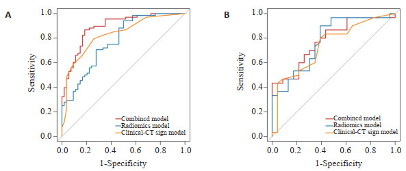
目的 探讨基于增强CT影像组学列线图在术前预测膀胱尿路上皮癌肌层浸润的价值。 方法 回顾性分析2018年8月~ 2023年4月于蚌埠医学院第一附属医院确诊为膀胱尿路上皮癌患者175例。将所有病例按7:3随机分为训练组(n=122)与验证组(n=53)。对增强CT多期图像进行手动勾画病灶感兴趣区并提取影像组学特征,通过最小绝对收缩和选择算子降维,采用支持向量机分类器对提取的特征进行机器学习,筛选出最优影像组学特征并构建影像组学评分模型。通过单因素分析及多因素二元Logistic回归分析筛选出膀胱尿路上皮癌肌层浸润的独立预测因素,构建临床-CT征象模型。将影像组学模型和临床-CT征象模型联合,构建联合模型。绘制ROC曲线,计算曲线下面积(AUC)、敏感度及特异性评估不同模型的预测效能,将最佳模型可视化构建列线图。 结果 联合模型的诊断效能最高(AUC=0.891),均高于影像组学模型(AUC=0.777)和临床-CT征象模型(AUC=0.829)。决策曲线分析及校正曲线证实了列线图有较高的预测性能。 结论 增强CT影像组学列线图在术前预测膀胱尿路上皮癌肌层浸润方面具有较高价值。 Abstract:Objective To investigate the value of enhanced CT imaging nomogram in preoperative prediction of muscular infiltration in urinary tract carcinoma of bladder. Methods A retrospective analysis was performed on 175 patients diagnosed with bladder urothelial carcinoma from August 2018 to April 2023 in the First Affiliated Hospital of Bengbu Medical College. All cases were randomly divided into training group (n=122) and verification group (n=53) according to 7:3. The region of interest was manually defined and the image omics features were extracted from the multi-phase enhanced CT images. The dimensionality was reduced by minimum absolute contraction and selection operator, and machine learning was performed on the extracted features using support vector machine classifier to screen out the optimal image omics features and construct the image omics scoring model. The independent predictors of muscular infiltration of bladder urothelial carcinoma were screened by univariate analysis and multivariate binary logistic regression analysis, and the clinical-CT signs model was constructed. Combining the imaging omics model with the clinical-CT sign model, the combined model was constructed. The ROC curve is plotted, the area under the curve (AUC), sensitivity, and specificity were calculated to evaluate the predictive efficacy of different models, and then the best model was visualized to construct a nomogram. Results The diagnostic efficacy of the combined model was the highest (AUC=0.891), which was higher than that of the imaging model (AUC=0.777) and the clinical-CT sign model (AUC=0.829). Decision curve analysis and correction curve confirm that the nomogram has high predictive performance. Conclusion Enhanced CT image nomogram has a high value in predicting myoinfiltration of bladder urothelial carcinoma before operation. -
Key words:
- bladder cancer /
- muscular invasion /
- radiomics /
- contrast-enhanced computed tomography
-

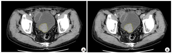
图 1 肿瘤最大层面ROI的勾画
Figure 1. Delineation of ROI on the maximum tumor level. A-B: Training group, a male patient, 75 years old, NMIBC, delineation of ROI in arterial and venous phases.

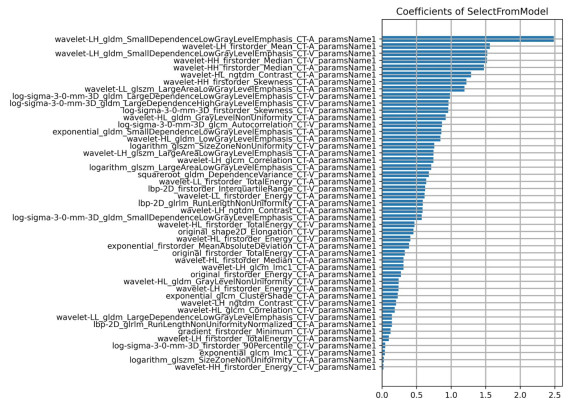
图 2 LASSO筛选出47个用于预测膀胱尿路上皮癌肌层浸润的最佳影像组学特征
Figure 2. LASSO selected 47 best image-omics features for predicting myographic infiltration of urinary tract carcinoma of bladder

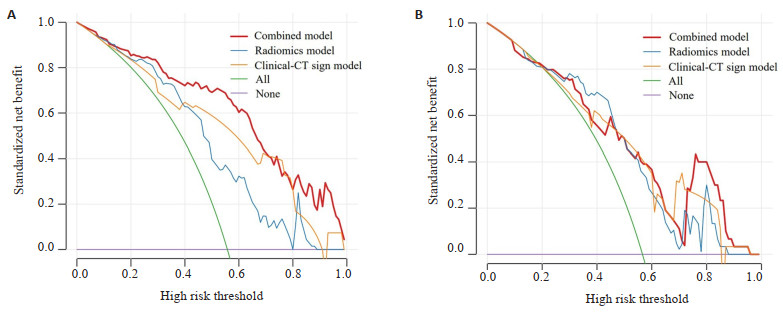
图 3 三组模型在训练组(A)和验证组(B)中的ROC曲线
Figure 3. ROC curves of three sets of models in training group (A) and validation group (B).

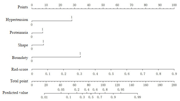
图 4 用于预测BCa肌层浸润的列线图
Figure 4. Nomogram for prediction of muscular infiltration in urothelial carcinoma of the bladder. Hypertension: 0 represents no, 1 represents yes; Proteinuria: 0 represents no, 1 represents yes; Shape: 0 represents Regular, 1 represents Non-regular; Boundary: 0 represents Clear, 1 represents Obscure.

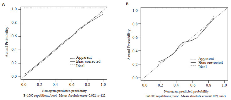
图 5 列线图的预测概率与理想概率之间的校准曲线
Figure 5. The calibration curve between the predicted probability and the ideal probability of a nomogram. A: Training group, B: Validation group.

图 6 训练组(A)和验证组(B)的DCA曲线
Figure 6. DCA curves for training group (A) and validation group (B).
表 1 训练组及验证组患者临床及影像资料单因素分析
Table 1. Univariate analysis of clinical and imaging data in training group and validation group
Index Training group(n=122) Validation group(n=53) NMIBC(n=54) MIBC(n=68) t/χ2 P NMIBC(n=23) MIBC(n=30) t/χ2 P Gender [n(%)] 0.603 0.437 0.541 0.462 Male 45(83.30) 60(88.20) 19(82.60) 21(70.00) Female 9(16.70) 8(11.80) 4(17.40) 9(30.00) Age [years, M(P25-P75)] 64(55.00-75.25) 68(58.25-74.75) -1.728 0.084 72(58.00-80.00) 72(58.75-74.50) -0.216 0.829 Smoke [n(%)] 0.519 0.471 0.981 0.322 No 41(75.90) 49(72.10) 19(82.60) 20(66.70) Yes 13(24.10) 19(27.90) 4(17.40) 10(33.30) Hematuresis [n(%)] 0.178 0.673 0.000 0.999 No 6(11.10) 6(8.80) 3(13.00) 4(13.30) Yes 48(88.90) 62(91.20) 20(87.00) 26(86.07) Hypertension [n(%)] 8.950 0.003 2.535 0.111 No 44(81.50) 38(55.90) 12(52.20) 22(73.30) Yes 10(18.50) 30(44.10) 11(47.80) 8(26.70) WBC [×109/L, M(P25-P75)] 6.09(5.37-6.85) 6.26(5.07-7.78) -0.866 0.387 5.71±0.24 6.75±0.35 -2.289 0.026 NEUT[×109/L, M(P25-P75)] 3.48(2.98-4.18) 3.87(3.02-4.77) -1.459 0.145 3.47±0.21 3.91±0.28 -1.132 0.263 LY [×109/L, M(P25-P75)] 1.71(1.48-2.34) 1.69(1.26-2.18) -0.941 0.347 1.63±0.07 2.02±0.12 -2.636 0.011 ALB(g/L, Mean±SD) 41.12±0.52 40.55±0.42 0.866 0.388 40.40(38.90-42.30) 41.75(38.45-44.50) -1.041 0.298 TG [n(%)] 0.233 0.630 0.541 0.462 < 1.7 mmol/L 41(75.90) 49(72.10) 19(82.60) 21(70.00) ≥1.7 mmol/L 13(24.10) 19(27.90) 4(17.40) 9(30.00) Proteinuria [n(%)] 9.696 0.002 6.273 0.012 No 31(57.40) 20(29.40) 14(60.90) 8(26.70) Yes 23(42.60) 48(70.60) 9(39.10) 22(73.30) Tumor number [n(%)] 1.298 0.255 0.275 0.600 One 43(79.60) 48(70.60) 17(73.90) 24(80.00) More than one 11(20.40) 20(29.40) 6(26.10) 6(20.00) Tumor length [n(%)] 4.599 0.032 4.658 0.030 < 3 cm 41(75.90) 39(57.40) 19(82.60) 15(50.00) ≥3 cm 13(24.10) 29(42.60) 4(17.40) 15(50.00) Shape [n(%)] 11.350 0.001 10.064 0.002 Regular 43(79.60) 34(50.00) 17(73.90) 9(30.00) Non-regular 11(20.40) 34(50.00) 6(26.10) 21(70.00) Boundary [n(%)] 13.536 <0.001 5.300 0.021 Clear 45(83.30) 35(51.50) 20(87.00) 16(53.30) Obscure 9(16.70) 33(48.50) 3(13.00) 14(46.70) Calcification [n(%)] 1.972 0.160 1.124 0.289 No 48(88.90) 54(79.40) 17(73.90) 18(60.00) Yes 6(11.40) 14(20.60) 6(26.10) 12(40.00) Enhancement mode [n(%)] 7.607 0.006 4.425 0.035 Homogeneous 43(79.60) 38(55.90) 18(78.30) 15(50.00) Heterogeneous 11(20.40) 30(44.10) 5(21.70) 15(50.00) Enhanced degree [n(%)] 1.002 0.606 0.596 0.742 Slight 6(11.10) 11(16.20) 1(4.30) 3(10.00) Moderate 23(42.60) 24(35.30) 9(39.10) 11(36.70) Obvious 25(46.30) 33(48.50) 13(56.50) 16(53.30) NMIBC: Non-muscle-invasive bladder cancer; MIBC: Muscle-invasive bladder cancer.  下载: 导出CSV
表 2 影像组学评分及临床、影像资料多因素Logistic回归分析
Table 2. Multivariate Logistic regression analysis of imaging scores and clinical and imaging data
Index OR(95% CI) P Hypertension 5.407(1.966-14.869) 0.001 Proteinuria 3.214(1.241-8.329) 0.016 Tumor length 0.812(0.265-2.491) 0.716 Shape 4.202(1.257-14.042) 0.020 Boundary 6.219(2.288-16.908) < 0.001 Enhancement mode 1.872(0.655-5.365) 0.242 Rad-score 516.989(41.873-6383.1228) < 0.001
下载: 导出CSV
表 3 3组模型比较
Table 3. Comparison of three groups of models
Groups AUC(95% CI) Sensitivity Specificity Accuracy Training group Radiomics model 0.777(0.694-0.859) 0.941 0.500 0.746 Clinical-CT sign model 0.829(0.756-0.902) 0.794 0.741 0.770 Combined model 0.891(0.834-0.948) 0.868 0.815 0.844 Validation group Radiomics model 0.780(0.653-0.908) 0.900 0.609 0.774 Clinical-CT sign model 0.740(0.604-0.876) 0.800 0.609 0.717 Combined model 0.781(0.657-0.905) 0.433 1.000 0.679
下载: 导出CSV
-
[1] Sung H, Ferlay J, Siegel RL, et al. Global cancer statistics 2020: GLOBOCAN estimates of incidence and mortality worldwide for 36 cancers in 185 countries[J]. CA A Cancer J Clinicians, 2021, 71 (3): 209-49. doi: 10.3322/caac.21660 [2] Lai AL, Law YM. VI-RADS in bladder cancer: overview, pearls and pitfalls[J]. Eur J Radiol, 2023, 160: 110666. doi: 10.1016/j.ejrad.2022.110666 [3] 卢文斌, 王尉, 聂海波, 等. 膀胱癌的诊疗研究进展[J]. 中国医药科学, 2022, 12(13): 62-5, 130. https://www.cnki.com.cn/Article/CJFDTOTAL-GYKX202213014.htm [4] Xu YS, Lou JH, Gao ZQ, et al. Computed tomography image features under deep learning algorithm applied in staging diagnosis of bladder cancer and detection on ceramide glycosylation[J]. Comput Math Meth Med, 2022, 2022: 1-8. [5] Lambin P, Rios- Velazquez E, Leijenaar R, et al. Radiomics: extracting more information from medical images using advanced feature analysis[J]. Eur J Cancer, 2012, 48(4): 441-6. doi: 10.1016/j.ejca.2011.11.036 [6] 王玫, 李宁, 张祁, 等. 基于影像组学的多参数磁共振定量指标在膀胱癌肌层浸润分析中的价值[J]. 中国临床医学影像杂志, 2022, 33 (8): 551-6. https://www.cnki.com.cn/Article/CJFDTOTAL-LYYX202208004.htm [7] Feng C, Zhou ZL, Huang QH, et al. Radiomics nomogram based on high-b-value diffusion-weighted imaging for distinguishing the grade of bladder cancer[J]. Life, 2022, 12(10): 1510. doi: 10.3390/life12101510 [8] Wu SX, Zheng JJ, Li Y, et al. Development and validation of an MRI- based radiomics signature for the preoperative prediction of lymph node metastasis in bladder cancer[J]. EBioMedicine, 2018, 34: 76-84. doi: 10.1016/j.ebiom.2018.07.029 [9] Qian J, Yang L, Hu S, et al. Feasibility study on predicting recurrence risk of bladder cancer based on radiomics features of multiphase CT images[J]. Front Oncol, 2022, 12: 899897. doi: 10.3389/fonc.2022.899897 [10] Zheng J, Kong J, Wu S, et al. Development of a noninvasive tool to preoperatively evaluate the muscular invasiveness of bladder cancer using a radiomics approach[J]. Cancer, 2019, 125(24): 4388-98. doi: 10.1002/cncr.32490 [11] Juri H, Narumi Y, Panebianco V, et al. Staging of bladder cancer with multiparametric MRI[J]. Br J Radiol, 2020, 93(1112): 20200116. doi: 10.1259/bjr.20200116 [12] Dobruch J, Oszczudłowski M. Bladder cancer: current challenges and future directions[J]. Medicina, 2021, 57(8): 749. doi: 10.3390/medicina57080749 [13] Elayat G, Punev I, Selim A. An overview of angiogenesis in bladder cancer[J]. Curr Oncol Rep, 2023, 25: 709-28. doi: 10.1007/s11912-023-01421-5 [14] Zhang XP, Zhang YC, Zhang GJ, et al. Deep learning with radiomics for disease diagnosis and treatment: challenges and potential[J]. Front Oncol, 2022, 12: 773840. doi: 10.3389/fonc.2022.773840 [15] 邹金钊, 叶靖, 徐圆. CT纹理特征结合机器学习对膀胱尿路上皮癌T分期及病理级别的诊断价值[J]. 临床放射学杂志, 2020, 39(8): 1553-8. https://www.cnki.com.cn/Article/CJFDTOTAL-LCFS202008022.htm [16] Zhang G, Wu Z, Xu LL, et al. Deep learning on enhanced CT images can predict the muscular invasiveness of bladder cancer[J]. Front Oncol, 2021, 11: 654685. doi: 10.3389/fonc.2021.654685 [17] Zhang X, Xu XP, Tian Q, et al. Radiomics assessment of bladder cancer grade using texture features from diffusion- weighted imaging[J]. J Magn Reson Imaging, 2017, 46(5): 1281-8. doi: 10.1002/jmri.25669 [18] Woldu SL, Bagrodia A, Lotan Y. Guideline of guidelines: nonmuscle-invasive bladder cancer[J]. BJU Int, 2017, 119(3): 371-80. doi: 10.1111/bju.13760 [19] Lai HD, Cheng XM, Liu Q, et al. Single-cell RNA sequencing reveals the epithelial cell heterogeneity and invasive subpopulation in human bladder cancer[J]. Int J Cancer, 2021, 149(12): 2099-115. doi: 10.1002/ijc.33794 [20] Kok VC, Zhang HW, Lin CT, et al. Positive association between hypertension and urinary bladder cancer: epidemiologic evidence involving 79, 236 propensity score-matched individuals[J]. Ups J Med Sci, 2018, 123(2): 109-15. doi: 10.1080/03009734.2018.1473534 [21] Connaughton M, Dabagh M. Association of hypertension and organ-specific cancer: a meta-analysis[J]. Healthcare, 2022, 10(6): 1074. doi: 10.3390/healthcare10061074 [22] Teleka S, Häggström C, Nagel G, et al. Risk of bladder cancer by disease severity in relation to metabolic factors and smoking: a prospective pooled cohort study of 800, 000 men and women[J]. Int J Cancer, 2018, 143(12): 3071-82. doi: 10.1002/ijc.31597 [23] Zhou Q, Zhang ZY, Ang XJ, et al. A nomogram combined with radiomics features, albuminuria, and metabolic syndrome to predict the risk of myometrial invasion of bladder cancer[J]. Transl Cancer Res, 2021, 10(7): 3177-91. doi: 10.21037/tcr-21-426 [24] Matsuoka S, Kaneko H, Okada A, et al. Association between proteinuria and incident colorectal cancer: analysis of a nationwide population-based database[J]. BMJ Open, 2022, 12(4): e056250. doi: 10.1136/bmjopen-2021-056250 [25] 陆亮, 徐圆, 袁为标, 等. 增强CT影像组学列线图预测膀胱尿路上皮癌肌层浸润[J]. 中国医学影像技术, 2021, 37(7): 1059-63. https://www.cnki.com.cn/Article/CJFDTOTAL-ZYXX202107029.htm [26] Tian ZJ, Meng LF, Wang X, et al. Predictive nomogram and risk factors for lymph node metastasis in bladder cancer[J]. Front Oncol, 2021, 11: 690324. doi: 10.3389/fonc.2021.690324 [27] Fang CY, An X, Li KJ, et al. A nomogram based on CT radiomics and clinical risk factors for prediction of prognosis of hypertensive intracerebral hemorrhage[J]. Comput Intell Neurosci, 2022, 2022: 9751988. -

 点击查看大图
计量
- 文章访问数: 72
- HTML全文浏览量: 31
- PDF下载量: 13
- 被引次数: 0