


Changes in amplitude of low-frequency fluctuation of functional magnetic resonance imaging in subjective cognitive decline
-
摘要:
目的 探究低频振幅(ALFF)在主观认知下降(SCD)患者中的改变及其与临床认知量表的相关性,为延缓和预防阿尔茨海默病的进展赢得宝贵时间窗。 方法 共招募45例SCD患者和40例匹配良好的健康对照组,最终纳入ALFF值分析的分别为35例和33例。所有受试者均进行静息态磁共振脑功能成像扫描和相关临床量表评估。采用ALFF分析方法比较两组间ALFF值存在差异的脑区,采用线性回归分析评估所有受试者各脑区ALFF值与临床认知评估量表之间的相关性。 结果 与健康对照组相比,SCD组左侧枕上回、左侧顶上回、左侧楔叶和楔前叶、左小脑以及右侧罗兰氏岛盖部的ALFF值均较低,差异有统计学意义(P<0.05)。SCD患者右侧罗兰氏岛盖部的ALFF值与韦氏逻辑记忆测试和波士顿命名测试评分呈正相关关系;左侧枕上回ALFF值与波士顿命名测试评分呈正相关关系,与连线测试A评分呈负相关关系(P<0.05)。 结论 SCD患者有异常的脑活动信号,且异常脑区的ALFF值与部分临床量表评估具有相关性,ALFF可能被认为是早期发现这些患者的特征性生物标志物。 -
关键词:
- 主观认知下降 /
- 静息态磁共振脑功能成像 /
- 低频振幅 /
- 阿尔茨海默病
Abstract:Objective To investigate the changes in amplitude of low-frequency (ALFF) and the correlations between ALFF and clinical cognitive evaluations in subjective cognitive decline (SCD) patients, in order to gain a valuable time window for delaying and preventing the progress of Alzheimer's disease. Methods Forty-five patients with SCD (SCD group) and forty well-matched healthy controls (healthy control group) were recruited in this study, thirty-five and thirth-three case were included in ALFF value analysis, respectively. All subjects were examined by functional MRI and assessed with multiple clinical scales. The changes of brain area between SCD and healthy control group were compared by ALFF analysis method. In addition, linear regression analysis was used to evaluate the correlation between ALFF values and clinical scores of all subjects. Results The ALFF values of the left superior occipital gyrus, left superior parietal gyrus, left cuneus and precuneus, left cerebellum and right Rolandic operculum were lower in the SCD group compared with healthy control group, the difference was statistically significant (P < 0.05). Among the SCD patients, the ALFF values of right Rolandic operculum were positively correlated with the Wechsler logical memory test and Boston naming test scores. The Boston naming test scores were also positively correlated with the ALFF values of left superior occipital gyrus. Additionally, the ALFF values of left superior occipital gyrus showed a significant negative correlation with trail making test A scores (P < 0.05). Conclusion SCD patients have abnormal brain activity signals, and the ALFF values of abnormal brain regions are correlated with some clinical scale evaluations. ALFF may be considered as a characteristic biomarker for early detection of these patients. -

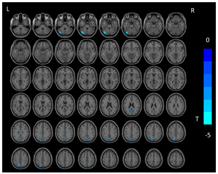
图 1 SCD组与HC组相比ALFF值差异脑区
Figure 1. Brain regions that demonstrate significant differences of ALFF values between SCD and HC groups.

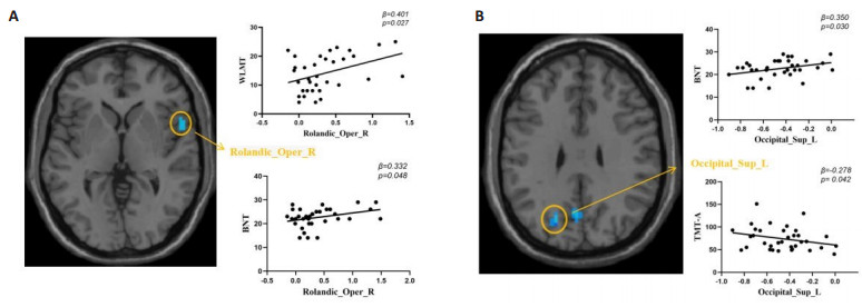
图 2 SCD患者ALFF值与临床认知评估量表相关性分析
Figure 2. Linear regression analysis between ALFF values and neuropsychological scales in SCD patients. A: The ALFF values of right Rolandic operculum were positively correlated with the WLMT scores and BNT scores; B: The ALFF values of left superior occipital gyrus were positively correlated with BNT scores and negatively correlated with TMT-A scores.
表 1 SCD组和HC组一般资料和神经心理学量表评估比较
Table 1. Baseline demographics and clinical characteristics of SCD and HC groups
Characteristics SCD(n=35) HC(n=33) P Gender(n, Male/Female) 11月24日 18/15 0.127 Age(years, Mean±SD) 70.05±6.90 71.24±6.40 0.738 Education(years, Mean±SD) 13.34±2.34 13.12±2.43 0.115 MMSE [M(Min, Max)] 28(26, 28) 29(28, 30) <0.001 MoCA [M(Min, Max)] 24(20.5, 25) 27(27, 28) <0.001 AVLT-H [M(Min, Max)] N5 4(3.5, 5.5) 4(3, 5) <0.001 N7 20(19, 22) 22(21, 23) <0.001 WLMT (Mean±SD) 14.06±6.29 19.51±6.57 <0.001 TMT-A [M(Min, Max)] 67(54.5, 92) 53(46, 65) <0.001 TMT-B (Mean±SD) 177.66±58.84 148.24±46.73 <0.001 SDMT (Mean±SD) 32.06±11.02 40.54±10.95 <0.001 BNT [M(Min, Max)] 23(21.5, 25.5) 25(22, 27) <0.001 VFT [M(Min, Max)] 17(14, 19.5) 20(19, 23) 0.004 SCD: Subjective cognitive decline; MMSE: Mini-mental state examination; MoCA: Montreal cognitive assessment; AVLT-H: Auditory verbal learning test-Huashan version; WLMT: Wechesler logical memory test; TMT-A/B: Trail making test A/B; SDMT: BNT: Boston naming test; VFT: Verbal fluency test.  下载: 导出CSV
表 2 SCD患者ALFF值与临床认知评估量表相关性分析
Table 2. Linear regression analysis between ALFF values and neuropsychological scales in SCD patients
Neuropsychological scales SC Beta t 95% CI P Lower Upper SCD WLMT Rolandic_Oper_R 0.401 2.328 0.799 12.201 0.027 BNT Rolandic_Oper_R 0.332 2.054 0.022 6.085 0.048 Occipital_Sup_L 0.350 2.274 0.619 11.356 0.030 TMT-A Occipital_Sup_L -0.278 -2.123 -60.04 -1.2 0.042
下载: 导出CSV
-
[1] Scheltens P, Blennow K, Breteler MMB, et al. Alzheimer's disease[J]. Lancet, 2016, 388(10043): 505-17. doi: 10.1016/S0140-6736(15)01124-1 [2] Karran E, De Strooper B. The amyloid cascade hypothesis: are we poised for success or failure?[J]. J Neurochem, 2016, 139(Suppl 2): 237-52. [3] 中国痴呆与认知障碍指南写作组, 中国医师协会神经内科医师分会认知障碍疾病专业委员会. 2018中国痴呆与认知障碍诊治指南(一): 痴呆及其分类诊断标准[J]. 中华医学杂志, 2018, 98 (13): 965-70. https://cpfd.cnki.com.cn/Article/CPFDTOTAL-ZHYX201505001554.htm [4] Jessen F, Amariglio RE, Van Boxtel M, et al. A conceptual framework for research on subjective cognitive decline in preclinical Alzheimer's disease[J]. Alzheimer's Dement, 2014, 10 (6): 844-52. doi: 10.1016/j.jalz.2014.01.001 [5] Molinuevo JL, Rabin LA, Amariglio R, et al. Implementation of subjective cognitive decline criteria in research studies[J]. Alzheimers Dement, 2017, 13(3): 296-311. doi: 10.1016/j.jalz.2016.09.012 [6] 韩璎. 中国阿尔茨海默病临床前期主观认知下降的诊治策略[J]. 中国临床医学影像杂志, 2018, 29(8): 534-8. https://www.cnki.com.cn/Article/CJFDTOTAL-LYYX201808004.htm [7] Reisberg B, Prichep L, Mosconi L, et al. The pre-mild cognitive impairment, subjective cognitive impairment stage of Alzheimer's disease[J]. Alzheimer's Dement, 2008, 4(1): S98-S108. [8] 盛灿, 刘芳, 韩璎. 临床前期阿尔茨海默病的功能MRI研究进展[J]. 中国临床医学影像杂志, 2018, 29(5): 359-62. https://www.cnki.com.cn/Article/CJFDTOTAL-LYYX201805017.htm [9] Yan CG, Wang XD, Zuo XN, et al. DPABI: data processing & analysis for (resting-state) brain imaging[J]. Neuroinform, 2016, 14 (3): 339-51. doi: 10.1007/s12021-016-9299-4 [10] 胡忠婕, 陈楠, 宋海庆, 等. 遗忘型轻度认知障碍和阿尔茨海默病的多模态MRI研究进展[J]. 中华放射学杂志, 2014(6): 517-20. [11] 胡忠婕, 盛灿, 孙宇, 等. 阿尔茨海默病高风险人群的结构和静息态功能MRI特征[J]. 中华神经科杂志, 2014, 47(12): 824-30. [12] Yang L, Yan Y, Wang YH, et al. Gradual disturbances of the amplitude of low-frequency fluctuations (ALFF) and fractional ALFF in alzheimer spectrum[J]. Front Neurosci, 2018, 12: 975. doi: 10.3389/fnins.2018.00975 [13] 沈柏, 张丽. 伴有认知障碍帕金森病患者的楔前叶影像学研究进展[J]. 临床神经病学杂志, 2016, 29(5): 392-4. https://www.cnki.com.cn/Article/CJFDTOTAL-LCSJ201605032.htm [14] Stoodley CJ, Valera EM, Schmahmann JD. Functional topography of the cerebellum for motor and cognitive tasks: an fMRI study[J]. NeuroImage, 2012, 59(2): 1560-70. [15] Sun Y, Dai ZJ, Li YX, et al. Subjective cognitive decline: mapping functional and structural brain changes-a combined resting-state functional and structural MR imaging study[J]. Radiology, 2016, 281(1): 185-92. [16] Kawagoe T, Onoda K, Yamaguchi S. Subjective memory complaints are associated with altered resting-state functional connectivity but not structural atrophy[J]. Neuroimage Clin, 2019, 21: 101675. [17] Sutoko S, Atsumori H, Obata A, et al. Lesions in the right Rolandic operculum are associated with self-rating affective and apathetic depressive symptoms for post-stroke patients[J]. Sci Rep, 2020, 10 (1): 20264. [18] Triarhou LC. Cytoarchitectonics of the rolandic operculum: morphofunctional ponderings[J]. Brain Struct Funct, 2021, 226(4): 941-50. [19] Gebauer L, Skewes J, Westphael G, et al. Intact brain processing of musical emotions in autism spectrum disorder, but more cognitive load and arousal in happy vs. sad music[J]. Front Neurosci, 2014, 8: 192. [20] Marshall CR, Hardy CJD, Russell LL, et al. The functional neuroanatomy of emotion processing in frontotemporal dementias[J]. Brain, 2019, 142(9): 2873-87. [21] Burgio L. Interventions for the behavioral complications of alzheimer's disease: behavioral approaches[J]. Int Psychogeriatr, 1996, 8(S1): 45-52. [22] Xia MS, Yang L, Sun GF, et al. Mechanism of depression as a risk factor in the development of Alzheimer's disease: the function of AQP4 and the glymphatic system[J]. Psychopharmacology, 2017, 234(3): 365-79. [23] Aalten P, de Vugt ME, Lousberg R, et al. Behavioral problems in dementia: a factor analysis of the neuropsychiatric inventory[J]. Dement Geriatr Cogn Disord, 2003, 15(2): 99-105. -

 点击查看大图
计量
- 文章访问数: 130
- HTML全文浏览量: 80
- PDF下载量: 13
- 被引次数: 0