


Global trans-lesional computed tomography-derived fractional flow reserve can predict abnormal myocardial blood flow: based on coronary artery CT angiography
-
摘要:
目的 通过比较全病变梯度冠状动脉电子计算机断层扫描血流储备分数(GlobalΔCT-FFR)与心肌血流量的关系,探讨GlobalΔCT-FFR在预测心肌血流异常方面的价值。 方法 回顾性纳入2019~2021年因疑似冠状动脉疾病行动态CT心肌灌注+冠状动脉CT血管造影“一站式”检查的患者76例共228支冠状动脉血管。采用Spearman相关分析GlobalΔCT-FFR与心肌血流量(MBF)的相关性。以MBF为参考标准,分别从患者水平和血管水平评价GlobalΔCT-FFR、CT-FFR和冠状动脉CT血管造影直径狭窄率(DS)对心肌血流异常的敏感度、特异性、诊断准确性、阳性预测值及阴性预测值,绘制ROC曲线并计算曲线下面积。 结果 在患者水平,GlobalΔCT-FFR与MBF呈中度负相关关系(r=-0.51,P<0.05),CT-FFR与MBF呈弱正相关关系(r=0.33,P<0.05);在血管水平,GlobalΔCT-FFR与MBF呈中度负相关关系(r=-0.47,P<0.05),CT-FFR与MBF呈弱正相关关系(r=0.39,P<0.05)。在患者水平,GlobalΔCT-FFR、CT-FFR、DS的曲线下面积分别为0.82(95% CI:0.72~0.90,P<0.05)、0.71(95% CI:0.59~0.81,P<0.05)、0.65(95% CI:0.53~0.76,P<0.05);在血管水平,GlobalΔCT-FFR、CT-FFR、DS的ROC曲线下面积分别为0.87(95% CI:0.75~0.86,P<0.05)、0.78(95% CI:0.72~0.83,P<0.05)、0.71(95% CI:0.65~0.77,P<0.05)。ROC曲线对比:在患者水平,GlobalΔCT-FFR诊断效能优于CT-FFR、DS(P=0.0471、P<0.0001);在血管水平,GlobalΔCT-FFR与CT-FFR、DS诊断效能的差异无统计学意义(P=0.5237、P=0.0530)。 结论 GlobalΔCT-FFR对心肌血流异常具有较好的诊断价值,有望成为心肌血流定量测量的替代指标。 Abstract:Objective To assess the predictive capability of GlobalΔCT-FFR in determining abnormal myocardial blood flow through an examination of the correlation between GlobalΔCT-FFR and myocardial blood flow. Methods A retrospective inclusion was conducted on a cohort of 76 patients who underwent dynamic computed tomographic myocardial perfusion + coronary computed tomographic angiography between 2019 and 2021, as a result of suspected coronary artery disease. The relationship between GlobalΔCT-FFR and myocardial blood flow (MBF) was assessed using Univariate Spearman correlation. We were assessed the sensitivity, specificity, diagnostic accuracy, positive predictive value, and negative predictive value of GlobalΔCT-FFR, CT-FFR and coronary computed tomographic angiography diameter stenosis (DS) for myocardial blood flow abnormalities at both per-patient and per-vessel level, utilizing MBF as a reference standard. The ROC curve was constructed, and the area under the curve (AUC) was calculated. Results At the per-patient level, there was a moderate negative correlation between GlobalΔCT-FFR and mean MBF (r=-0.51, P < 0.05), CT-FFR exhibited a weak positive correlation with mean MBF (r=0.33, P < 0.05). At the per-vessel level, GlobalΔCT-FFR showed a moderate negative correlation with mean MBF (r=-0.47, P < 0.05), CT-FFR demonstrated a weak positive correlation with mean MBF (r=0.39, P < 0.05). At the per-patient level, the AUC for GlobalΔCT-FFR, CT-FFR, and DS was found to be 0.82(95%CI: 0.72-0.90, P < 0.05), 0.71(95% CI: 0.59-0.81, P < 0.05) and 0.65(95% CI: 0.53-0.76, P < 0.05), respectively. At the per-vessel level, the AUC for GlobalΔCT-FFR, CT-FFR, and DS was determined to be 0.81(95% CI: 0.75-0.86, P < 0.05), 0.78(95% CI: 0.72-0.83, P < 0.05) and 0.71(95% CI: 0.65-0.77: P < 0.05), respectively. In the evaluation of ROC curves, GlobalΔCT-FFR exhibited superior performance compared to CT-FFR and DS in per-patient analysis (P=0.0471, P < 0.0001). In per-vessel analysis, GlobalΔCT-FFR demonstrated similar performance to CT-FFR and DS (P=0.5237, P=0.0530). Conclusion The diagnostic performance of GlobalΔCT-FFR in predicting abnormal myocardial blood flow has been confirmed, suggesting its potential as an alternative indicator for quantitatively measuring myocardial blood flow. -

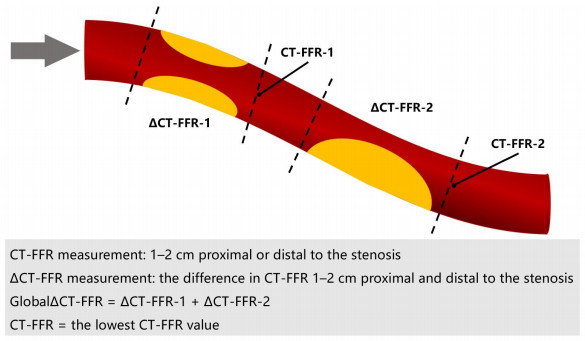
图 1 血管水平GlobalΔCT-FFR、ΔCT-FFR、CT-FFR示意图
Figure 1. GlobalΔCT-FFR, ΔCT-FFR, CT-FFR in per-vessel level schematic diagram.

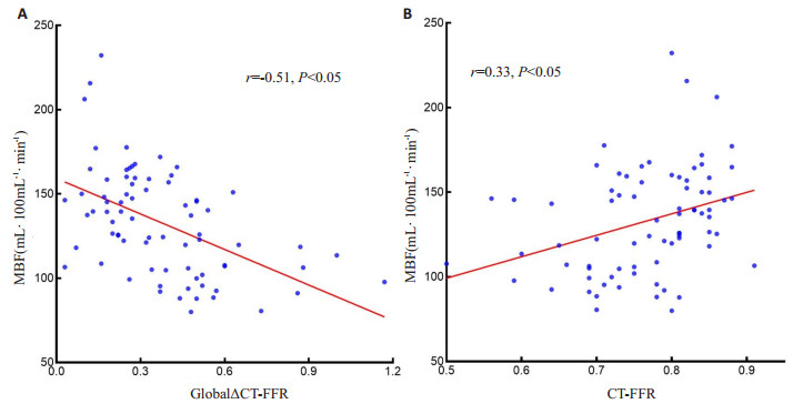
图 2 患者水平GlobalΔCT-FFR、CT-FFR与MBF相关性散点图
Figure 2. Scatter diagram of the correlation between CT-FFR, CT-FFR, and MBF in per-patient level. A: GlobalΔCT-FFR and MBF, B: CT-FFR and MBF.

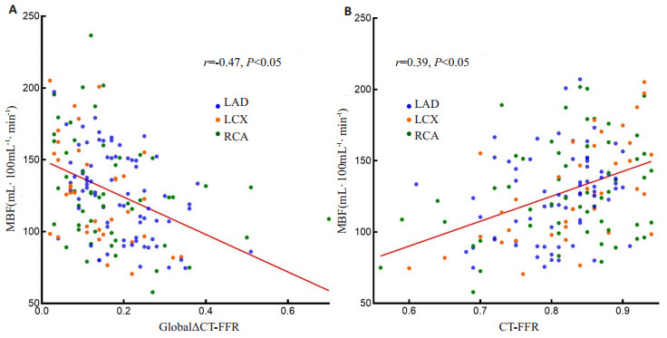
图 3 血管水平GlobalΔCT-FFR、CT-FFR与MBF相关性散点图
Figure 3. Scatter diagram of the correlation between CT-FFR, CT-FFR, and MBF in per-vessel level. A: GlobalΔCT-FFR and MBF, B: CT-FFR and MBF.

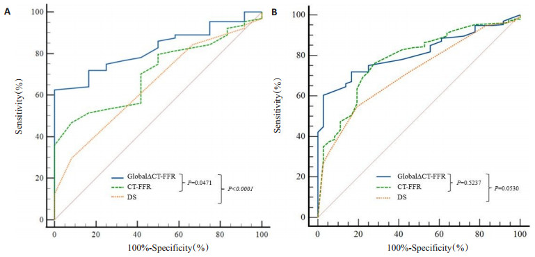
图 4 GlobalΔCT-FFR、CT-FFR及DS的ROC曲线
Figure 4. Comparison of ROC curves of GlobalΔCT-FFR, CT-FFR and DS. A: Per-patient level; B: Per-vessel level.

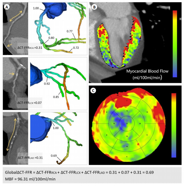
图 5 患者男性,46岁,初步诊断为冠心病
Figure 5. A 46- year- old male patient with a preliminary diagnosis of coronary artery disease. A: CCTA curved surface reconstruction image using B26f core reconstruction and computer simulation diagram of CT-FFR; B, C: Schematic diagrams of left ventricular myocardial MBF and bull's-eye diagram.
表 1 研究对象的一般临床情况及用药情况
Table 1. Clinical characteristics and medication of the patients
Characteristic n(%) Male 57 (75.0) Cardiac risk factors Hypertension 45 (59.2) Diabetes 15 (19.7) Hyperlipidemia 23 (30.3) Current smoking 30 (39.5) Family history of CAD 11 (14.5) Medication Aspirin 72 (92.7) Clopidogrel 52 (68.4) Statin 63 (82.9) Beta blocker 51 (67.1) Calcium channel blocker 28 (36.8) ACEI/ARB 22 (28.9) ACEI: Angiotensin-converting enzyme inhibitor; ARB: Angiotonin receptor blocker.  下载: 导出CSV
表 2 GlobalΔCT-FFR、CT-FFR和DS的诊断效能
Table 2. Diagnostic performance of GlobalΔCT-FFR, CT-FFR and DS
Method Sensitivity (95% CI) Specificity (95% CI) Accuracy (95% CI) Positive predictive value (95% CI) Negative predictive value (95% CI) True positive True negative False positive False negative AUC (95% CI) P Per-patient level GlobalΔCT-FFR 0.63(0.50-0.74) 0.86(0.65-1.07) 0.66(0.55-0.77) 1.00(1.00-1.00) 0.33(0.27-0.41) 38 12 2 24 0.82(0.72-0.90) <0.05 CT-FFR 0.47(0.34-0.60) 0.92(0.92-1.00) 0.54(0.42-0.65) 0.95(0.88-1.02) 0.24(0.20-0.30) 30 11 1 34 0.71(0.59-0.81) <0.05 DS 0.30(0.19-0.42) 0.92(0.62-1.00) 0.26(0.16-0.36) 0.95(0.74-0.99) 0.20(0.16-0.24) 8 12 0 56 0.65(0.53-0.76) <0.05 Per-vessel level GlobalΔCT-FFR 0.60(0.53-0.67) 0.97(0.86-1.00) 0.66(0.60-0.72) 0.99(0.94-1.00) 0.32(0.28-0.36) 116 35 1 76 0.81(0.75-0.86) <0.05 CT-FFR 0.76(0.69-0.82) 0.72(0.55-0.86) 0.75(0.69-0.81) 0.94(0.90-0.96) 0.49(0.40-0.57) 145 26 10 47 0.78(0.22-0.83) <0.05 DS 0.55(0.47-0.62) 0.81(0.64-0.92) 0.59(0.52-0.65) 0.94(0.88-0.97) 0.25(0.21-0.29) 105 29 7 87 0.71(0.65-0.77) <0.05 AUC: Area under the curve; MBF: Myocardial blood flow; CT-FFR: Computed tomography-derived fractional flow reserve; GlobalΔCT-FFR: Global trans-lesional computed tomography-derived fractional flow reserve; DS: Diameter stenosis.
下载: 导出CSV
-
[1] Lawton JS, Tamis-Holland JE, Bangalore S, et al. 2021 ACC/AHA/SCAI guideline for coronary artery revascularization: a report of the American college of cardiology/American heart association joint committee on clinical practice guidelines[J]. Circulation, 2022, 145(11): e18-e114. [2] Min JK, Taylor CA, Achenbach S, et al. Noninvasive fractional flow reserve derived from coronary CT angiography: clinical data and scientific principles[J]. JACC Cardiovasc Imaging, 2015, 8(10): 1209-22. doi: 10.1016/j.jcmg.2015.08.006 [3] Nakamura S, Kitagawa K, Goto Y, et al. Incremental prognostic value of myocardial blood flow quantified with stress dynamic computed tomography perfusion imaging[J]. JACC Cardiovasc Imaging, 2019, 12(7): 1379-87. doi: 10.1016/j.jcmg.2018.05.021 [4] Zhuang BY, Wang SL, Zhao SH, et al. Computed tomography angiography-derived fractional flow reserve (CT-FFR) for the detection of myocardial ischemia with invasive fractional flow reserve as reference: systematic review and meta-analysis[J]. Eur Radiol, 2020, 30(2): 712-25. doi: 10.1007/s00330-019-06470-8 [5] Rasoul H, Fyyaz S, Noakes D, et al. NHS England-funded CT fractional flow reserve in the era of the ISCHEMIA trial[J]. Clin Med, 2021, 21(2): 90-5. doi: 10.7861/clinmed.2020-0691 [6] Kogame N, Ono M, Kawashima H, et al. The impact of coronary physiology on contemporary clinical decision making[J]. JACC Cardiovasc Interv, 2020, 13(14): 1617-38. doi: 10.1016/j.jcin.2020.04.040 [7] Fournier S, Collet C, Xaplanteris P, et al. Global fractional flow reserve value predicts 5-year outcomes in patients with coronary atherosclerosis but without ischemia[J]. J Am Heart Assoc, 2020, 9 (24): e017729. doi: 10.1161/JAHA.120.017729 [8] Takagi H, Leipsic JA, McNamara N, et al. Trans-lesional fractional flow reserve gradient as derived from coronary CT improves patient management: advance registry[J]. J Cardiovasc Comput Tomogr, 2022, 16(1): 19-26. doi: 10.1016/j.jcct.2021.08.003 [9] Coenen A, Rossi A, Lubbers MM, et al. Integrating CT myocardial perfusion and CT-FFR in the work-up of coronary artery disease[J]. JACC Cardiovasc Imaging, 2017, 10(7): 760-70. doi: 10.1016/j.jcmg.2016.09.028 [10] Hamon M, Geindreau D, Guittet L, et al. Additional diagnostic value of new CT imaging techniques for the functional assessment of coronary artery disease: a meta-analysis[J]. Eur Radiol, 2019, 29 (6): 3044-61. doi: 10.1007/s00330-018-5919-8 [11] Cury RC, Leipsic J, Abbara S, et al. CAD-RADSTM 2.0-2022 coronary artery disease-reporting and data system an expert consensus document of the society of cardiovascular computed tomography (SCCT), the American college of cardiology (ACC), the American college of radiology (ACR) and the North America society of cardiovascular imaging (NASCI)[J]. Radiol Cardiothorac Imaging, 2022, 4(5): e220183. doi: 10.1148/ryct.220183 [12] Pontone G, Andreini D, Guaricci AI, et al. Incremental diagnostic value of stress computed tomography myocardial perfusion with whole-heart coverage CT scanner in intermediate-to high-risk symptomatic patients suspected of coronary artery disease[J]. JACC Cardiovasc Imaging, 2019, 12(2): 338-49. doi: 10.1016/j.jcmg.2017.10.025 [13] Coenen A, Kim YH, Kruk M, et al. Diagnostic accuracy of a machine-learning approach to coronary computed tomographic angiography-based fractional flow reserve: result from the MACHINE consortium[J]. Circ Cardiovasc Imaging, 2018, 11(6): e007217. doi: 10.1161/CIRCIMAGING.117.007217 [14] Rabbat MG, Berman DS, Kern M, et al. Interpreting results of coronary computed tomography angiography-derived fractional flow reserve in clinical practice[J]. J Cardiovasc Comput Tomogr, 2017, 11(5): 383-8. doi: 10.1016/j.jcct.2017.06.002 [15] Nozaki YO, Fujimoto S, Kawaguchi YO, et al. Prognostic value of the optimal measurement location of on-site CT-derived fractional flow reserve[J]. J Cardiol, 2022, 80(1): 14-21. doi: 10.1016/j.jjcc.2022.02.019 [16] Tanabe Y, Kurata A, Matsuda T, et al. Computed tomographic evaluation of myocardial ischemia[J]. Jpn J Radiol, 2020, 38(5): 411-33. doi: 10.1007/s11604-020-00922-8 [17] Kalykakis GE, Antonopoulos AS, Pitsargiotis T, et al. Relationship of endothelial shear stress with plaque features with coronary CT angiography and vasodilating capability with PET[J]. Radiology, 2021, 300(3): 549-56. doi: 10.1148/radiol.2021204381 [18] Siasos G, Sara JD, Zaromytidou M, et al. Local low shear stress and endothelial dysfunction in patients with nonobstructive coronary atherosclerosis[J]. J Am Coll Cardiol, 2018, 71(19): 2092-102. doi: 10.1016/j.jacc.2018.02.073 [19] Kumar A, Hung OY, Piccinelli M, et al. Low coronary wall shear stress is associated with severe endothelial dysfunction in patients with nonobstructive coronary artery disease[J]. JACC Cardiovasc Interv, 2018, 11(20): 2072-80. doi: 10.1016/j.jcin.2018.07.004 [20] Gutiérrez-Chico JL. Endothelial function and shear stress[J]. J Am Coll Cardiol, 2018, 71(19): 2103-5. doi: 10.1016/j.jacc.2018.03.452 [21] Assen MV, Vonder M, Pelgrim GJ, et al. Computed tomography for myocardial characterization in ischemic heart disease: a state-of-theart review[J]. Eur Radiol Exp, 2020, 4(1): 1-13. doi: 10.1186/s41747-019-0127-0 [22] Wardziak Ł, Kruk M, Pleban W, et al. Coronary CTA enhanced with CTA based FFR analysis provides higher diagnostic value than invasive coronary angiography in patients with intermediate coronary stenosis[J]. J Cardiovasc Comput Tomogr, 2019, 13(1): 62-7. doi: 10.1016/j.jcct.2018.10.004 -

 点击查看大图
计量
- 文章访问数: 140
- HTML全文浏览量: 50
- PDF下载量: 31
- 被引次数: 0