


Predictive value of CT mixed sign, island sign combined with NPR for the hematoma enlargement of spontaneous basal ganglia hemorrhage
-
摘要:
目的 分析CT混合征、岛征结合中性粒细胞与血小板比值(NPR)对自发性基底节脑出血(sBGICH)血肿增大的预测价值。 方法 回顾性分析本院急诊科及神经外科2020年2月~2022年11月收治的200例sBGICH患者的资料。根据其发病24 h内出血是否血肿增大,将患者分为出血血肿增大组(n=43)和出血无血肿增大组(n=157)。比较两组的CT混合征、岛征、卫星征、漩涡征、黑洞征、NPR和预后评分。 结果 两组的性别、年龄、发病时间、CT漩涡征、破入脑室的差异均无统计学意义(P > 0.05);出血血肿增大组的CT混合征、岛征、卫星征、黑洞征和NPR均高于出血无血肿增大组,预后评分低于出血无血肿增大组(P < 0.05)。经多因素Logistic回归性分析,CT混合征、岛征、卫星征、黑洞征、NPR > 5均是sBGICH发病24 h内出血血肿增大的危险因素(P < 0.05)。以sBGICH发病24 h内是否出血血肿增大进行分组,绘制ROC曲线,CT混合征、岛征、卫星征、黑洞征、NPR联合诊断预测的AUC为0.873,敏感度、特异性分别为72.30%、91.50%。 结论 CT混合征、岛征等结合NPR对sBGICH血肿增大有较高的预测价值,可根据临床实际情况灵活运用。 -
关键词:
- 混合征 /
- 岛征 /
- 中性粒细胞与血小板比值 /
- 自发性基底节脑出血 /
- 血肿增大
Abstract:Objective To analyze the predictive value of CT mixed sign, island sign combined with neutrophil to platelet ratio (NPR) for the hematoma enlargement of spontaneous basal ganglia hemorrhage (sBGICH). Methods We retrospectively reviewed the data of 200 patients with sBGICH admitted to the Department of Emergency and Department of Neurosurgery of our hospital from February 2020 to November 2022. The patients were grouped as bleeding hematoma enlargement group (n=43) and no bleeding hematoma enlargement group (n=157) according to whether the bleeding had enlarged within 24 hours of onset. The CT mixed sign, island sign, satellite sign, swirl sign, black hole sign, NPR and prognosis scores were compared between the two groups. Results There were no statistically significant differences in gender, age, onset time, CT swirl sign, and intraventricular rupture between the two groups (P > 0.05). The CT mixed sign, island sign, satellite sign, black hole sign, and NPR were significantly higher in the bleeding hematoma enlargement group than in the no bleeding hematoma enlargement group, while the prognosis score was significantly lower (P < 0.05). Multivariate logistic regression analysis showed that CT mixed sign, island sign, satellite sign, black hole sign, and NPR > 5 were risk factors for bleeding hematoma enlargement within 24 hours of sBGICH onset (P < 0.05). The ROC curve was drawn according to whether bleeding hematoma enlargement occurred within 24 h of sBGICH onset, and the area under the curve of the joint diagnosis and prediction of CT mixed sign, island sign, satellite sign, black hole sign and NPR was 0.873, with a sensitivity and specificity of 72.30% and 91.50%, respectively. Conclusion CT mixed sign, island sign combined with NPR have a high predictive value for the hematoma enlargement of sBGICH and it can be flexibly used according to the actual clinical situation. -

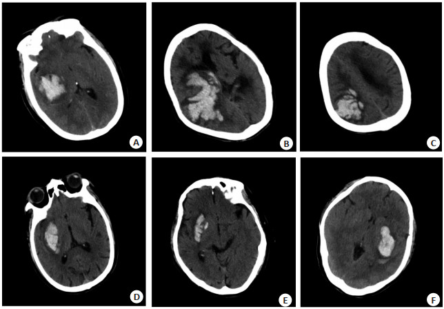
图 1 脑出血血肿增大各类CT影像
Figure 1. Various CT images with enlarged hematoma of cerebral hemorrhage.
A: CT mixed sign; B: CT island sign/black hole sign/CT swirl sign; C: CT island sign/CT swirl sign; D: CT mixed sign; E: CT satellite sign; F: CT swirl sign.

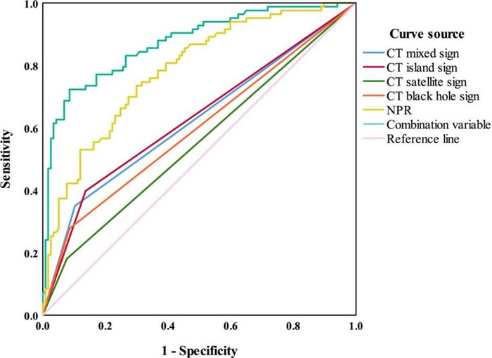
图 2 CT征象与NPR预测sBGICH发病24 h内出血血肿增大的ROC曲线
Figure 2. ROC curve of CT signs and NPR prediction of hematoma enlargement within 24 h of onset of sBGICH.
表 1 两组临床资料比较
Table 1. Comparison of general data between the two groups (n)
Index HE group (n=43) Non-HE group (n=157) t/χ2 P Gender 0.021 0.884 Male 26 93 Female 17 64 Age (years, Mean±SD) 56.29±5.94 57.53±6.81 1.086 0.279 Onset time (h, Mean±SD) 3.22±0.65 3.31±0.58 0.878 0.381 Mixed sign 27.692 < 0.001 Yes 17 12 No 26 145 Island sign 18.887 < 0.001 Yes 13 10 No 30 147 Satellite sign 16.477 < 0.001 Yes 11 8 No 32 149 Swirl sign 2.928 0.087 Yes 7 12 No 36 145 Black hole sign 21.923 < 0.001 Yes 14 10 No 29 147 Break into encephalocoele 2.370 0.124 Yes 5 8 No 38 149 NPR (Mean±SD) 5.68±1.07 4.89±0.87 5.010 < 0.001 Prognostic score (point, Mean±SD) 2.74±0.55 3.85±0.63 10.505 < 0.001 NPR: Neutrophil-to-platelets ratio; HE: Hematoma enlargement.  下载: 导出CSV
表 2 变量赋值表
Table 2. Variable assignment table
Variable Assignment Mixed sign 0:No;1:Yes Island sign 0:No;1:Yes Satellite sign 0:No;1:Yes Black hole sign 0:No;1:Yes NPR 0:≤5;1: > 5
下载: 导出CSV
表 3 sBGICH发病24 h内出血血肿增大的多因素Logistic回归性分析
Table 3. Logistic regression analysis of the increase of haemorrhagic hematoma within 24 hours after the onset of sBGICH
Factor β SE Wald χ2 OR P 95% CI Mixed sign 2.131 0.533 7.501 8.420 < 0.001 3.124-17.274 Island sign 1.463 0.464 6.795 4.321 0.001 1.772-12.913 Satellite sign 1.296 0.619 3.382 3.655 0.026 1.154-8.322 Black hole sign 1.599 0.509 6.172 4.947 0.021 1.189-10.744 NPR > 5 1.479 0.256 22.568 4.389 < 0.001 2.681-27.321
下载: 导出CSV
表 4 各指标敏感度、特异性及AUC
Table 4. Sensitivity, specificity and AUC of each index.
Index Sensitivity(%) Specificity(%) AUC AUC 95% CI Mixed sign 34.90 89.70 0.632 0.559-0.688 Island sign 39.80 86.30 0.630 0.566-0.695 Satellite sign 18.10 92.30 0.552 0.485-0.619 Black hole sign 27.70 81.50 0.596 0.530-0.661 NPR 73.50 70.10 0.784 0.730-0.837 Combined index 72.30 91.50 0.873 0.832-0.914
下载: 导出CSV
-
[1] Tsai HH, Lee BC, Chen YF, et al. Cerebral venous reflux and dilated basal Ganglia perivascular space in hypertensive intracerebral hemorrhage[J]. J Stroke, 2022, 24(3): 363-71. doi: 10.5853/jos.2022.01004 [2] Perosa V, Arts T, Assmann A, et al. Pulsatility index in the basal Ganglia arteries increases with age in elderly with and without cerebral small vessel disease[J]. AJNR Am J Neuroradiol, 2022, 43(4): 540-6. doi: 10.3174/ajnr.A7450 [3] 何敏, 陈后勤, 邵凌云, 等. CT平扫非均一密度血肿与原发性基底节区脑出血患者预后的关系[J]. 中国医药导报, 2019, 16(27): 184-8. https://www.cnki.com.cn/Article/CJFDTOTAL-YYCY201927044.htm [4] 王婷, 苗重昌, 张永刚. CT检查对自发性脑出血患者早期血肿扩大的评估作用分析[J]. 医学影像学杂志, 2022, 32(9): 1466-70. https://www.cnki.com.cn/Article/CJFDTOTAL-XYXZ202209003.htm [5] Dienel A, Peeyush KT, Blackburn SL, et al. Role of platelets in the pathogenesis of delayed injury after subarachnoid hemorrhage[J]. J Cereb Blood Flow Metab, 2021, 41(11): 2820-30. doi: 10.1177/0271678X211020865 [6] 王志花, 宣兆博, 姜晓雪, 等. 头颅平扫CT"岛征"对HICH患者早期血肿扩大的预测价值研究[J]. 脑与神经疾病杂志, 2019, 27(12): 764-767 https://www.cnki.com.cn/Article/CJFDTOTAL-LYSJ201912010.htm [7] 王希, 仲艳, 颜伟, 等. CT平扫岛征和黑洞征对原发性脑出血早期血肿扩大的预测价值[J]. 中华神经外科杂志, 2021, 37(6): 557-561 https://cpfd.cnki.com.cn/Article/CPFDTOTAL-SJWK202206001223.htm [8] 孔祥宇, 席宇君, 钱志远. 脑出血患者血肿增大的预测因素[J]. 国际脑血管病杂志, 2018, 26(1): 51-6. https://www.cnki.com.cn/Article/CJFDTOTAL-HNSJ202114007.htm [9] Marazziti D, Torrigiani S, Carbone MG, et al. Neutrophil/lymphocyte, platelet/lymphocyte, and monocyte/lymphocyte ratios in mood disorders[J]. Curr Med Chem, 2022, 29(36): 5758-81. [10] Lattanzi S, Brigo F, Trinka E, et al. Neutrophil-to-lymphocyte ratio in acute cerebral hemorrhage: a system review[J]. Transl Stroke Res, 2019, 10(2): 137-45. [11] 毛宝杰, 王明, 万曙. 血小板衍生生长因子及其受体在脑出血中的作用[J]. 浙江大学学报(医学版), 2022, 51(5): 634-9. https://www.cnki.com.cn/Article/CJFDTOTAL-ZJYB202205012.htm [12] 中华医学会神经外科学分会, 中国医师协会急诊医师分会, 国家卫生和计划生育委员会脑卒中筛查与防治工程委员会. 自发性脑出血诊断治疗中国多学科专家共识[J]. 中华神经外科杂志, 2015, 31(12): 1189-94. [13] 徐运, 刘鸣, 崔丽英. 中国脑血管病影像应用指南2019[J]. 中华神经科杂志, 2020, 53(4): 250-2, 253. https://www.cnki.com.cn/Article/CJFDTOTAL-YYCY202019002.htm [14] 王琮智, 许梓璧, 马祥园, 等. 基于数据扩增和迁移学习的Mask RCNN脑CT图像自动分割研究[J]. 中国生物医学工程学报, 2021, 40 (4): 410-8 https://www.cnki.com.cn/Article/CJFDTOTAL-ZSWY202104004.htm [15] Goyal N, Tsivgoulis G, Malhotra K, et al. Minimally invasive endoscopic hematoma evacuation vs best medical management for spontaneous basal- Ganglia intracerebral hemorrhage[J]. J Neurointerv Surg, 2019, 11(6): 579-83. [16] Rzepliński R, Sługocki M, Tarka S, et al. Mechanism of spontaneous intracerebral hemorrhage formation: an anatomical specimens-based study[J]. Stroke, 2022, 53(11): 3474-80. [17] Haußmann R, Homeyer P, Haußmann M, et al. Intrazerebrale blutungen unter plättchenaggregationshemmung und oraler antikoagulation Bei patienten mit zerebraler amyloidangiopathie[J]. Nervenarzt, 2022, 93(6): 599-604. [18] 黄萌, 喻晓刚, 刘玲. 自发性脑出血CT影像分割在老年脑梗死合并脑出血病灶中的计算效果[J]. 神经损伤与功能重建, 2022, 17(11) 666-667, 674 https://www.cnki.com.cn/Article/CJFDTOTAL-GWKF202211012.htm [19] Coulibaly A, Gartman W, Swank V, et al. RAR- related orphan receptor gamma T (RoRγt)-related cytokines play a role in neutrophil infiltration of the central nervous system after subarachnoid hemorrhage[J]. Neurocritical Care, 2019, 33: 140-151. [20] Mengel A, Stefanou MI, Hadaschik KA, et al. Early administration of desmopressin and platelet transfusion for reducing hematoma expansion in patients with acute antiplatelet therapy associated intracerebral hemorrhage[J]. Crit Care Med, 2020, 48(7): 1009-17. -

 点击查看大图
计量
- 文章访问数: 124
- HTML全文浏览量: 62
- PDF下载量: 8
- 被引次数: 0