


Diagnostic value of ultrasound and digital breast tomosynthesis in breast complex cystic and solid mass
-
摘要:
目的 分析乳腺囊实性肿块声像图及数字乳腺断层摄影(DBT)图像特征,探讨超声(US)及DBT及二者联合对乳腺囊实性肿块良恶性判别的诊断价值。 方法 回顾性分析乳腺囊实性肿块251例患者共257个肿块。根据超声声像图特征将肿块分为4型,计算各型阳性预测值。分析声像图特征、DBT图像特征与恶性的相关性;以病理结果为金标准,比较US、DBT及US+DBT的诊断效能。 结果 乳腺囊实性肿块恶性率为30.4%。Ⅰ~Ⅳ型囊实性肿块阳性预测值分别为0、22.2%、19.3%和38.0%,差异有统计学意义(P < 0.001)。年龄及US特征中的肿块大小、分型、形态、生长方式、边缘、钙化、肿块内血流、异常腋窝淋巴结以及DBT特征中的肿块大小、形态、边缘、密度、钙化、小梁结构、腺体分型差异有统计学意义(P < 0.05)。US、DBT、US+DBT的AUC值分别为0.806、0.880、0.903。US与DBT、US+DBT的AUC值对比,差异均有统计学意义(P < 0.05),US+DBT与DBT的AUC值对比,差异无统计学意义(P > 0.05)。 结论 DBT对乳腺囊实性肿块良恶性判别的诊断效能优于US,两者联合可明显提高诊断的准确性,增加诊断的敏感度和特异性。 Abstract:Objective To analyze the ultrasonographic and DBT features of breast complex cystic and solid mass, and to explore the diagnostic value of ultrasound (US), digital breast tomosynthesis (DBT) and their combination in differentiating benign and malignant breast complex cystic and solid mass. Methods A total of 257 masses in 251 patients with breast complex cystic and solid mass were analyzed retrospectively. According to the sonographic features, the masses were divided into 4 types, and the positive predictive values of each type were calculated. The correlation between the features of sonogram, DBT and malignancy was analyzed. The diagnostic efficacy of US, DBT and US + DBT was compared according to the pathological results as the gold standard. Results The malignant rate of breast complex cystic and solid mass was 30.4%. The positive predictive values of Ⅰ-Ⅳ type complex cystic and solid mass were 0, 22.2%, 19.3% and 38.0%, respectively(P < 0.001). There were significant differences in age, tumor size, classification, morphology, growth mode, margin, calcification, blood flow in the tumor, abnormal ALN in ultrasonic characteristics, and tumor size, morphology, margin, density, calcification, trabecular structure and gland classification in DBT characteristics (P < 0.05). The AUC values for US, DBT and US+DBT were 0.806, 0.880 and 0.903, respectively. There was significant difference in AUC between US and DBT, US + DBT (P < 0.05), but there was no significant difference in AUC between US+DBT and DBT (P < 0.05). Conclusion The diagnostic efficacy of DBT in differentiating benign and malignant breast complex cystic and solid mass is better than that of US. The combination of DBT and US can significantly improve the diagnostic accuracy, sensitivity and specificity. -
Key words:
- breast /
- complex cystic and solid mass /
- ultrasound /
- digital breast tomosynthesis
-

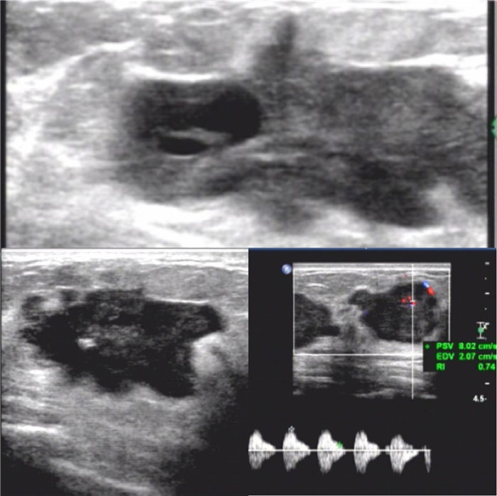
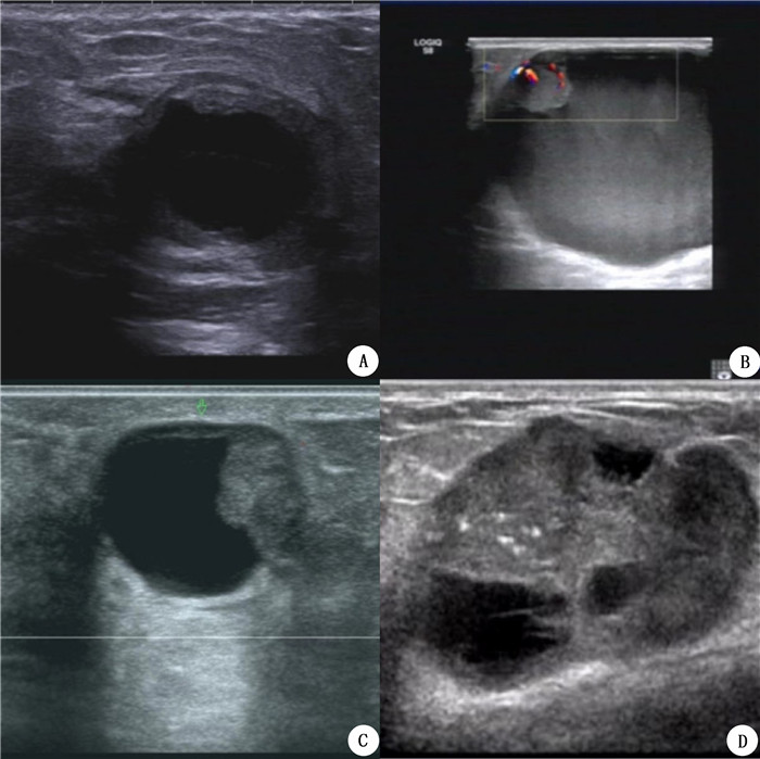
图 1 乳腺囊实性肿块超声图像
Figure 1. Ultrasound image of a cystic solid breast mass. A-D: Cystic solid masses of types Ⅰ, Ⅱ, Ⅲ and Ⅳ, respectively.

图 2 浸润性导管癌超声图像
Figure 2. Ultrasound image of infiltrating ductal carcinoma. The lesion is a type Ⅳ cystic solid mass with irregular shape and angular edges, presenting as a " crab foot like" change. Multiple punctate strong echoes can be seen inside, and CDFI shows blood flow inside and around the mass, with a blood flow resistance index > 0.7.

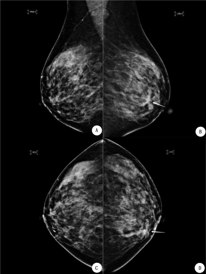
图 3 浸润性导管癌DBT图像
Figure 3. DBT image of invasive ductal carcinoma. A-D: images of RMLO bit, LMLO bit, RCC bit and LCC bit, respectively. DBT shows a slightly high-density mass (arrow) on the inner side of the left breast, with irregular morphology, spiculated edges, and several small pleomorphic calcifications visible inside. Pathology: Invasive ductal carcinoma.

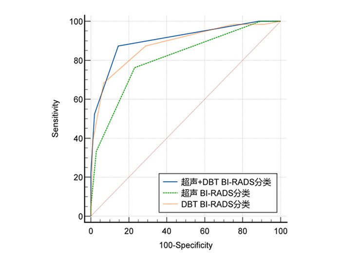
图 4 US、DBT及US+DBT诊断乳腺囊实性肿块的ROC曲线
Figure 4. ROC curve of US, DBT and US+ DBT in diagnosis of breast complex cystic and solid mass.

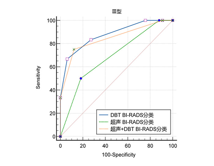
图 5 US、DBT、US + DBT诊断Ⅲ型乳腺囊实性肿块的ROC曲线
Figure 5. ROC curve of US, DBT, and US + DBT in diagnosis of Type Ⅲ breast complex cystic and solid mass.

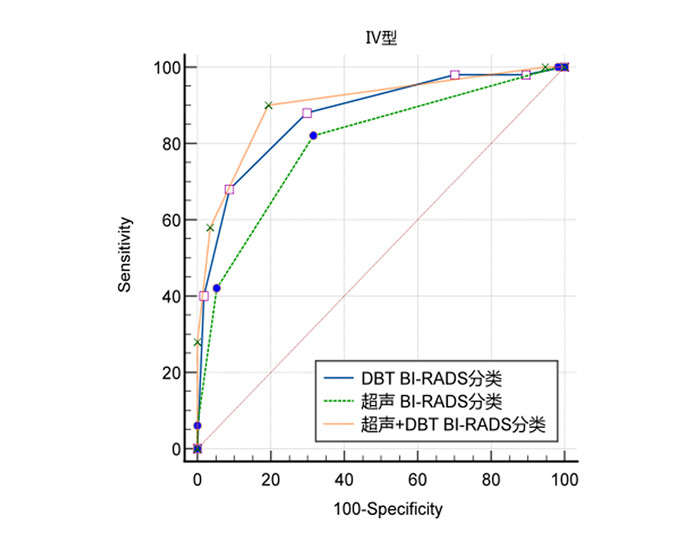
图 6 US、DBT及US+DBT诊断Ⅳ型乳腺囊实性肿块的ROC曲线
Figure 6. ROC curve of US, DBT, and US+DBT in diagnosis of type Ⅳ breast complex cystic and solid mass.
表 1 乳腺囊实性肿块的病理类型和超声分型对照
Table 1. Comparison of pathologic type and ultrasonic classification of breast complex cystic and solid mass (n)
Findings Type Ⅰ Type Ⅱ Type Ⅲ Type Ⅳ Total Benign Fibrocystic change 6 3 14 24 47 Fibroadenoma 2 32 34 Intraductal papilloma 2 12 16 30 Breast adenosis 3 1 9 11 24 Galactocele 8 3 3 14 Mastitis 2 9 11 Abscess 1 1 1 3 6 Boundary phyllodes tumor 2 3 5 Usual ductal hyperplasia 1 3 4 Benign phyllodes tumor 1 1 Fat necrosis 1 1 Prosthesis leakage 1 1 Atypical ductal hyperplasia 1 1 Total 20 7 46 106 179 Malignant Invasive ductal carcinoma 1 7 41 49 Intraductal papillary carcinoma 1 1 4 6 Encapsulated papillary carcinoma 2 4 6 Ductal carcinoma in situ 5 5 Mucinous carcinoma 5 5 Solid papillary carcinoma 1 4 5 Malignant phyllodes tumor 1 1 Invasive lobular carcinoma 1 1 Total 0 2 11 65 78  下载: 导出CSV
表 2 乳腺囊实性肿块超声特征与良恶性相关性分析
Table 2. Ultrasonographic characteristics of breast complex cystic and solid mass and its correlation with benign and malignant (n)
Factor Univariate Total P Benign Malignant Age (years) < 0.001 ≤50 156 37 193 >50 23 41 64 Mass size (mm) 0.003 ≤20 89 23 112 >20 90 55 145 Mass type < 0.001 Ⅰ 20 0 20 Ⅱ 7 2 9 Ⅲ 46 11 57 Ⅳ 106 65 171 Shape < 0.001 Regular 130 26 156 Irregular 49 52 101 Orientation 0.010 Parallel 173 69 242 Not parallel 6 9 15 Margin < 0.001 Circumscribed 130 22 152 Not circumscribed 49 56 105 Posterior features 0.905 None 55 26 81 Enhancement 121 51 172 Shadowing 3 1 4 Combined 0 0 0 Calcification < 0.001 Absent 124 33 157 Present 55 45 100 Structure distortion 0.091 Absent 179 76 255 Present 0 2 2 Duct changes 0.728 Absent 171 76 247 Present 8 2 10 Skin changes 0.219 Absent 178 76 254 Present 1 2 3 Edema 0.437 Absent 175 75 250 Present 4 3 7 Vascularity < 0.001 Absent 127 36 163 Present 52 42 94 Abnormal ALN < 0.001 Absent 174 53 227 Present 5 25 30
下载: 导出CSV
表 3 乳腺囊实性肿块DBT征象与良恶性相关性分析
Table 3. Correlation analysis of DBT signs with benign and malignant breast complex cystic and solid mass(n)
Factor Univariate P Benign Malignant Total Age (years) < 0.001 ≤50 83 28 111 >50 15 33 48 Mass size (mm) 0.040 ≤20 45 18 63 >20 53 43 96 Shape < 0.001 Regular 62 18 79 Irregular 37 43 80 Margin 0.001 Circumscribed 24 8 32 Obscured 26 6 32 Microlobulated 47 41 88 Indistinct 1 6 7 Spiculated 0 0 0 Density < 0.001 High density 29 52 81 Equal density 63 9 72 Low density 0 0 0 Fat-containing 6 0 6 Associated calcification 0.001 Absent 80 38 118 Benign 9 3 12 Suspected malignant 9 20 29 Trabecular < 0.001 No changes 50 14 64 Thickening, disorder 48 47 95 Nipple retraction 0.349 Absent 91 54 145 Present 7 7 14 Breast composition < 0.001 Fatty 1 4 5 Scattered fibroglandular 2 12 14 Heterogeneously dense 86 43 129 Extremely dense 9 2 11
下载: 导出CSV
表 4 US、DBT及US+DBT对乳腺囊实性肿块诊断效能对比
Table 4. Comparison of US, DBT and US+DBT in the diagnosis of breast complex cystic and solid mass
Methods AUC (95% CI) Sensitivity(95% CI, %) Specificity(95% CI, %) US 0.806(0.738-0.863) 76.19(63.8-86.0) 76.92(67.6-84.6) DBT 0.880(0.821-0.925) 87.30(76.5-94.4) 71.15(61.4-79.6) US+DBT 0.903(0.848-0.943) 87.30(76.5-94.4) 85.58(77.3-91.7) US: Ultrasound; DBT: Digital breast tomography.
下载: 导出CSV
表 5 US、DBT及US+DBT对Ⅲ、Ⅳ型乳腺囊实性肿块诊断效能对比
Table 5. Comparison of the diagnostic efficacy of US, DBT, US+DBT in breast complex cystic and solid mass of Type Ⅲ and Type Ⅳ
Mass type AUC (95% CI) Sensitivity (95% CI, %) Specificity (95% CI, %) Ⅲ(n=45) US 0.689(0.534-0.819) 50.00(21.1-78.9) 81.82(64.5-93.0) DBT 0.876(0.744-0.955) 83.33(51.6-97.9) 72.73(54.5-86.7) US+DBT 0.846(0.707-0.936) 75.00(42.8-94.5) 87.88(71.8-96.6) Ⅳ(n=107) US 0.800(0.712-0.871) 82.00(68.6-91.4) 68.42(54.8-80.1) DBT 0.874(0.796-0.930) 88.00(75.7-95.5) 70.18(56.6-81.6) US+DBT 0.901(0.828-0.950) 90.00(78.2-96.7) 80.70(68.1-90.0)
下载: 导出CSV
-
[1] Athanasiou A, Aubert E, Vincent Salomon A, et al. Complex cystic breast masses in ultrasound examination[J]. Diagn Interv Imaging, 2014, 95(2): 169-79. doi: 10.1016/j.diii.2013.12.008 [2] 于洋, 薛改琴. 超声检查与乳腺X线摄影对乳腺复杂囊性病变诊断价值分析[J]. 医学影像学杂志, 2022, 32(07): 1178-82. https://www.cnki.com.cn/Article/CJFDTOTAL-XYXZ202207021.htm [3] Hsu HH, Yu JC, Lee HS, et al. Complex cystic lesions of the breast on ultrasonography: feature analysis and BI-RADS assessment[J]. Eur J Radiol, 2011, 79(1): 73-9. doi: 10.1016/j.ejrad.2009.12.037 [4] Makki J. Diversity of Breast Carcinoma: Histological Subtypes and Clinical Relevance[J]. Clin Med Insights Pathol, 2015, 8: 23-31. [5] Xiang H, Tang G, Li Y, et al. Value of Hand-held Ultrasound in the Differential Diagnosis and Accurate Breast Imaging Reporting and Data System Subclassification of Complex Cystic and Solid Breast Lesions[J]. Ultrasound Med Biol, 2020, 46(5): 1111-8. doi: 10.1016/j.ultrasmedbio.2020.01.006 [6] Rao AA, Feneis J, Lalonde C, et al. A Pictorial Review of Changes in the BI-RADS Fifth Edition[J]. Radiographics, 2016, 36(3): 623-39. doi: 10.1148/rg.2016150178 [7] Yao JP, Hao YZ, Chang Q, et al. Value of Ultrasonographic Features for Assessing Malignant Potential of Complex Cystic Breast Lesions [J]. J Ultrasound Med, 2017, 36(4): 699-704. doi: 10.7863/ultra.16.05012 [8] 王玉敏, 红华, 王芳, 等. 超声对乳腺囊实性复合回声肿块的诊断价值[J]. 中国超声医学杂志, 2019, 3(12): 1127-1130. https://www.cnki.com.cn/Article/CJFDTOTAL-ZGCY201912023.htm [9] Chen M, Zhan WW, Wang WP. Cystic breast lesions by convention-al ultrasonography: sonographic subtype-pathologic correlation and BI-RADS Assessment[J]. Arch Med Sci, 2014, 10(1): 76-83. [10] 张和庆, 彭玉兰, 赵海娜, 等. 乳腺囊实性肿块的BI-RADS-US图像特征及良恶性鉴别[J]. 中国超声医学杂志, 2015, 31(09): 777-80. https://www.cnki.com.cn/Article/CJFDTOTAL-ZGCY201509004.htm [11] Porembka JH, Baydoun S, Mootz AR, et al. Choice of imaging method in the work-up of non-calcified breast lesions identified on tomosynthesis screening[J]. Eur J Radiol, 2020, 131: 109203. doi: 10.1016/j.ejrad.2020.109203 [12] Li X, Qin G, He Q, et al. Digital breast tomosynthesis versus digital mammography: integration of image modalities enhances deep learning-based breast mass classification[J]. Eur Radiol, 2020, 30(2): 778-788. doi: 10.1007/s00330-019-06457-5 [13] Haas BM, Kalra V, Geisel J, et al. Comparison of tomosynthesis plus digital mammography and digital mammography alone for breast cancer screening[J]. Radiology, 2013, 269(3): 694-700. doi: 10.1148/radiol.13130307 [14] Nakashima K, Uematsu T, Itoh T, et al. Comparison of visibility of circumscribed masses on Digital Breast Tomosynthesis (DBT) and 2D mammography: are circumscribed masses better visualized and assured of being benign on DBT?[J]. Eur Radiol, 2017, 27(2): 570-77. doi: 10.1007/s00330-016-4420-5 [15] 黄新玲, 胡汉金. 数字乳腺断层合成摄影和全视野数字化乳腺摄影在乳房肿块型病变中的诊断价值[J]. 安徽医药, 2021, 25(02): 296-299. https://www.cnki.com.cn/Article/CJFDTOTAL-AHYY202102021.htm [16] D'Angelo A, Orlandi A, Bufi E, et al. Automated breast volume scanner (ABVS) compared to handheld ultrasound (HHUS) and contrast-enhanced magnetic resonance imaging (CE-MRI) in the early assessment of breast cancer during neoadjuvant chemotherapy: an emerging role to monitoring tumor response?[J]. Radiol Med, 2021, 126(4): 517-26. doi: 10.1007/s11547-020-01319-3 [17] Liu J, Zhao H, Huang Y, et al. Genome-wide cell-free DNA methylation analyses improve accuracy of non-invasive diagnostic imaging for early-stage breast cancer[J]. Mol Cancer, 2021, 20(1): 36. doi: 10.1186/s12943-021-01330-w [18] Carbognin G, Girardi V, Calciolari C, et al. Utility of second-look ultrasound in the management of incidental enhancing lesions detected by breast MR imaging[J]. Radiol Med, 2010, 115(8): 1234-1245. doi: 10.1007/s11547-010-0561-9 [19] Lee WK, Chung J, Cha ES, et al. Digital breast tomosynthesis and breast ultrasound: Additional roles in dense breasts with category 0 at conventional digital mammography[J]. Eur J Radiol, 2016, 85(1): 291-296. doi: 10.1016/j.ejrad.2015.09.026 [20] 代晓倩, 张伟. 数字乳腺断层摄影与超声对致密型乳腺内病变的诊断效能对比研究[J]. 中国医药, 2019, 14(05): 738-741. https://www.cnki.com.cn/Article/CJFDTOTAL-ZGYG201905024.htm [21] 胡丹婷, 郑晓静, 尤超, 等. 数字乳腺摄影联合超声在小乳腺癌诊断中的应用[J]. 中国超声医学杂志, 2022, 38(03): 267-270. https://www.cnki.com.cn/Article/CJFDTOTAL-ZGCY202203007.htm [22] Goh JHL, Tan TL, Aziz S, et al. Comparative Study of Digital Breast Tomosynthesis (DBT) with and without Ultrasound versus Breast Magnetic Resonance Imaging (MRI) in Detecting Breast Lesion[J]. Int J Environ Res Public Health, 2022, 19(2): 759. doi: 10.3390/ijerph19020759 [23] 韩金花, 张正福, 王海燕, 等. 超声及X线摄影BI-RADS诊断乳腺囊性及以囊性为主病变的价值[J]. 医学影像学杂志, 2016, 26(02): 250-253. https://www.cnki.com.cn/Article/CJFDTOTAL-XYXZ201602022.htm -

 点击查看大图
计量
- 文章访问数: 150
- HTML全文浏览量: 74
- PDF下载量: 15
- 被引次数: 0