


Value of individualized scan in CT imaging of small pulmonary nodules ≤ 1 cm
-
摘要:
目的 比较个性化和常规两种扫描方案对≤1 cm肺小结节的CT成像效果,探索最佳扫描方案。 方法 按照纳入及排除标准,选择2020年1月~2022年6月在我院行肺小结节CT成像的患者,按随机对照表以平衡对照法随机分为个性化组(n=67)和常规组(n=70),个性化组进行严格的过度吸气训练、个性化体位、个性化扫描方向制定,获得的图像与常规扫描组进行图像质量比较。 结果 个性化组的CT平扫、肺动脉期、肺静脉期图像质量均优于常规扫描组,差异有统计学意义(χ2=8.204,P=0.017;χ2= 6.625,P=0.036;χ2=7.139,P=0.028);两组组内CT平扫、肺动脉期、肺静脉期图像质量比较,常规组组内CT平扫和肺静脉期图像质量差异有统计学意义(χ2=9.188,P=0.010),常规组其余各期、个性化组组内比较图像质量差异均无统计学意义(P > 0.05)。 结论 个性化扫描方案成像质量稳定,能提高≤1cm肺小结节CT成像的图像质量。 Abstract:Objective To compare the CT imaging effects of personalized and conventional scanning schemes on pulmonary nodules≤1 cm, and explore the best scanning scheme. Methods A total of 142 patients were included from January 2020 to June 2022 according to inclusion and exclusion criteria. According to the random control table, they were randomly divided into personalized group and conventional group by balanced control method. Patients in the personalized group were subjected to rigorous hyperinspiratory training, develop personalized position and personalized scanning direction. The obtained images were compared with the conventional scan group for image quality. Results The image quality of CT plain scan, pulmonary artery phase and pulmonary vein phase in the personalized group was better than that in the conventional scan group, the difference was statistically significant (χ2=8.204, P=0.017, χ2=6.625, P=0.036, χ2=7.139, P=0.028). The image quality of CT plain scan, pulmonary artery phase and pulmonary vein phase was compared within the two groups. In the conventional group, there was a statistically significant difference in image quality between CT plain scan and pulmonary venous stage (χ2=9.188, P=0.010), but there was no statistical significance between the rest phases in the conventional group, also between all the phases in the personalized group(P > 0.05). Conclusion The personalized scanning scheme can provide stable imaging quality, and improve the image quality of pulmonary nodules≤1cm CT imaging. -

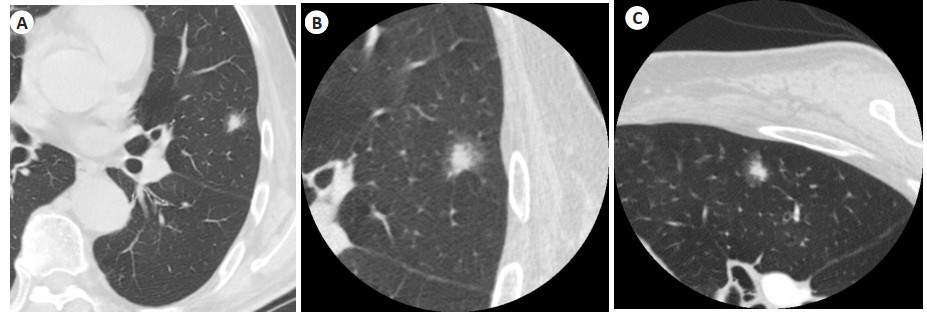
图 1 左肺上叶舌段胸膜下直径约8 mm实性结节
A: 常规组0.625 mm轴位图像; B: 常规组基础上的靶重建图像; C: 个性化组的靶重建图像.
Figure 1. Solid nodule with a diameter of about 8 mm in the subpleural segment of the upper lobe of the left lung.

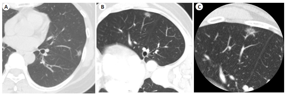
图 2 左肺上叶舌段胸膜下直径约10 mm混合磨玻璃密度结节
A: 常规组0.625 mm轴位图像; B: 常规组基础上的靶重建图像; C: 个性化组的靶重建图像.
Figure 2. Mixed ground-glass density nodule with a diameter of about 10 mm in the subpleural segment of the upper lobe of the left lung.

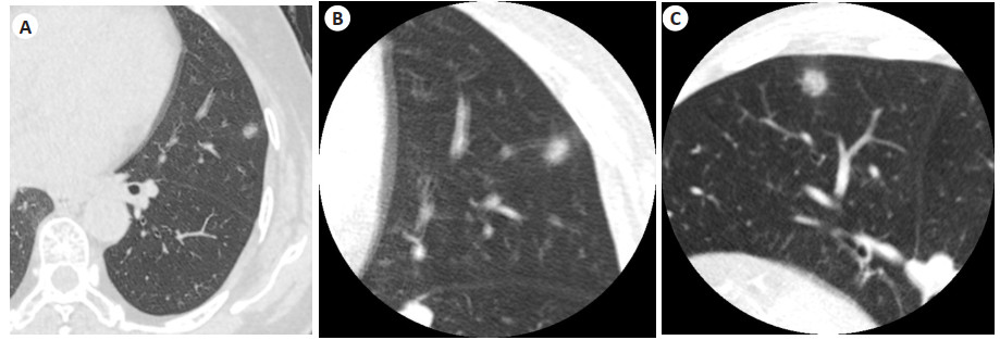
图 3 左肺上叶舌段胸膜下直径约9 mm纯磨玻璃密度结节
A: 常规组0.625 mm轴位图像; B: 个性化组0.625 mm轴位图像; C: 个性化组的靶重建图像.
Figure 3. Pure ground-glass density nodule with a diameter of about 9 mm in the subpleural segment of the upper lobe of the left lung.
表 1 个性化组与常规组一般资料
Table 1. General characteristics of individualized group and conventional group
组别 年龄(岁,Mean±SD) 男性[n(%)] 结节直径(mm) 实性结节[n(%)] 单发[n(%)] 个性化组(n=67) 53.8±17.8 35(52.2) 7.0(6.0,8.0) 19(28.4) 52(77.2) 常规组(n=70) 52.3±18.8 31(44.3) 7.0(6.0,8.0) 16(22.9) 59(84.3) t/χ2 0.467 0.867 -0.442 0.545 0.992 P 0.641 0.352 0.658 0.461 0.319  下载: 导出CSV
表 2 个性化组与常规组肺小结节分布
Table 2. Distribution of pulmonary nodules in individualized group and conventional group (n)
组别 右肺上叶 右肺中叶 右肺下叶 左肺上叶 左肺下叶 个性化组(n=67) 11 12 14 16 14 常规组(n=70) 14 11 15 15 15 χ2 0.439 P 0.979
下载: 导出CSV
表 3 个性化组和常规组图像质量比较
Table 3. Comparison of image quality between individualized group and conventional group (n)
组别 平扫 增强肺动脉期 增强肺静脉期 优秀 合格 差 优秀 合格 差 优秀 合格 差 个性化组(n=67) 23 38 6 30 35 2 38 26 3 常规组(n=70) 13 45 17 22 38 10 28 30 12 χ2 8.204 6.625 7.139 P 0.017 0.036 0.028
下载: 导出CSV
表 4 组内CT平扫、肺动脉期、肺静脉期图像质量比较
Table 4. Comparison of image quality of non-enhanced CT scan, pulmonary arterial phase, and pulmonary venous phase within the group (n)
分组及期相 图像质量 检验值 P 优秀 合格 差 个性化组 平扫 23 38 6 3.048 0.218 肺动脉期 30 35 2 平扫 23 38 6 6.939 0.031 肺静脉期 38 26 3 肺动脉期 30 35 2 2.469 0.291 肺静脉期 38 26 3 常规组 平扫 13 45 17 4.552 0.103 肺动脉期 22 38 10 平扫 13 45 17 9.188 0.010 肺静脉期 28 30 12 肺动脉期 22 38 10 1.843 0.398 肺静脉期 28 30 12
下载: 导出CSV
-
[1] Siegel RL, Miller KD, Fuchs HE, et al. Cancer statistics, 2021[J]. CAA Cancer J Clin, 2021, 71(1): 7-33. doi: 10.3322/caac.21654 [2] Howlader N, Noone AM, Krapcho M, et al. SEER Cancer Statistics Review[M]. Bethesda (MD): National Cancer Institute, 2021. [3] Jonna S, Subramaniam DS. Molecular diagnostics and targeted therapies in non- small cell lung cancer (NSCLC): an update[J]. Discov Med, 2019, 27(148): 167-70. [4] Goldstraw P, et al. The IASLC lung cancer staging project: proposals for revision of the TNM stage groupings in the forthcoming (eighth) edition of the TNM classification for lung cancer[J]. J Thorac Oncol, 2016, 11(1): 39-51. doi: 10.1016/j.jtho.2015.09.009 [5] Gould M, Donington J, Lynch W, et al. Evaluation of individuals with pulmonary nodules: when is it lung cancer? Diagnosis and management of lung cancer, 3rd ed: American College of Chest Physicians evidence-based clinical practice guidelines[J]. Chest, 2013, 143(5): 93-120. doi: 10.1378/chest.12-2351 [6] Lachenbruch PA, Cohen J. Statistical power analysis for the behavioral sciences (2nd ed. )[J]. J Am Stat Assoc, 1989, 84(408): 1096. [7] Singh S, Kalra MK, Moore MA, et al. Dose reduction and compliance with pediatric CT protocols adapted to patient size, clinical indication, and number of prior studies[J]. Radiology, 2009, 252(1): 200-8. doi: 10.1148/radiol.2521081554 [8] 刘士远, 范丽, 萧毅. 加强肺内纯磨玻璃密度结节的影像学研究, 提升临床处理水平[J]. 中华放射学杂志, 2017, 51(7): 481-3. doi: 10.3760/cma.j.issn.1005-1201.2017.07.001 [9] 马硕, 杜华阳, 宋兰, 等. 不同重建矩阵对薄层胸部CT图像质量及磨玻璃结节显示的影响研究[J]. 中国医学装备, 2022, 19(7): 30-4. https://www.cnki.com.cn/Article/CJFDTOTAL-YXZB202207007.htm [10] 陈子敏, 向子云, 王毅, 等. 三维重建在孤立性肺结节血管集束征的诊断价值[J]. CT理论与应用研究, 2017, 26(1): 69-76. https://www.cnki.com.cn/Article/CJFDTOTAL-CTLL201701012.htm [11] 程晓伟, 赖仕宇, 缪显龙, 等. CT靶重建联合MRI诊断孤立性肺结节中早期周围型肺癌[J]. 分子影像学杂志, 2021, 44(2): 346-9. doi: 10.12122/j.issn.1674-4500.2021.02.26 [12] 刘士远, 肖湘生. 孤立性肺结节的处理策略[J]. 中华放射学杂志, 2005, 39(1): 6-8. https://www.cnki.com.cn/Article/CJFDTOTAL-ZHGS200501001.htm [13] 叶峰, 陈城, 张永奎. 直径≤1 cm肺结节CT定性诊断的可行性分析[J]. 实用放射学杂志, 2016, 32(5): 699-702. https://www.cnki.com.cn/Article/CJFDTOTAL-HCYX202205032.htm [14] 叶爱华, 孙岳, 于洋, 等. 高分辨率CT在最大径1 cm以下肺实性小结节诊断中的应用价值[J]. 中国血液流变学杂志, 2017, 27(2): 226-8, 236. [15] 王梅, 曹捍波, 许华权. MSCT对最大径≤1cm肺腺癌亚型分型的诊断价值[J]. 医学影像学杂志, 2017, 27(8): 1466-70. https://www.cnki.com.cn/Article/CJFDTOTAL-XYXZ201708013.htm [16] 寇介丽, 冯刚, 蒋浩, 等. MPR联合VR对肺小结节(直径≤1 cm)的早期诊断价值[J]. 实用临床医药杂志, 2017, 21(11): 128-9. https://www.cnki.com.cn/Article/CJFDTOTAL-XYZL201711041.htm [17] 陈小宇, 陈进军, 陈瑞莹, 等. 靶扫描与多维重建对肺小结节密度分析在早期肺癌诊断中的价值[J]. 临床肺科杂志, 2021, 26(8): 1251-4. https://www.cnki.com.cn/Article/CJFDTOTAL-LCFK202108026.htm [18] 望云, 范丽, 刘士远, 等. 改变体位联合CT靶扫描对特殊部位肺结节的诊断价值[J]. 实用放射学杂志, 2016, 32(5): 694-8. [19] 揭磊明. 规范化呼吸训练在多层螺旋CT胸部检查中的应用效果[J]. 中国当代医药, 2019, 26(31): 166-8. https://www.cnki.com.cn/Article/CJFDTOTAL-ZGUD201931050.htm [20] 李晓冬, 李传富, 唐业斌, 等. CT反向扫描对消除慢阻肺患者呼吸运动伪影的应用价值[J]. 实用放射学杂志, 2010, 26(1): 120-2. https://cpfd.cnki.com.cn/Article/CPFDTOTAL-ZHYX200909001396.htm [21] 石海兵, 申斌. 改良胸部CT扫描法在去除伪影中的应用[J]. 中国CT和MRI杂志, 2008, 6(2): 76. https://www.cnki.com.cn/Article/CJFDTOTAL-CTMR200802027.htm -

 点击查看大图
计量
- 文章访问数: 184
- HTML全文浏览量: 120
- PDF下载量: 10
- 被引次数: 0