Inflvencing factors of hematoma enlargement in cerebral hemorrhage based on CT plain scan imaging findings and hematoma volume evaluation
-
摘要:
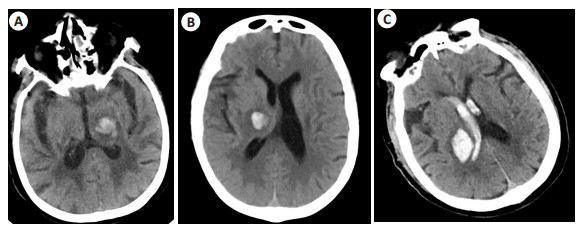
目的 分析多层螺旋CT(MSCT)肺动脉造影相关参数诊断急诊肺动脉栓塞的临床价值。 方法 选取2021年10月~2022年1月于本院接受CT平扫影像学检查的80例脑出血患者的临床资料进行回顾性研究,依据血肿是否扩大将其分为血肿扩大组(n=32)和非血肿扩大组(n=48)。记录所有患者的CT平扫影像学征象及血肿体积,比较两组比较CT平扫影像学征象及血肿体积的差异,采用Spearman相关性分析单一征象、血肿体积与脑出血患者血肿扩大的相关性,以ROC曲线分析CT平扫影像学征象、血肿体积评估血肿扩大的价值,采用Logistic多因素回归分析血肿扩大的相关影响因素。 结果 血肿扩大组的CT平扫出现“岛征”、“黑洞征”、“混杂征”占比均高于非血肿扩大组(P < 0.05);血肿扩大组的血肿体积大于非血肿扩大组(P < 0.05)。Spearman相关性分析显示,“岛征”、“黑洞征”、“混杂征”、血肿体积与脑出血患者血肿扩大均呈正相关关系(r=0.423、0.456、0.427、0.516,P < 0.05)。ROC曲线分析结果显示,“岛征”、“黑洞征”、“混杂征”联合血肿体积评估血肿扩大的曲线下面积为0.934,敏感度为87.5%,特异性为89.6%,均高于单一征象及血肿体积。Logistic多因素回归分析显示,岛征、黑洞征、混合征、初诊血肿体积增大是血肿扩大的独立危险因素(P < 0.05)。 结论 基于CT平扫影像学表现及血肿体积对脑出血患者血肿扩大具有一定的评估价值,且联合评估价值更高。 Abstract:Objective To analyze the clinical value of multi-slice spiral CT (MSCT) pulmonary angiography parameters in the diagnosis of emergency pulmonary embolism. Methods The clinical data of 80 patients with intracerebral hemorrhage who received plain CT imaging examination in our hospital from October 2021 to January 2022 were retrospectively studied. According to whether the hematoma was enlarged, they were divided into the enlarged hematoma group (n=32) and the enlarged non-hematoma group (n=48). The CT imaging signs and hematoma volume of all patients were recorded, and the differences of CT imaging signs and hematoma volume were compared between the two groups. Spearman correlation was used to analyze the correlation between single sign, hematoma volume and hematoma enlargement in patients with intracerebral hemorrhage. ROC curve was used to analyze CT imaging signs and hematoma volume to evaluate the value of hematoma enlargement, and Logistic multifactor regression was used to analyze the related influencing factors of hematoma enlargement. Results The proportion of "island sign", "black hole sign" and "mixed sign" in CT plain scan of hematoma expansion group was significantly higher than that of non-hematoma expansion group (P < 0.05). The volume of hematoma in the enlarged group were significantly higher than that in the non-enlarged group (P < 0.05). Spearman correlation analysis showed that "island sign", "black hole sign", "mixed sign" and hematoma volume were positively correlated with hematoma enlargement in patients with intracerebral hemorrhage (r=0.423, 0.456, 0.427, 0.516, P < 0.05). ROC curve analysis showed that the area under the curve of "island sign", "black hole sign" and "hybrid sign" combined with hematoma volume for evaluation of hematoma enlargement was 0.934, the sensitivity was 87.5%, and the specificity was 89.6%, which were all higher than single sign and hematoma volume. Logistic multifactor regression analysis showed that island sign, black hole sign, mixed sign and newly diagnosed hematoma volume increase were independent risk factors for hematoma enlargement (P < 0.05). Conclusion CT imaging findings and hematoma volume have certain evaluation value for hematoma enlargement in patients with cerebral hemorrhage, and the combined evaluation value is higher. -
表 1 血肿扩大组及非血肿扩大组的征象及血肿体积比较
Table 1. Comparison of signs and hematoma volume between hematoma enlargement group and non hematoma enlargement group
指标 血肿扩大组(n=32) 非血肿扩大组(n=48) χ2/t P 岛征[n(%)] 16(50.00) 7(14.58) 11.757 0.001 混杂征[n(%)] 20(62.50) 8(16.67) 17.729 < 0.001 黑洞征[n(%)] 13(40.62) 6(12.50) 8.387 0.004 血肿体积(mm3, Mean±SD) 30.89±6.84 24.56±5.74 4.473 < 0.001 表 2 CT平扫影像学征象、血肿体积对血肿扩大的评估价值分析
Table 2. Analysis of evaluation value of CT plain scan imaging signs and hematoma volume on hematoma enlargement
变量 面积 标准误 P 95% CI 下限 上限 岛征 0.677 0.052 0.001 0.563 0.777 混杂征 0.729 0.051 <0.001 0.618 0.823 黑洞征 0.641 0.050 0.005 0.526 0.745 血肿体积 0.773 0.058 <0.001 0.665 0.859 联合诊断 0.934 0.030 <0.001 0.855 0.977 表 3 影响血肿扩大的独立危险因素分析
Table 3. Analysis of independent risk factors affecting hematoma enlargement
相关因素 回归系数 标准误 P OR 95% CI 年龄 0.033 3.341 0.421 2.033 1.452~2.435 性别 0.013 3.152 1.301 2.574 1.785~3.198 破入脑室 0.101 4.452 0.068 3.737 1.657~4.463 岛征 0.328 4.654 0.031 4.665 1.208~8.985 混杂征 0.304 6.168 0.019 6.621 2.711~10.523 黑洞征 0.311 5.322 0.017 5.134 3.374~12.258 血肿体积 0.344 3.578 0.023 4.642 2.243~13.156 -
[1] Whiteley WN, Emberson J, Lees KR, et al. Risk of intracerebral haemorrhage with alteplase after acute ischaemic stroke: a secondary analysis of an individual patient data meta- analysis[J]. Lancet Neurol, 2016, 15(9): 925-33. doi: 10.1016/S1474-4422(16)30076-X [2] Anderson CS. Intracerebral hemorrhage[M]//Stroke. Amsterdam: Elsevier, 2022: 422-428.e2. [3] Tanaka K, Toyoda K. Clinical strategies against early hematoma expansion following intracerebral hemorrhage[J]. Front Neurosci, 2021, 15: 677744. doi: 10.3389/fnins.2021.677744 [4] Law ZK, Ali A, Krishnan K, et al. Noncontrast computed tomography signs as predictors of hematoma expansion, clinical outcome, and response to tranexamic acid in acute intracerebral hemorrhage[J]. Stroke, 2020, 51(1): 121-8. doi: 10.1161/STROKEAHA.119.026128 [5] 杨荣, 石力涛, 申亚凡. 早期颅内血肿微创清除术对轻中度基底核区高血压脑出血患者神经功能及认知功能的影响[J]. 临床误诊误治, 2019, 32(6): 92-5. doi: 10.3969/j.issn.1002-3429.2019.06.022 [6] Yu F, Yang YL, He YL, et al. Establishment and evaluation of a nomogram model for predicting hematoma expansion in hypertensive intracerebral hemorrhage based on clinical factors and plain CT scan signs[J]. Ann Palliat Med, 2021, 10(12): 12789-800. doi: 10.21037/apm-21-3569 [7] Liu R, Gong JP, Zhu JT, et al. Predictor measures on CT for hematoma expansion following acute intracerebral hemorrhage[J]. Zhonghua Yi Xue Za Zhi, 2016, 96(9): 720-3. [8] 李子聪, 孔祥宇, 黄鸿翔, 等. 基于平扫CT征象的脑出血早期血肿扩大预测量表[J]. 临床神经外科杂志, 2021, 18(4): 375-80. doi: 10.3969/j.issn.1672-7770.2021.04.004 [9] 王希, 仲艳, 颜伟, 等. CT平扫岛征和黑洞征对原发性脑出血早期血肿扩大的预测价值[J]. 中华神经外科杂志, 2021, 37(6): 557-61. doi: 10.3760/cma.j.cn112050-20201230-00652 [10] 彭佳华, 龙少好, 黄兰青, 等. 自发性脑出血患者血肿形态分析对早期血肿扩大的预测与诊断价值[J]. 中华急诊医学杂志, 2020, 29(4): 565-72. doi: 10.3760/cma.j.issn.1671-0282.2020022.012-1 [11] 王娟, 郭龙军, 李昌, 等. 基于CT评估脑出血征象和血肿体积、高低密度差预测血肿增大及软化灶的价值研究[J]. 影像科学与光化学, 2021, 39(2): 298-304. https://www.cnki.com.cn/Article/CJFDTOTAL-GKGH202102024.htm [12] 中华医学会神经病学分会, 中华医学会神经病学分会脑血管病学组. 中国脑出血诊治指南(2019) [J]. 中华神经科杂志, 2019, 52(12): 994-1005. doi: 10.3760/cma.j.issn.1006-7876.2019.12.003 [13] 曾春. 高血压脑出血早期血肿扩大的临床特点和CT表现[J]. 中国实用神经疾病杂志, 2016, 19(7): 19-20. doi: 10.3969/j.issn.1673-5110.2016.07.011 [14] Singh SD, Pasi M, Schreuder FHBM, et al. Computed tomography angiography spot sign, hematoma expansion, and functional outcome in spontaneous cerebellar intracerebral hemorrhage[J]. Stroke, 2021, 52(9): 2902-9. doi: 10.1161/STROKEAHA.120.033297 [15] Guo DC, Gu J, He J, et al. External validation study on the value of deep learning algorithm for the prediction of hematoma expansion from noncontrast CT scans[J]. BMC Med Imaging, 2022, 22(1): 45. doi: 10.1186/s12880-022-00772-y [16] Wang JL, Chen Y, Liang JJ, et al. Study of the pathology and the underlying molecular mechanism of tissue injury around hematoma following intracerebral hemorrhage[J]. Mol Med Rep, 2021, 24(4): 702. doi: 10.3892/mmr.2021.12341 [17] 余锦刚, 陈汉民, 廖圣芳, 等. 急性脑出血患者超急性期血肿扩大速率与预后不良相关性研究[J]. 河北医药, 2020, 42(12): 1780-3, 1788. doi: 10.3969/j.issn.1002-7386.2020.12.004 [18] 汤奉琼, 李强, 白崔魏, 等. 超急性期血肿扩大速度对脑出血血肿扩大的预测性研究[J]. 中国CT和MRI杂志, 2018, 16(1): 8-11. doi: 10.3969/j.issn.1672-5131.2018.01.003 [19] Ng D, Churilov L, Mitchell P, et al. The CT swirl sign is associated with hematoma expansion in intracerebral hemorrhage[J]. AJNR Am J Neuroradiol, 2018, 39(2): 232-7. doi: 10.3174/ajnr.A5465 [20] 贾维, 石长青, 刘亚龙, 等. CT平扫岛征和混合征对自发性脑出血患者早期血肿扩大的预测作用[J]. 中华神经外科杂志, 2019, 35(10): 1036-40. doi: 10.3760/cma.j.issn.1001-2346.2019.10.015 [21] Li Q, Liu QJ, Yang WS, et al. Island sign: an imaging predictor for early hematoma expansion and poor outcome in patients with intracerebral hemorrhage[J]. Stroke, 2017, 48(11): 3019-25. doi: 10.1161/STROKEAHA.117.017985 [22] 马凯伦, 汤若琪, 徐金娥, 等. 混杂征与卫星征预测成人自发性脑血肿扩大的比较[J]. 影像诊断与介入放射学, 2021, 30(2): 97-102. doi: 10.3969/j.issn.1005-8001.2021.02.003 [23] 宋杨君. CT平扫影像学征象预测自发性脑出血患者早期血肿扩张的临床价值分析[J]. 中国CT和MRI杂志, 2021, 19(11): 20-2. doi: 10.3969/j.issn.1672-5131.2021.11.007 -


 下载:
下载:


