


Evaluation of staging and thrombolytic efficacy of lower extremity deep vein thrombosis by shear wave elastography
-
摘要:
目的 探讨实时剪切波弹性成像技术在评价下肢深静脉血栓(DVT)分期及溶栓疗效中的应用价值。 方法 选取212例DVT患者为研究对象,结合发病时间长短与彩色多普勒超声检查结果将其分为急性期组(n=97)、亚急性期组(n=68)、慢性期组(n=47),全部患者均接受溶栓治疗,根据治疗结果将其分为治愈组(n=83)、有效组(n=97)、无效组(n=32),采用剪切波弹性成像技术测定不同分期、不同疗效患者杨氏模量值,通过绘制ROC曲线评估治疗后血栓杨氏模量对DVT患者溶栓疗效的诊断效能。 结果 治疗前,不同分期DVT患者血栓杨氏模量的差异有统计学意义(P<0.05),其中急性期组<亚急性期组<慢性期组(P<0.001);治疗后,各组血栓杨氏模量低于治疗前(P<0.05),且各组治疗后血栓杨氏模量差异有统计学意义(P<0.05),其中急性期<亚急性期<慢性期(P<0.001)。治疗后,不同预后DVT患者血栓杨氏模量差异有统计学意义(P<0.05),其中治愈组<有效组<无效组(P<0.001)。ROC分析结果显示,当Youden指数取最大值0.632时,血栓杨氏模量截断值为6.825 kPa,此时诊断DVT患者溶栓治疗无效的敏感度为93.8%,特异性为69.4%,曲线下面积为0.885。 结论 实时剪切波弹性成像技术测定杨氏模量,可作为评价DVT分期及溶栓疗效的辅助手段。 Abstract:Objective To investigate the application value of shear wave elastography in the staging and thrombolytic efficacy of deep venous thrombosis (DVT) of lower extremities. Methods A total of 212 patients with DVT were selected as the research objects.The patients were divided into acute phase group (n=97), subacute phase group (n=68), chronic phase group (n=47) according to the time of onset and the results of color Doppler ultrasonography. According to treatment outcome, all patients received thrombolytic therapy and were divided into cure group (n=83), effective group (n=97), ineffective group (n=32). The shear wave elastography technique was used to measure the Young's modulus of patients with different stages and different therapeutic effects. The ROC curve was drawn to evaluate the diagnostic efficacy of Young's modulus of thrombolysis in patients with DVT after treatment. Results Before treatment, there were statistically significant differences in young's modulus of thrombus in patients with different stages of DVT (P<0.05), including acute stage group<subacute stage group<chronic stage group (P<0.001). After treatment, the young's modulus of thrombus in all groups was lower than before (P<0.05), and the difference was statistically significant (P<0.05), among which the acute stage<subacute stage<chronic stage (P<0.001). After treatment, young's modulus of thrombus in DVT patients with different prognosis had statistical significance (P<0.05), among which cured group<effective group<invalid group (P<0.001). ROC analysis results showed that when Youden index was 0.632, the cut-off value of Young's modulus of thrombolysis was 6.825 kPa, and the sensitivity, specificity and AUC for diagnosing ineffective thrombolytic therapy in DVT patients were 93.8%, 69.4% and 0.885 respectively. Conclusion The measurement of Young's modulus by shear wave elastography technique can be used as an auxiliary method to evaluate stage of DVT and the efficacy of thrombolysis. -

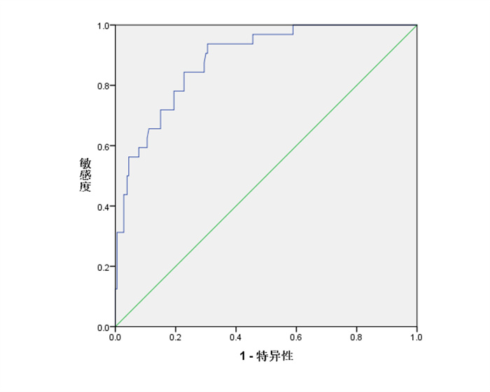
图 1 不同分期DVT患者治疗前后短轴切面弹性图
A~C: 依次为治疗前急性期(约4 d)腘静脉血栓短轴切面弹性图、亚急性期(约98 d)腘静脉血栓短轴切面弹性图、慢性期(约1年)胫后静脉血栓短轴切面弹性图; D~F: 依次为对应治疗后静脉血栓短轴切面弹性图.
Figure 1. Elastic diagram of short axis section of DVT patients in different stages before and after treatment.

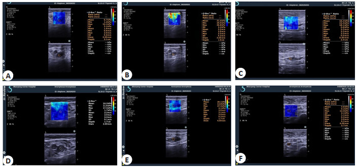
图 2 治疗后血栓杨氏模量值诊断DVT患者溶栓疗效的ROC曲线图
Figure 2. ROC curve of thrombolytic efficacy of DVT patients diagnosed by Young's modulus after treatment.
表 1 不同分期DVT患者基线资料比较
Table 1. Comparison of baseline data of DVT patients in different stages (n)
组别 性别 年龄(岁, Mean±SD) 患侧 男 女 单侧 双侧 急性期(n=97) 49 48 47.94±11.26 61 36 亚急性期(n=68) 32 36 46.49±10.58 46 22 慢性期(n=47) 22 25 48.21±10.60 31 16 χ2/F 0.267 0.472 0.419 P 1.210 0.624 1.035  下载: 导出CSV
表 2 不同分期DVT患者治疗前血栓杨氏模量值比较
Table 2. Comparison of Young's modulus of thrombosis in patients with DVT of different stages before treatment (kPa, Mean±SD)
组别 治疗前 治疗后 t P 急性期(n=97) 7.96±2.33 5.30±1.98 8.568 < 0.001 亚急性期(n=68) 14.58±2.61 6.92±2.43 17.713 < 0.001 慢性期(n=47) 26.02±6.71 8.86±3.86 18.280 < 0.001 F 354.897 30.180 P < 0.001 < 0.001
下载: 导出CSV
-
[1] Nicklas JM, Gordon AE, Henke PK. Resolution of deep venous thrombosis: proposed immune paradigms[J]. Int J Mol Sci, 2020, 21(6): 2080. doi: 10.3390/ijms21062080 [2] 黄金, 陈希玲, 黄智华. 血栓弹力图与常规凝血相关指标预测颅脑损伤术后产生深静脉血栓的价值[J]. 川北医学院学报, 2022, 37(6): 712-5. https://www.cnki.com.cn/Article/CJFDTOTAL-NOTH202206007.htm [3] 陈慧娇, 孙晓红, 张喆, 等. 出血性卒中患者下肢深静脉血栓形成风险预测模型的构建与验证[J]. 中华神经外科杂志, 2021, 37(3): 255-9. doi: 10.3760/cma.j.cn112050-20201014-00537 [4] Igneri LA, Hammer JM. Systemic thrombolytic therapy for massive and submassive pulmonary embolism[J]. J Pharm Pract, 2020, 33(1): 74-89. doi: 10.1177/0897190018767769 [5] 罗剑木, 曹奔. DWI-ASPECTS评分在静脉溶栓治疗急性脑梗死患者预后评估中的价值[J]. 中南医学科学杂志, 2022, 50(1): 113-6. https://www.cnki.com.cn/Article/CJFDTOTAL-HYYY202201027.htm [6] Lim WTH, Ooi EH, Foo JJ, et al. Shear wave elastography: a review on the confounding factors and their potential mitigation in detecting chronic kidney disease[J]. Ultrasound Med Biol, 2021, 47(8): 2033-47. doi: 10.1016/j.ultrasmedbio.2021.03.030 [7] 时瑞霞. 二维彩色多普勒超声联合实时剪切波弹性成像对乳腺癌的诊断效能研究[J]. 实用中西医结合临床, 2022, 22(4): 86-8. https://www.cnki.com.cn/Article/CJFDTOTAL-SZXL202204026.htm [8] 张惠敏, 叶新华, 刘辉, 等. 经阴道实时剪切波弹性成像技术联合阴道镜检查在宫颈癌中的诊断应用[J]. 河北医药, 2022, 44(2): 272-4. https://www.cnki.com.cn/Article/CJFDTOTAL-HBYZ202202029.htm [9] 慕鹏莺, 刘晓荷, 王梦碧, 等. 早期应用替罗非班对急性缺血性卒中尿激酶静脉溶栓后的疗效及对血小板指标的影响[J]. 血栓与止血学, 2022, 28(1): 54-5, 58. https://www.cnki.com.cn/Article/CJFDTOTAL-XSZX202201025.htm [10] Parker RI. Catheter- associated deep venous thrombosis prevention: which path to choose?[J]. Crit Care Med, 2021, 49(3): 537-40. [11] Piscaglia F, Salvatore V, Mulazzani L, et al. Ultrasound shear wave elastography for liver disease. A critical appraisal of the many actors on the stage[J]. Ultraschall Med, 2016, 37(1): 1-5. [12] Jonkman AH, de Korte CL. Shear wave elastography of the diaphragm: good vibrations?[J]. Am J Respir Crit Care Med, 2021, 204(7): 748-50. [13] Lehoux MC, Sobczak S, Cloutier F, et al. Shear wave elastography potential to characterize spastic muscles in stroke survivors: literature review[J]. Clin Biomech, 2020, 72: 84-93. [14] van der Bijl P, Delgado V, Bax JJ. Shear wave elastography to evaluate hepatic damage in heart failure[J]. ESC Heart Fail, 2021, 8(3): 1735-7. [15] 沈涛, 李娜, 刘晓娜, 等. 超声剪切波弹性成像定量指标对深静脉血栓分期价值的实验研究[J]. 医学影像学杂志, 2018, 28(4): 683-5. https://www.cnki.com.cn/Article/CJFDTOTAL-XYXZ201804049.htm [16] 夏晴, 孙顺吉, 马桂凤, 等. 探究实时剪切波弹性成像技术预测静脉血栓硬度与溶栓疗效的相关性[J]. 中国医疗设备, 2017, 32(11): 82-5. https://www.cnki.com.cn/Article/CJFDTOTAL-YLSX201711022.htm [17] Leong SS, Wong JHD, Md Shah MN, et al. Shear wave elastography accurately detects chronic changes in renal histopathology[J]. Nephrology (Carlton), 2021, 26(1): 38-45. [18] Bedewi MA, Elsifey AA, Alfaifi T, et al. Shear wave elastography of the tibial nerve in healthy subjects[J]. Medicine, 2021, 100(3): e23999. [19] 朱颖慧, 曾凡祎, 孙医学. 剪切波弹性成像在下肢深静脉血栓分期中的应用价值[J]. 包头医学院学报, 2022, 38(4): 13-7. https://www.cnki.com.cn/Article/CJFDTOTAL-BTYX202204004.htm [20] 洪登科, 杨嘉嘉, 薛恩生, 等. 实时剪切波弹性成像应用于股总静脉血栓临床分期[J]. 中国医学影像技术, 2019, 35(8): 1200-4. https://www.cnki.com.cn/Article/CJFDTOTAL-ZYXX201908032.htm [21] 王真. 不同时期下肢深静脉血栓超声弹性成像评价的初步临床研究[M]. 西安: 第四军医大学, 2011: 1-61. -

 点击查看大图
计量
- 文章访问数: 194
- HTML全文浏览量: 88
- PDF下载量: 14
- 被引次数: 0