


MRI features of pediatric diffuse midline glioma, H3K27-altered
-
摘要:
目的 探讨儿童弥漫性中线胶质瘤伴H3K27M改变的MRI特点。 方法 回顾性分析上海市儿童医院2017年7月~2021年11月经手术病理证实为儿童弥漫性中线胶质瘤伴H3K27M改变的7例患儿临床及影像资料,所有患儿术前均行MRI检查,对肿瘤MRI特征进行分析。 结果 7例均为单发病灶,其中5例位于脑干,1例位于丘脑,1例位于胸髓。4例肿瘤形态不规则,2例呈类圆形,边界均较清楚;1例胸髓病变呈长条形,边界不清。2例出现囊变、坏死,均未见出血。6例肿瘤实性成分T1WI呈低信号、T2WI呈稍高-高信号、T2-FLAIR呈高信号;1例胸髓内肿瘤实性成分T1WI呈稍高信号、T2WI呈高信号、T2-FLAIR呈高信号。6例肿瘤实性成分扩散加权成像扩散受限,1例未见明显扩散受限。增强扫描,6例呈明显不均匀强化,其中3例呈环形强化,2例呈结节状强化,1例呈斑片状强化;1例未见明显强化。7例未见瘤周水肿,均未发现远处转移及脑脊液播散。 结论 儿童弥漫性中线胶质瘤伴H3K27M改变的MRI表现具有一定的特征性,综合分析其发病部位、扩散有无受限和强化方式及范围等情况,有助于提高对该病影像学诊断与鉴别诊断水平。 -
关键词:
- 弥漫性中线胶质瘤伴H3K27M改变 /
- 磁共振成像 /
- 儿童
Abstract:Objective To investigate the MRI features of pediatric diffuse midline gliomas, H3K27-altered. Methods A retrospective study was carried out to analyze 7 pediatric patients of diffuse midline gliomas, H3K27-altered confirmed by surgery and pathology in Shanghai Children's Hospital from July 2017 to November 2021. The clinical and MRI data were abstracted from their electronic medical records. All the 7 patients underwent plain and enhanced MRI scan before operation, and the MRI features of the tumors were observed and analyzed. Results All the patients showed a unilateral mass. 5 cases were located in brainstem, 1 case was located in thalamus, and 1 case was located in thoracic spinal cord. 4 cases were irregular and 2 cases were round-like with clear boundaries, but the case in thoracic spinal cord was strip with unclear boundary. 2 cases had cystic or necrosis, and all cases had no hemorrhage. In 6 cases, the solid components of tumor showed low signal on T1WI, slightly-high signal on T2WI and high signal on T2- FLAIR; The solid components of thoracic spinal cord tumor showed slightly higher signal on T1WI, high signal on T2WI and high signal on T2-FLAIR. 6 cases of DWI had diffusion restriction and 1 case had no diffusion restriction.MRI enhanced scans showed 6 cases had obvious uneven enhancement and 1 case had no obvious enhancement, of which 3 cases showed circular enhancement, 2 cases showed nodular enhancement and 1 case showed patchy enhancement. There was no obvious edema around the tumors. Distant metastasis and cerebrospinal fluid dissemination were no found in all cases. Conclusion There are certain MRI features of pediatric diffuse midline gliomas, H3K27-altered. The location of the tumor, diffusion restriction, and the mode and scope of enhancement will help to improve the level of imaging diagnosis and differential diagnosis of the disease. -
Key words:
- diffuse midline glioma, H3K27-altered /
- magnetic resonance imaging /
- child
-

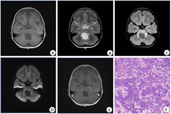
图 1 女,6岁,行走不稳10 d,肿瘤位于脑干
MRI示脑干可见一囊性异常信号影, T1WI(A)呈低信号, T2WI(B)呈高信号, T2-FLAIR(C)囊壁呈高信号、囊内成分呈低信号, 囊壁高b值DWI呈高信号(D), 提示肿瘤实性成分扩散受限,增强扫描(E)可见环形强化; F: 病理(HE染色, ×200), 肿瘤细胞部分区域高密度排列, 部分呈菊形团样结构, 灶性出血及坏死.
Figure 1. A 6-year-old female patient with unstable walking for 10 d, the tumor was located in the brain stem.

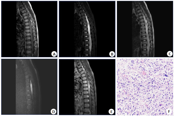
图 2 男,11岁4月,行走不稳1月伴肢体无力10 d,肿瘤位于胸髓
A~E: MRI平扫矢状位T1WI、T2WI、STIR、DWI及T1WI增强图像. 胸髓MRI示胸6-10段胸髓内可见条片状异常信号影, T1WI(A)呈稍高信号, T2WI(B)高信号, STIR(C)呈高信号, 肿瘤高b值DWI呈高信号(D), 提示肿瘤扩散受限, 增强扫描(E)可见明显不均匀强化; F: 病理(HE染色, ×200), 肿瘤细胞大小一致, 核深染, 呈弥漫性片状分布伴灶状性出血.
Figure 2. A male patient at the age of 11 years and 4 months, with unstable walking accompanied by limb weakness for 10 d, the tumor was located in the thoracic medulla.
-
[1] Louis DN, Perry A, Reifenberger G, et al. The 2016 World Health Organization classification of tumors of the central nervous system: a summary[J]. Acta Neuropathol, 2016, 131(6): 803-20. doi: 10.1007/s00401-016-1545-1 [2] Solomon DA, Wood MD, Tihan T, et al. Diffuse midline gliomas with histone H3-K27M mutation: a series of 47 cases assessing the spectrum of morphologic variation and associated genetic alterations [J]. Brain Pathol, 2016, 26(5): 569-80. doi: 10.1111/bpa.12336 [3] Mittal PK, AbdallaAS, ChatterjeeA, et al. Spectrum of extratesticular and testicular pathologic conditions at scrotal MR imaging[J]. Radiographics, 2018, 38(3): 806-30. doi: 10.1148/rg.2018170150 [4] 申楠茜, 张佳璇, 甘桐嘉, 等. 2021年WHO中枢神经系统肿瘤分类概述[J]. 放射学实践, 2021, 36(7): 818-31. doi: 10.13609/j.cnki.1000-0313.2021.07.001 [5] Schreck KC, Ranjan S, Skorupan N, et al. Incidence and clinicopathologic features of H3 K27M mutations in adults with radiographically-determined midline gliomas[J]. J Neurooncol, 2019, 143(1): 87-93. doi: 10.1007/s11060-019-03134-x [6] 杨豪, 邵剑波, 彭雪华, 等. 儿童弥漫性中线胶质瘤伴H3K27M突变型的影像学特征与分析[J]. 临床放射学杂志, 2020, 39(11): 2310-4. doi: 10.13437/j.cnki.jcr.2020.11.036 [7] 李海南, 山常国, 范冲竹, 等. H3K27M突变型弥漫性中线胶质瘤30例临床病理学特征和预后分析[J]. 中华病理学杂志, 2019, 48(3): 192-8. doi: 10.3760/cma.j.issn.0529-5807.2019.03.005 [8] 王佳, 许朗宁, 葛明, 等. 伴H3 K27M突变的儿童弥漫性中线胶质瘤的临床特点、治疗及预后[J]. 中华神经外科杂志, 2021, 37(2): 118-22. https://www.cnki.com.cn/Article/CJFDTOTAL-YXYQ202208015.htm [9] Roux A, Pallud J, Saffroy R, et al. High-grade gliomas in adolescents and young adults highlight histomolecular differences from their adult and pediatric counterparts[J]. Neuro Oncol, 2020, 22(8): 1190-202. doi: 10.1093/neuonc/noaa024 [10] Karremann M, Gielen GH, Hoffmann M, et al. Diffuse high- grade gliomas with H3 K27M mutations carry a dismal prognosis independent of tumor location[J]. Neuro Oncol, 2017, 20(1): 123-31. [11] 李娟, 马阳阳, 冯佳燕, 等. 儿童H3K27变异型弥漫性中线胶质瘤41例临床病理学分析[J]. 中华病理学杂志, 2022, 51(4): 319-25. [12] Funata N, Nobusawa S, Nakata S, et al. A case report of adult cerebellar high-grade glioma with H3.1 K27M mutation: a rare example of an H3 K27M mutant cerebellar tumor[J]. Brain Tumor Pathol, 2018, 35(1): 29-35. [13] Qiu T, Chanchotisatien A, Qin Z, et al. Imaging characteristics of adult H3 K27M-mutant gliomas[J]. J Neurosurg, 2019: 1-9. doi: 10.3171/2019.9.JNS191920. [14] 丁茗, 郑慧, 冯赟, 等. 儿童弥漫性中线胶质瘤伴H3K27M突变MRI表现[J]. 实用放射学杂志, 2020, 36(3): 444-7. doi: 10.3969/j.issn.1002-1671.2020.03.026 [15] Jung JS, Choi YS, Ahn SS, et al. Differentiation between spinal cord diffuse midline glioma with histone H3 K27M mutation and wild type: comparative magnetic resonance imaging[J]. Neuroradiology, 2019, 61(3): 313-22. [16] Aboian MS, Solomon DA, Felton E, et al. Imaging characteristics of pediatric diffuse midline gliomas with histone H3 K27M mutation[J]. AJNRAm J Neuroradiol, 2017, 38(4): 795-800. [17] 曹亚先, 王芮, 陈臻, 等. 儿童弥漫性中线胶质瘤, H3K27M突变型的MRI表现[J]. 影像诊断与介入放射学, 2020, 29(5): 343-8. https://www.cnki.com.cn/Article/CJFDTOTAL-YXZD202005008.htm [18] 邓达标, 郭珺, 梁倩雯, 等. 弥漫性中线胶质瘤伴H3K27M突变的MRI表现[J]. 中华放射学杂志, 2019, 53(7): 545-8. https://www.cnki.com.cn/Article/CJFDTOTAL-FSXS202111008.htm [19] 陆苑婷, 艾斌, 刘鸿圣. 儿童H3K27M突变型弥漫性中线胶质瘤的MRI表现[J]. 中国中西医结合影像学杂志, 2021, 19(1): 75-7. https://www.cnki.com.cn/Article/CJFDTOTAL-JHYX202101021.htm [20] Su XR, Liu YH, Wang HY, et al. Multimodal MR imaging signatures to identify brain diffuse midline gliomas with H3 K27M mutation[J]. Cancer Med, 2022, 11(4): 1048-58. [21] 李楠, 葛明, 张建. 儿童弥漫性中线胶质瘤的研究进展[J]. 中华神经外科杂志, 2021, 37(7): 737-9. https://www.cnki.com.cn/Article/CJFDTOTAL-XDJB202004010.htm -

 下载:
下载:
 点击查看大图
计量
- 文章访问数: 310
- HTML全文浏览量: 119
- PDF下载量: 16
- 被引次数: 0