


Imaging features of magnetic resonance enterography in the diagnosis of radiation enteriti
-
摘要:
目的 分析磁共振小肠造影诊断放射性肠炎(RE)的影像学特征。 方法 回顾性分析2016年1月~2019年6月本院行妇科盆腔肿瘤放疗后疑似的RE患者108例磁共振小肠造影检查资料,以患者肠镜病理检查结果以及随访资料作为RE的诊断参考,总结RE在磁共振小肠造影中的影像学特点,分析磁共振小肠造影在诊断RE中的价值。 结果 所有患者后期均行肠镜检查,且接受随访复诊,根据检查结果及随访情况,确诊108例患者中共81例为RE,其27例排除RE。以最终诊断作为“金标准”,磁共振小肠造影在诊断RE中的敏感度及特异性分别为87.65%与74.07%。RE在磁共振小肠造影增强扫描及DWI扫描图像中有“同心圆”分层状高信号表现,肠壁呈强化信号,同时存在肠管周围改变以及肠壁增厚表现。 结论 RE在磁共振小肠造影中具有特殊的影像学表现,磁共振小肠造影可用于临床RE的诊断。 Abstract:Objective To investigate the imaging features of magnetic resonance enterography in the diagnosis of radiation enteritis (RE). Methods The imaging data of magnetic resonance enterography in 108 patients with suspected RE after gynecological pelvic tumor radiotherapy in our hospital from January 2016 to June 2019 were analyzed. We put the pathological examination results of patients' enteroscopy and follow- up data as the diagnosis reference of RE. The imaging characteristics of RE in magnetic resonance enterography was summarized. We analyzed the value of magnetic resonance enterography in diagnosing RE. Results All patients underwent colonoscopy in the later stage and were followed up. According to the examination results and follow-up, a total of 81 of 108 patients were diagnosed as RE, and 27 of them were excluded from RE. Taking the final diagnosis as the "gold standard", statistics showed that the sensitivity and specificity of magnetic resonance enterography in diagnosing RE were 87.65% and 74.07%. RE had "concentric circle" layered high signal performance in enhanced MR enterography and DWI scanning images. The intestinal wall showed enhanced signal, and there were changes around the intestinal tube and thickening of the intestinal wall. Conclusion RE has special imaging features in MR enterography, which can be used for clinical diagnosis of RE. -
Key words:
- magnetic resonance enterography /
- radiation enteritis /
- imaging features /
- T2WI /
- DWI sequence
-

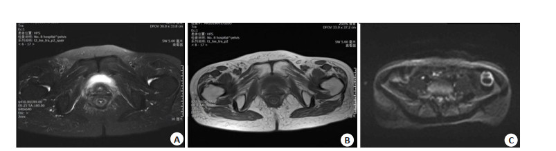
图 1 某宫颈癌放疗后RE患者磁共振小肠造影检查图
磁共振小肠造影提示患者肌层薄弱部分缺损紧贴阴道后壁, 肠壁均匀增厚, 可见直肠壁水肿. A: T1WI呈稍高信号; B: T2WI呈高信号; C: DWI上病变肠管和增大淋巴结呈高信号, 后经肠镜及随访确诊为RE.
Figure 1. Magnetic resonance enterography of RE patient with cervical cancer after radiotherapy

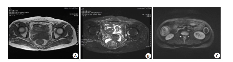
图 2 某子宫内膜癌患者放疗后RE患者磁共振小肠造影检查图
常规MRI提示: A: T1WI呈稍高信号; B: T2WI呈高信号,增强后呈分层样强化, 提示直肠及乙状结肠远端肠壁肿胀且水肿; C: DWI上病变肠管和增大淋巴结呈高信号,后经肠镜及随访确诊为RE.
Figure 2. Magnetic resonance enterography of RE patient after radiotherapy in a endometrial cancer patient
表 1 磁共振小肠造影与临床诊断结果分析
Table 1. Analysis of magnetic resonance enterography and clinical diagnosis results
诊断方法 临床诊断(n) 总计(n) 敏感度(%) 特异性(%) 阳性 阴性 磁共振小肠造影 87.65 74.07 阳性 71 2 96 阴性 10 25 12 T1WI 79.01 81.48 阳性 64 5 86 阴性 17 22 22 T2WI 76.25 74.07 阳性 62 7 69 阴性 19 20 39 DWI 76.54 85.19 阳性 62 4 66 阴性 19 23 42 总计 81 27 108  下载: 导出CSV
-
[1] Zhang XM, Hu X, Ou JY, et al. Glycyrrhizin ameliorates radiation enteritis in mice accompanied by the regulation of the HMGB1/TLR4 pathway[J]. Evid Based Complementary Altern Med, 2020, 2020: 8653783. [2] Gong XF, Yu GH, Song ZC, et al. Patients with radiation enteritis present regulatory T cell impairment associated with CTLA-4[J]. Immunol Res, 2020, 68(4): 179-88. doi: 10.1007/s12026-020-09142-8 [3] 彭明洋, 智婷婷, 张卫东, 等. 磁共振小肠造影弥散加权成像对炎症性肠病的诊断价值[J]. 中国医疗设备, 2018, 33(11): 70-2, 82. doi: 10.3969/j.issn.1674-1633.2018.11.019 [4] 王英杰, 王顺金, 龚良庚, 等. 急性放射性肠炎模型研究: 两种造模方法的对比与评价[J]. 中华放射肿瘤学杂志, 2016, 25(6): 628-33. doi: 10.3760/cma.j.issn.1004-4221.2016.06.019 [5] Greer MLC. Paediatric magnetic resonance enterography in inflammatory bowel disease[J]. Eur J Radiol, 2018, 102: 129-37. doi: 10.1016/j.ejrad.2018.02.029 [6] 张明明, 方文佳, 沈萍萍, 等. 肠镜与多层螺旋CT小肠造影对炎症性肠病的诊断价值评估[J]. 中国内镜杂志, 2020, 26(8): 31-6. doi: 10.3969/j.issn.1007-1989.2020.08.006 [7] Yu H, Shen YQ, Tan FQ, et al. Quantitative diffusion-weighted magnetic resonance enterography in ileal Crohn's disease: a systematic analysis of intra and interobserver reproducibility[J]. World J Gastroenterol, 2019, 25(27): 3619-33. doi: 10.3748/wjg.v25.i27.3619 [8] Wang HS, Du K, Qu J, et al. Dosimetric evaluation of magnetic resonance-generated synthetic CT for radiation treatment of rectal cancer[J]. PLoS One, 2018, 13(1): e0190883. doi: 10.1371/journal.pone.0190883 [9] 赵本琦, 乔建, 张晨, 等. CT增强成像评估慢性放射性肠炎严重程度的价值[J]. 中国医学影像学杂志, 2019, 27(9): 687-90. doi: 10.3969/j.issn.1005-5185.2019.09.013 [10] Hall WA, Paulson ES, van der Heide UA, et al. The transformation of radiation oncology using real-time magnetic resonance guidance: a review[J]. Eur J Cancer, 2019, 122: 42-52. doi: 10.1016/j.ejca.2019.07.021 [11] Odéen H, de Bever J, Hofstetter LW, et al. Multiple-point magnetic resonance acoustic radiation force imaging[J]. Magn Reson Med, 2019, 81(2): 1104-17. doi: 10.1002/mrm.27477 [12] 华龙, 王炳平, 张凡, 等. 分析MRI在宫颈癌放疗后放射性直肠损伤中的诊断价值[J]. 影像研究与医学应用, 2021, 5(11): 72-3. https://www.cnki.com.cn/Article/CJFDTOTAL-YXYY202111035.htm [13] 马志鸿, 王翠祥, 刘铜锁, 等. 宫颈癌放疗后的放射性直肠损伤患者MRI影像学特征及诊断价值[J]. 医药论坛杂志, 2021, 42(17): 143-5. https://www.cnki.com.cn/Article/CJFDTOTAL-HYYX202117039.htm [14] 王吕斌, 钱军, 陈杰, 等. 放射性肠炎内镜与影像诊断进展[J]. 肿瘤学杂志, 2018, 24(10): 1008-13. doi: 10.11735/j.issn.1671-170X.2018.10.B014 [15] Gach HM, Curcuru AN, Mutic S, et al. B0 field homogeneity recommendations, specifications, and measurement units for MRI in radiation therapy[J]. Med Phys, 2020, 47(9): 4101-14. doi: 10.1002/mp.14306 [16] Lukovic J, Henke L, Gani CH, et al. MRI-based upper abdominal organs-at-risk atlas for radiation oncology[J]. Int J Radiat Oncol, 2020, 106(4): 743-53. doi: 10.1016/j.ijrobp.2019.12.003 [17] Hirsch FW, Sorge I, Vogel-Claussen J, et al. The Current status and further prospects for lung magnetic resonance imaging in pediatric radiology[J]. Pediatr Radiol, 2020, 50(5): 734-49. doi: 10.1007/s00247-019-04594-z [18] Schernberg A, Kumar T, Achkar S, et al. Incorporating magnetic resonance imaging (MRI) based radiation therapy response prediction into clinical practice for locally advanced cervical cancer patients[J]. Semin Radiat Oncol, 2020, 30(4): 291-9. [19] Arai TJ, Yang DM, Campbell JW Ⅲ, et al. Oxygen-sensitive MRI: a predictive imaging biomarker for tumor radiation response?[J]. Int J Radiat Oncol, 2021, 110(5): 1519-29. -

 点击查看大图
计量
- 文章访问数: 143
- HTML全文浏览量: 89
- PDF下载量: 5
- 被引次数: 0