


Value of localization and qualitative diagnosis of malignant biliary obstruction by MRCP+DWI combined with enhanced CT scan
-
摘要:
目的 分析探讨磁共振胰胆管成像(MRCP)与弥散加权成像(DWI)联合CT增强扫描对恶性胆道梗阻的定位定性诊断价值。 方法 选择2015年6月~2020年12月在我院收治的疑似恶性胆道梗阻患者80例,所有患者均接受MRCP、DWI、CT增强扫描检查与病理学诊断; 以病理学诊断为“金标准”,对比所有患者MRCP形态,比较MRCP+DWI单独检查与CT增强扫描检查对胆道梗阻定位、定性诊断价值。 结果 病理组织学结果显示,80例患者中良性肝外胆道梗阻患者33例(41.25%),其中胆管结石27例,胆管炎性狭窄6例; 恶性肝外胆道梗阻患者47例,占比58.75%,其中胆总管癌34例、壶腹癌4例、胰头癌9例; MRCP影像学特征显示,良性梗阻患者主要表现为“枯枝状”,恶性梗阻患者主要表现为“软藤状”; 两种检测方法中,MRCP+DWI联合CT增强扫描对胆道梗阻的定位诊断准确率高于MRCP+DWI诊断(P < 0.05);MRCP+DWI检查胆道梗阻对胆管结石、胆管炎性狭窄、胆管癌、胰头癌的诊断符合率均低于MRCP+DWI联合CT增强扫描,两种检查方法对壶腹癌的诊断符合率均为100%;MRCP+ DWI联合CT增强扫描定性诊断总符合率高于MRCP+DWI检查(P < 0.05)。 结论 MRCP+DWI联合CT增强扫描可对恶性胆道梗阻进行准确定位,还可提高定性诊断恶性胆道梗阻的准确率,值得临床推广使用。 Abstract:Objective To investigate the value of localization and qualitative diagnosis of malignant biliary obstruction by magnetic resonance cholangiopancreatography (MRCP) and diffusion-weighted imaging (DWI) combined with enhanced CT. Methods Patients with suspected malignant biliary obstruction admitted to our hospital from June 2015 to December 2020 were selected. All patients received MRCP, DWI, CT enhanced scanning examination and pathological diagnosis. We took pathological diagnosis as the "gold standard", and the morphology of MRCP in all patients was compared. The value of MRCP+ DWI alone and enhanced CT scan in the localization and qualitative diagnosis of biliary obstruction were compared. Results Histopathological results showed that among the 80 patients, 33 (41.25%) had benign extrahepatic biliary obstruction, including 27 bile duct stones, 6 cholangitis strictures. There had 47 malignant extrahepatic biliary obstruction, accounting for 58.75%, including 34 cases of bile total duct cancer, 4 cases of ampullary carcinoma, and 9 cases of pancreatic head carcinoma. MRCP imaging features showed that patients with benign obstruction were mainly "withered branches shape", and patients with malignant obstruction were mainly "soft vine shape". Among the two detection methods, MRCP + DWI combined with enhanced CT scanning had higher accuracy in diagnosing biliary obstruction than MRCP + DWI (P < 0.05). The diagnostic coincidence rates of MRCP + DWI for biliary obstruction in bile duct stones, cholangitic stenosis, cholangiocarcinoma, and pancreatic head cancer were lower than those for MRCP + DWI combined with enhanced CT scanning. The diagnostic coincidence rate of the two methods for ampullary carcinoma was 100%. The overall coincidence rate of qualitative diagnosis of MRCP + DWI combined with enhanced CT scan was higher than that of MRCP + DWI (P < 0.05). Conclusion MRCP + DWI combined with enhanced CT scanning can accurately locate malignant biliary obstruction and improve the accuracy of qualitative diagnosis of malignant biliary obstruction. -

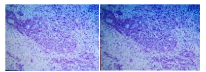
图 1 恶性胆道梗阻病理图片(HE染色, ×100)
病理: (胆总管)中分化, 腺癌.
Figure 1. Pathological picture of malignant biliary obstruction (HE staining, ×100).

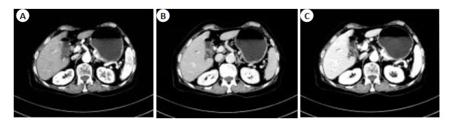
图 2 恶性胆道梗阻患者CT增强图
A: 动脉期; B: 门脉期; C: 平衡期.
Figure 2. Enhanced CT imagings of the patient with malignant biliary obstruction.

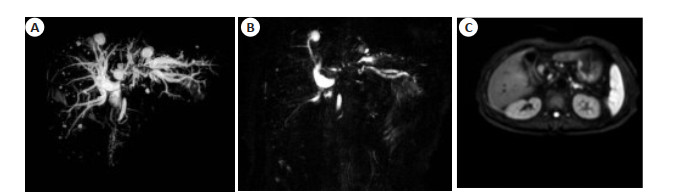
图 3 恶性胆道梗阻患者ERCP及DWI影像学表现
A~B: ERCP图显示肝内胆管呈软藤状扩张,胆总管近肝门处截断; DWI显示胆总管近肝门管腔内结节呈高信号.
Figure 3. ERCP and DWI imagings of the patient with malignant biliary obstruction.
表 1 胆管在MRCP上的形态
Table 1. Morphology of bile duct on MRCP [n(%)]
组别 例数 胆道扩张的形态 软藤状 枯枝状 双管征 四管征 良性梗阻 33 胆管结石 27 7(25.93) 19(70.37) 1(3.70) 0(0.00) 胆管炎性狭窄 6 1(16.67) 4(66.67) 1(16.67) 0(0.00) 恶性梗阻 47 胆管癌 34 14(41.48) 11(32.35) 5(14.71) 4(11.76) 壶腹癌 4 2(50.00) 0(0.00) 1(25.00) 1(25.00) 胰头癌 9 0(0.00) 0(0.00) 0(0.00) 9(100.00)  下载: 导出CSV
表 2 定位诊断结果与病理学检查结果对比
Table 2. Comparison of localization diagnosis results with pathological examination results [n(%)]
病灶部位 例数 MRCP+DWI MRCP+DWI联合CT增强扫描 P 胆总管 33 32(40.00)* 33(41.25)* 1.000 肝门区 18 16(20.00)* 18(22.50)* 0.486 胰上区 14 12(15.00)* 14(17.50)* 0.481 胰腺区 8 6(7.50)* 7(8.75)* 1.000 壶腹区 7 7(8.75)* 7(8.75)* 1.000 合计 80 73(91.25)* 79(98.75)* 0.030 *均采用Fisher精确检验, 以总例数为基础计算占比.
下载: 导出CSV
表 3 定性诊断与病理学检查结果对比
Table 3. Comparison of qualitative diagnosis and pathological examination results [n(%)]
病灶部位 MRCP+DWI MRCP+DWI联合CT增强扫描 P 胆管结石(n=27) 19(70.37) 26(96.30) 0.024 胆管炎性狭窄(n=6) 3(50.00) 6(100.00) 0.182 胆管癌(n=34) 33(97.06) 34(100.00) 1.000 壶腹(n=4) 4(100.00) 4(100.00) 1.000 胰头癌(n=9) 4(44.44) 6(66.67) 0.637 合计(n=80) 63(78.75) 76(95.00) 0.004 MRCP: 磁共振胰胆管成像; DWI: 弥散加权成像.
下载: 导出CSV
-
[1] 彭祖祥, 唐永梁, 梁先春, 等. 超声内镜引导下胆道引流术治疗恶性胆道梗阻临床研究进展[J]. 中国实用外科杂志, 2021, 41(4): 455-8. https://www.cnki.com.cn/Article/CJFDTOTAL-ZGWK202104032.htm [2] Aziz H, Segalini N, Ahmed Z, et al. National trends in cholecystectomy and endoscopic retrograde cholangiopancreatography during index hospitalization for mild gallstone pancreatitis[J]. World J Surg, 2022, 46(3): 524-30. doi: 10.1007/s00268-021-06389-6 [3] Zheng HL, Zheng YF, Zhang GH, et al. Diagnosis of the gallbladder duplication coinciding with a gallbladder carcinoma by magnetic resonance cholangio-pancreatography: a case report[J]. Asian J Surg, 2021, 44(7): 1012-3. doi: 10.1016/j.asjsur.2021.04.028 [4] 高磊. 磁共振胰胆管成像联合MR增强扫描对肝外胆管梗阻定位和定性诊断的研究[J]. 肝脏, 2019, 24(8): 966-7, 974. doi: 10.3969/j.issn.1008-1704.2019.08.045 [5] Xu X, Zhang XN. The application of intravoxel incoherent motion diffusion-weighted imaging in the diagnosis of hilar obstructive jaundice[J]. J ComputAssist Tomogr, 2019, 43(2): 228-34. doi: 10.1097/RCT.0000000000000837 [6] 朱望舒, 石思雅, 王东烨, 等. MRI检查弥散加权成像对肝门部胆管癌侵袭性的预测价值[J]. 中华消化外科杂志, 2018, 17(3): 310-7. doi: 10.3760/cma.j.issn.1673-9752.2018.03.017 [7] 滕才钧, 邓燕云, 韦建林. 18氟-氟代脱氧葡萄糖正电子发射断层显像/计算机断层成像联合磁共振胰胆管造影异机融合显像在梗阻性黄疸诊断中的应用[J]. 广西医学, 2019, 41(11): 1356-9. https://www.cnki.com.cn/Article/CJFDTOTAL-GYYX201911005.htm [8] Liang XH, Xue CQ, Huang XY, et al. Value of energy spectrum CT parameters in the differential diagnosis of high-grade clear cell renal cell carcinoma and type Ⅱ papillary renal cell carcinoma[J]. Acta Radiol, 2022, 63(4): 545-52. doi: 10.1177/02841851211002830 [9] 陶洁, 黄波, 邓勋伟, 等. MRCP技术联合血清CEA、CA19-9及CA242检测在胆道恶性梗阻中的诊断价值[J]. 解放军医药杂志, 2021, 33(12): 79-82, 87. doi: 10.3969/j.issn.2095-140X.2021.12.019 [10] 徐小满, 赵悦竹, 宋吉涛, 等. ERCP胆道金属支架置入在恶性胆道梗阻中的研究进展[J]. 现代消化及介入诊疗, 2021, 26(9): 1193-7. doi: 10.3969/j.issn.1672-2159.2021.09.028 [11] Berrad S, Oualla K, Nouikh L, et al. Palliative management of malignant biliary obstruction in patients with advanced biliary tract cancers[J]. Ann Oncol, 2019, 30: iv103. [12] 王怡鑫, 石喻, 郭启勇. 胆道梗阻患者基于磁共振弹性成像上的肝脏硬度值分析[J]. 中国临床医学影像杂志, 2021, 32(4): 267-71. https://www.cnki.com.cn/Article/CJFDTOTAL-LYYX202104013.htm [13] Danila M, Sirli R, Popescu A, et al. A rare cause of biliary obstruction-intraductal neuroendocrine tumor of the right hepatic biliary duct: a case report[J]. Med Ultrason, 2021, 23(2): 235-7. [14] 张丰灵, 李慧敏, 任亮. 彩超联合MRI对胆道梗阻定位和定性诊断的临床应用[J]. 中国CT和MRI杂志, 2021, 19(10): 167-70. https://www.cnki.com.cn/Article/CJFDTOTAL-CTMR202110053.htm [15] Hacım NA, Akbas A, Meric S, et al. Predictive value of ultrasonography and magnetic resonance cholangiopancreatography in the diagnosis of biliary obstruction[J]. Ann Ital Chir, 2020, 91: 277-82. [16] Jin Z, Zhang XF. Malignant biliary obstruction: endoscopic ultrasound-guided versus endoscopic retrograde cholangiopancreatography-guided biliary drainage[J]. Endoscopy, 2019, 51(12): 1185. [17] 蔡悦, 宋彬. 胆道梗阻病变定位和定性诊断的多模态影像学研究[J]. 中国普外基础与临床杂志, 2021, 28(5): 669-72. https://www.cnki.com.cn/Article/CJFDTOTAL-ZPWL202105020.htm [18] 张厚强, 张伟, 樊英. 磁共振胰胆管造影联合不同序列成像诊断恶性胆道梗阻性疾病的效果研究[J]. 中国医学装备, 2019, 16(6): 46-9. https://www.cnki.com.cn/Article/CJFDTOTAL-YXZB201906014.htm [19] Saito H, Iwagoi Y, Noda K, et al. Dual-layer spectral detector computed tomography versus magnetic resonance cholangiopancrea tography for biliary stones[J]. Eur J Gastroenterol Hepatol, 2021, 33 (1): 32-9. [20] Zhu JG, Su W, Guo W, et al. Accuracy of magnetic resonance cholangiopancreatography for the diagnosis of choledocholithiasis in clinical practice[J]. HPB, 2019, 21: S268. [21] Hu GW, Liang W, Wu MX, et al. Comparison of T1 mapping and T1rho values with conventional diffusion-weighted imaging to assess fibrosis in a rat model of unilateral ureteral obstruction[J]. Acad Radiol, 2019, 26(1): 22-9. [22] Togao O, DoiS, Kuro-o M, et al. Assessment of renal fibrosis with diffusion-weighted MR imaging: study with murine model of unilateral ureteral obstruction[J]. Radiology, 2010, 255(3): 772-80. [23] Cayet S, Pasco J, Dujardin F, et al. Diagnostic performance of contrast-enhanced CT-scan in sinusoidal obstruction syndrome induced by chemotherapy of colorectal liver metastases: radiopathological correlation[J]. Eur J Radiol, 2017, 94: 180-90. [24] Dai LJ, Shi GF, Li Y, et al. Values of thoracic contrast-enhanced computed tomography in detecting incidental pulmonary thromboembolism in patients with malignant tumors[J]. Oncol Lett, 2019, 17(1): 355-9. -

 点击查看大图
计量
- 文章访问数: 151
- HTML全文浏览量: 66
- PDF下载量: 6
- 被引次数: 0