


Musculoskeletal ultrasonographic features of noninsertional Achilles tendinopathy
-
摘要:
目的 分析非止点性跟腱病患者肌骨超声显像特征。 方法 收集2020年11月~2022年4月共60例患者82足跟腱超声结果,将其分为健康对照组(A组,15例30足)和非止点性跟腱病组(B组,45例52足),应用肌骨超声和彩色多普勒血流显像观察并分析比较各组:(1)跟骨后结节上2 cm处跟腱厚度; (2)跟腱止点上2 cm处跟腱横截面积; (3)跟骨上缘上1 cm处Kager脂肪垫前后径; (4)跟骨后滑囊积液检出率; (5)跟腱内血流信号检出率。总结归纳非止点性跟腱病的超声显像特征。 结果 (1) 跟骨后结节上2 cm处跟腱厚度:A组小于B组(0.43±0.06 cm vs 0.55±0.17 cm,P < 0.05);(2)跟腱止点上2 cm处跟腱横截面积:A组小于B组(0.52±0.11 cm2 vs 0.74±0.23 cm2,P < 0.05);(3)跟骨上缘上1 cm处Kager脂肪垫前后径:A组小于B组(1.01±0.21 cm vs1.49±0.26 cm,P < 0.05);(4)跟骨后滑囊积液检出率:A组未检出,B组38.46%,差异有统计学意义(P < 0.05);(5)跟腱内血流信号检出率:A组未检出,B组51.92%,差异有统计学意义(P < 0.05)。 结论 非止点性跟腱病超声显像特征为跟腱厚度增厚,跟腱横截面积增大,跟腱前Kager脂肪垫前后径增宽,跟骨后滑囊积液产生或增加,以及跟腱内部血管增生。 Abstract:Objective To summarize the musculoskeletal ultrasonographic features of noninsertional Achilles tendinopathy. Methods Ultrasound results of 82 Achilles tendons in 60 cases were collected from November 2020 to April 2022, and 15 cases including 30 feet were set as healthy control group (group A), 45 cases including 52 feet as noninsertional Achilles tendon group (group B). Musculoskeletal ultrasound and color Doppler flow imaging were used to observe and compare the features of the noninsertional Achilles tendon: (1) The thickness of Achilles tendon at 2 cm above the posterior calcaneal tubercle; (2) The cross-sectional area of Achilles tendon at 2cm above the Achilles tendon insertion point; (3)The anterior-posterior diameter of Kager fat pad at 1cm above the upper edge of calcaneus; (4)The detection rate of retrocalcaneal bursal effusion; (5)The detection rate of blood flow signal in Achilles tendon. The features of noninsertional Achilles tendinopathy were summarized. Results (1) The thickness of Achilles tendon at 2 cm above the posterior calcaneal tubercle: group A was less than group B (0.43± 0.06 cm vs 0.55±0.17 cm, P < 0.05). (2)The cross-sectional area of Achilles tendon at 2 cm above the Achilles tendon insertion point: group A was less than group B (0.52±0.11 cm2 vs 0.74±0.23 cm2, P < 0.05). (3)The anterior-posterior diameter of Kager fat pad at 1 cm above the upper edge of calcaneus: group A was smaller than group B (1.01±0.21 cm vs 1.49±0.26 cm, P < 0.05). (4) The detection rate of retrocalcaneal bursal effusion: negative in group A and 38.46% in group B, the difference was statistically significant (P < 0.05). (5)The detection rate of blood flow signal in Achilles tendon: group A was not detected and 51.92% in group B, the difference was statistically significant (P < 0.05). Conclusion Noninsertional Achilles tendinopathy was characterized by thickening of Achilles tendon thickness, increasing of cross- sectional area of Achilles tendon, widening of anterior-posterior diameter of Kager fat pad, increasing of retrocalcaneal bursal effusion and the blood flow signal in Achilles tendon. -

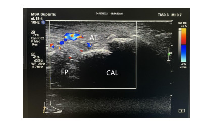
图 1 超声设备及跟腱测量方法
A: 彩色超声诊断仪; B: 跟横纵轴扫描; C: 跟腱横轴扫描.
Figure 1. Ultrasonic equipment and methods of Achilles tendon measurement.

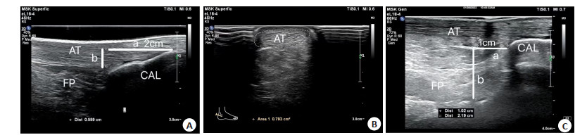
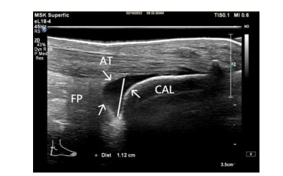
图 2 跟腱测量数据
A: 纵轴扫描测量跟腱厚度, a线为跟骨后结节上2 cm, b为所测量的跟腱厚度; B: 横轴测量跟腱CAS; C: 纵轴测量Kager脂肪垫前后径, a线为跟骨上缘前方1cm, b为所测量的Kager脂肪垫前后径. AT: 跟腱; CAL: 跟骨; FP: Kager脂肪垫.
Figure 2. Achilles tendon measurements.

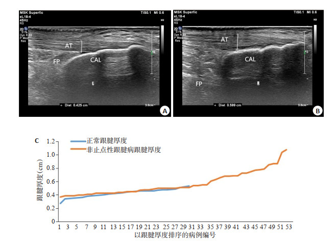
图 3 跟骨后结节上2 cm跟腱厚度比较
A: 正常跟腱; B: 非止点性跟腱病跟腱; C: 两组病例跟腱厚度对比.
Figure 3. Comparison of Achilles tendon thickness at 2 cm above the posterior calcaneal tubercle.

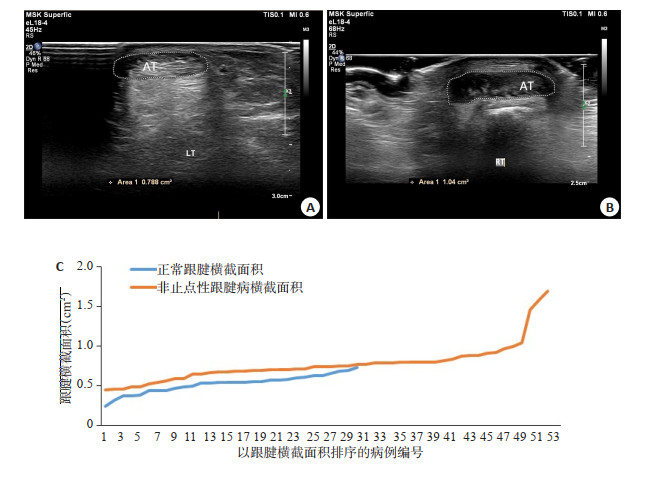
图 4 跟腱止点上2 cm处跟腱CAS比较
A: 正常跟腱; B: 非止点性跟腱病跟腱; C: 两组病例跟腱横截面积对比.
Figure 4. Comparison of Achilles tendon CAS at 2 cm above Achilles tendon insertion point.

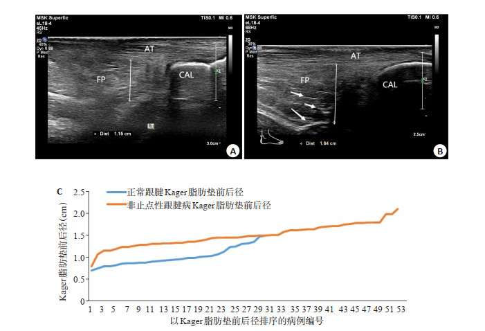
图 5 跟骨上缘上1 cm处Kager脂肪垫前后径比较
A: 正常跟腱; B: 非止点性跟腱病跟腱; C: 两组病例Kager脂肪垫前后径对比.
Figure 5. Comparison of anterior-posterior diameters of Kager's fat pad 1 cm above the upper edge of calcaneus.
表 1 两组一般资料比较
Table 1. Comparison of general data between the two groups
分组 足数 性别(男/女,n) 性别(男/女,足数) 分布(左/右,足数) 年龄(岁,Mean±SD) A组(n=15) 30 8/7 16/14 15/15 35.33±14.90 B组(n=45) 52 29/16 33/19 27/25 39.51±11.76 χ2/Z 0.818 1.137 0.038 -1.778 P 0.366 0.286 0.846 0.075 A组: 健康对照组; B组: 非止点性跟腱病组.  下载: 导出CSV
表 2 两组跟腱超声表现
Table 2. Ultrasonic manifestations of Achilles tendon in two groups
组别 跟腱厚度(cm,Mean±SD) 横截面积(cm2,Mean±SD) Kager前后径(cm,Mean±SD) 跟骨后滑囊(n) 跟腱血流(n) 有 无 有 无 A组(n=30) 0.43±0.06 0.52±0.11 1.01±0.21 0 30 0 30 B组(n=52) 0.55±0.17 0.74±0.23 1.49±0.26 20 32 27 25 Z/χ2 -3.826 -5.783 -6.85 10.714 18.117 P < 0.001 < 0.001 < 0.001 0.01 < 0.001
下载: 导出CSV
-
[1] 林武杰, 庄汝杰. 止点性跟腱炎的研究进展[J]. 中医正骨, 2020, 32 (1): 60-3. doi: 10.3969/j.issn.1001-6015.2020.01.017 [2] 王玉仲, 温树正, 郝江慧, 等. 非止点性跟腱病临床治疗策略的研究与分析[J]. 实用手外科杂志, 2018, 32(2): 210-4. doi: 10.3969/j.issn.1671-2722.2018.02.024 [3] Masci L, Spang C, van Schie HTM, et al. How to diagnose plantaris tendon involvement in midportion Achilles tendinopathy-clinical and imaging findings[J]. BMC Musculoskelet Disord, 2016, 17(1): 97. doi: 10.1186/s12891-016-0955-5 [4] 唐康来. 曼氏足踝外科学(上下册) [M]. 北京: 人民卫生出版社, 2015. [5] 陈仕宇, 臧国礼, 许伟莹, 等. 高频超声对类风湿性跟腱病的诊断价值[J]. 中国医学影像学杂志, 2017, 25(9): 702-6. doi: 10.3969/j.issn.1005-5185.2017.09.018 [6] Molyneux P, Carroll M, Stewart S, et al. Ultrasound characteristics of the mid-portion of the Achilles tendon in runners: a systematic review protocol[J]. Syst Rev, 2017, 6(1): 1-4. doi: 10.1186/s13643-016-0385-3 [7] 方建强, 朱文峰, 李维芝, 等. 声触诊组织成像量化技术在诊断急性跟腱炎中的应用价值[J]. 临床超声医学杂志, 2017, 19(2): 99-101. https://www.cnki.com.cn/Article/CJFDTOTAL-LCCY201702010.htm [8] Romero-Morales C, Martín-Llantino PJ, Calvo-Lobo C, et al. Comparison of the sonographic features of the Achilles Tendon complex in patients with and without Achilles tendinopathy: a casecontrol study[J]. Phys Ther Sport, 2019, 35: 122-6. doi: 10.1016/j.ptsp.2018.12.003 [9] Balint PV, Kane D, Wilson H, et al. Ultrasonography of entheseal insertions in the lower limb in spondyloarthropathy[J]. Ann Rheum Dis, 2002, 61(10): 905-10. doi: 10.1136/ard.61.10.905 [10] Bakkegaard M, Johannsen FE, Højgaard B, et al. Ultrasonography as a prognostic and objective parameter in Achilles tendinopathy: a prospective observational study[J]. Eur J Radiol, 2015, 84(3): 458-62. doi: 10.1016/j.ejrad.2014.11.028 [11] 程浩, 陆伟萍, 高献忠, 等. 超声引导下高容量注射与冲击波治疗慢性非止点跟腱腱病的比较[J]. 临床麻醉学杂志, 2019, 35(12): 1201-4. doi: 10.12089/jca.2019.12.014 [12] Kim DH, Choi JH, Park CH, et al. The diagnostic significance of ultrasonographic measurement of the Achilles tendon thickness for the insertional Achilles tendinopathy in patients with heel pain[J]. J Clin Med, 2021, 10(10): 2165. doi: 10.3390/jcm10102165 [13] He LL, Genin J, Delzell P. Ultrasound diagnosis and percutaneous treatment of Achilles tendon tethering: a case series[J]. Skeletal Radiol, 2016, 45(9): 1293-8. doi: 10.1007/s00256-016-2416-5 [14] Malagelada F, Stephen J, Dalmau-Pastor M, et al. Pressure changes in the Kager fat pad at the extremes of ankle motion suggest a potential role in Achilles tendinopathy[J]. Knee Surg Sports TraumatolArthrosc, 2020, 28(1): 148-54. doi: 10.1007/s00167-019-05585-1 [15] 林馥纯, 袁胜超, 林家东, 等. 高频彩超评估活动期强直性脊柱炎患者肌腱端病变的价值[J]. 临床医学研究与实践, 2021, 6(33): 121-3. https://www.cnki.com.cn/Article/CJFDTOTAL-YLYS202133038.htm [16] Mascarenhas S. A narrative review of the classification and use of diagnostic ultrasound for conditions of the Achilles tendon[J]. Diagnostics (Basel), 2020, 10(11): 944. doi: 10.3390/diagnostics10110944 [17] Sunding K, Fahlström M, Werner S, et al. Evaluation of Achilles and patellar tendinopathy with greyscale ultrasound and colour Doppler: using a four-grade scale[J]. Knee Surg Sports Traumatol Arthrosc, 2016, 24(6): 1988-96. doi: 10.1007/s00167-014-3270-4 [18] 龚海燕, 杨雅婷, 王平. 超声在跟腱疾病诊疗中的应用进展[J]. 临床超声医学杂志, 2021, 23(7): 533-6. https://www.cnki.com.cn/Article/CJFDTOTAL-LCCY202107013.htm [19] Mahlfeld K, Kayser R, Mahlfeld A, et al. Value of ultrasound in diagnosis of bursopathies in the area of the Achilles tendon[J]. Ultraschall Med, 2001, 22(2): 87-90. doi: 10.1055/s-2001-12854 [20] Vinod Kumar K, Ravikumar R, Sujai S, et al. Surgical management of refra ctory retro-calcaneal bursitis evaluation of its results[J]. Jemds, 2015, 4(50): 8712-5. -

 点击查看大图

计量
- 文章访问数: 241
- HTML全文浏览量: 126
- PDF下载量: 7
- 被引次数: 0