


Value of PET myocardial perfusion-metabolism mismatch in diagnosis of obstructive coronary artery disease
-
摘要:
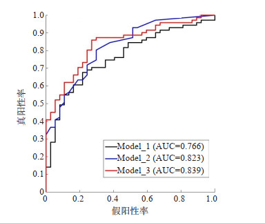
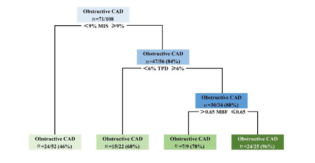
目的 探讨定量PET心肌灌注-代谢不匹配(MIS)能否进一步提升阻塞性冠心病(CAD)诊断的准确性。 方法 回顾性收集阻塞性CAD疑似患者97例,将其按照5∶3的比例随机划分为训练集(n=61)和测试集(n=36),并将阻塞性CAD定义为冠脉血管狭窄≥75%。单变量和多变量的Logistic回归分析用于分类模型的训练和性能评估。ROC曲线用于确定诊断阻塞性CAD的最佳截断值以及比较模型间的差异性。 结果 单变量Logistic回归模型的AUC值分别为0.735、0.758和0.823,MIS的最佳截断值为9%;多变量Logistic回归模型的AUC值分别为0.766、0.823和0.839,以包含MIS指标的Model_3最高,且高于对照模型(P= 0.0034);基于Model_3构建的决策树,在测试集中的准确度最高可达96%。 结论 PET MIS是阻塞性CAD的有效预测因子,能有效提升诊断准确性,对疑似病例快速、准确的鉴别诊断具有重要的临床价值。 -
关键词:
- PET /
- 心肌灌注-代谢不匹配 /
- 阻塞性冠心病 /
- Logistic回归分析 /
- 决策树
Abstract:Objective To investigate whether quantitative PET myocardial perfusion-metabolism mismatch can further improve the diagnosis accuracy in predicting obstructive coronary artery disease (CAD). Methods A total of 97 patients with suspected obstructive CAD were retrospectively collected, and randomly divided into training set (61 patients) and validation set (36 patients) according to 5∶3. Obstructive CAD was defined as coronary artery ≥75% stenosis. Univariate and multivariate logistic regression analysis were used for model training and performance evaluation. The ROC curve analysis was used to determine the best trade-off for diagnosing obstructive CAD and compare the differences between models. Results The areas under the ROC curve (AUC) of three univariate logistic regression models were 0.735, 0.758 and 0.823, respectively, and the best trade-off of MIS was 9%. The AUC values of three multivariate logistic regression models were 0.766, 0.823 and 0.839, respectively. The model including TPD/MBF/MIS achieved the highest AUC and it was significantly higher than the control model including TPD/MBF (P=0.0034). The decision tree based on this model had an accuracy of up to 96% in the validation set. Conclusion PET myocardial perfusion- metabolism mismatch is an effective predictor of obstructive CAD, which can effectively improve the diagnostic accuracy and has important clinical value for the rapid and accurate differential diagnosis of suspected cases. -

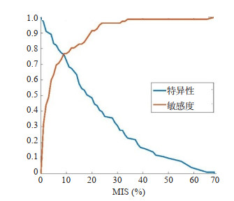
图 1 MIS在鉴别阻塞性CAD时的敏感度和特异性曲线
Figure 1. Sensitivity and specificity for the identification of obstructive CAD using regional MIS.

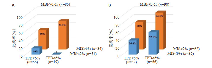
图 3 当冠脉血管的MBF > 0.65 mL/min/g(A)及MBF≤0.65 mL/min/g(B)时,阻塞性CAD在不同TPD和MIS组别中的发病率
Figure 3. Prevalence of obstructive CAD across different categories of TPD and MIS in vessels with MBF > 0.65 mL/min/g (A) and MBF≤0.65 mL/min/g (B).

图 4 基于TPD、MBF和MIS的最佳截断值,构建的冠脉血管分类的决策树
Figure 4. Decision tree for the identification of obstructive CAD based on the best trade- off values of TPD, MBF and MIS.
表 1 97例患者的临床特征
Table 1. Clinical characteristics of 97 patients (Mean±SD)
临床资料 所有患者(n=97) 非CAD组(n=10) CAD组(n=87) P 年龄(岁) 58±11 57±17 58±10 0.87 男性[n(%)] 92(95) 8(80) 84(97) < 0.05 体质量(kg) 66.0±9.8 63.8±10.5 66.2±9.7 0.47 心率(次/min) 78±14 73±10 79±14 0.20 舒张压(mmHg) 74±11 66±6 75±11 < 0.05 收缩压(mmHg) 120±17 105±20 121±17 < 0.05 吸烟[n(%)] 35(36) 3(30) 32(37) 0.68 CAD: 冠心病.  下载: 导出CSV
表 2 定量指标TPD、MBF和MIS在训练集两类血管中的差异性比较
Table 2. Comparison of the three indicators (TPD, MBF and MIS) in vessels with and without obstructive CAD on the training set (Mean±SD)
指标 所有血管(n=183) 非CAD组(n=82) CAD组(n=101) P TPD 8.40±8.91 4.07±5.70 11.92±9.49 < 0.001 MBF 0.66±0.28 0.78±0.29 0.56±0.23 < 0.001 MIS 15.04±16.32 6.41±10.16 22.04±17.05 < 0.001 TPD: 灌注总缺陷; MBF: 心肌血流量; MIS: 灌注-代谢不匹配.
下载: 导出CSV
-
[1] Zhou MG, Wang HD, Zeng XY, et al. Mortality, morbidity, and risk factors in China and its provinces, 1990-2017: a systematic analysis for the Global Burden of Disease Study 2017[J]. Lancet, 2019, 394 (10204): 1145-58. doi: 10.1016/S0140-6736(19)30427-1 [2] Litmeier S, Meinel TR, von Rennenberg R, et al. Coronary angiography in acute ischemic stroke patients: frequency and determinants of pathological findings in a multicenter cohort study [J]. J Neurol, 2022, 269(7): 3745-51. doi: 10.1007/s00415-022-11001-5 [3] Hasselbalch RB, Pries-Heje M, Engstrøm T, et al. Coronary risk stratification of patients with newly diagnosed heart failure[J]. Open Heart, 2019, 6(2): e001074. doi: 10.1136/openhrt-2019-001074 [4] Mirshahvalad SA, Chavoshi M, Hekmat S. . Diagnostic performance of prone-only myocardial perfusion imaging versus coronary angiography in the detection of coronary artery disease: a systematic review and meta-analysis[J]. J Nucl Cardiol, 2022, 29(3): 1339-51. doi: 10.1007/s12350-020-02376-x [5] Rosenkrans ZT, Massey CF, Bernau K, et al. 68 Ga]Ga-FAPI-46 PET for non-invasive detection of pulmonary fibrosis disease activity[J]. Eur J Nucl Med Mol Imaging, 2022, 49(11): 3705-16. doi: 10.1007/s00259-022-05814-9 [6] Papp L, Spielvogel CP, Grubmüller B, et al. Supervised machine learning enables non-invasive lesion characterization in primary prostate cancer with[68Ga]Ga-PSMA-11 PET/MRI[J]. Eur J Nucl Med Mol Imaging, 2021, 48(6): 1795-805. doi: 10.1007/s00259-020-05140-y [7] Kawakubo M, Nagao M, Yamamoto A, et al. 13 N-ammonia PETderived right ventricular longitudinal strain and myocardial flow reserve in right coronary artery disease[J]. Eur J Nucl Med Mol Imaging, 2022, 49(6): 1870-80. doi: 10.1007/s00259-021-05647-y [8] Tanja Kero MD P, Antti Saraste MD P, Bo Lagerqvist MD P, et al. Quantitative myocardial perfusion response to adenosine and regadenoson in patients with suspected coronary artery disease[J]. J Nucl Cardiol, 2022, 29(1): 24-36. doi: 10.1007/s12350-021-02731-6 [9] Miura S, Naya M, Kumamaru H, et al. Prognostic value of modified coronary flow capacity by 13N-ammonia myocardial perfusion positron emission tomography in patients without obstructive coronary arteries[J]. J Cardiol, 2022, 79(2): 247-56. doi: 10.1016/j.jjcc.2021.09.001 [10] Wang FH, Xu WP, Lv WB, et al. Evaluation of the diagnostic value of joint PET myocardial perfusion and metabolic imaging for vascular stenosis in patients with obstructive coronary artery disease[J]. J Nucl Cardiol, 2021, 28(6): 3070-80. doi: 10.1007/s12350-020-02160-x [11] Promteangtrong C, Jantarato A, Kunawudhi A, et al. Clinical impact of quantitative[15O]H2O PET/CT myocardial perfusion imaging on decision-making regarding invasive management of coronary artery disease[J]. J Nucl Cardiol, 2022, 29(4): 1887-99. doi: 10.1007/s12350-021-02604-y [12] Zampella E, Acampa W, Assante R, et al. Combined evaluation of regional coronary artery calcium and myocardial perfusion by 82Rb PET/CT in the identification of obstructive coronary artery disease [J]. Eur J Nucl Med Mol Imaging, 2018, 45(4): 521-9. doi: 10.1007/s00259-018-3935-1 [13] Moody JB, Poitrasson-Rivière A, Hagio T, et al. Added value of myocardial blood flow using 18F-flurpiridaz PET to diagnose coronary artery disease: the flurpiridaz 301 trial[J]. J Nucl Cardiol, 2021, 28(5): 2313-29. doi: 10.1007/s12350-020-02034-2 [14] Terrence D Ruddy MD M, Md AT, Viviany R Taqueti MD M. Role of nuclear cardiology in diagnosis and risk stratification of coronary microvascular disease[J]. J Nucl Cardiol, 2022: 1-14. [15] Goel A, Bandyopadhyay D, He Z, et al. Cardiac 18F-FDG imaging for direct myocardial ischemia imaging[J]. J Nucl Cardiol, 2022: 1-5. [16] Yang X, Zhang F, Chen Y, et al. Value of rest 18F-FDG myocardial imaging in the diagnosis of obstructive coronary artery disease in Chinese patients with suspected unstable angina: a prospective realworld clinical study[J]. J Nucl Cardiol, 2022. doi: 10.1007/s12350-022-03068-4. [17] de Oliveira Brito JB, deKemp RA, Ruddy TD. Evolving use of PET viability imaging[J]. J Nucl Cardiol, 2022, 29(3): 1000-2. doi: 10.1007/s12350-020-02460-2 [18] Sun X, Xu WC. Fast implementation of DeLong's algorithm for comparing the areas under correlated receiver operating characteristic curves[J]. IEEE Signal Process Lett, 2014, 21(11): 1389-93. doi: 10.1109/LSP.2014.2337313 [19] Nakajima K, Kudo T, Nakata T, et al. Diagnostic accuracy of an artificial neural network compared with statistical quantitation of myocardial perfusion images: a Japanese multicenter study[J]. Eur J Nucl Med Mol Imaging, 2017, 44(13): 2280-9. doi: 10.1007/s00259-017-3834-x [20] Nakajima K, Matsuo S, Wakabayashi H, et al. Diagnostic performance of artificial neural network for detecting ischemia in myocardial perfusion imaging[J]. Circ J, 2015, 79(7): 1549-56. doi: 10.1253/circj.CJ-15-0079 [21] Assante R, Zampella E, Arumugam P, et al. Quantitative relationship between coronary artery calcium and myocardial blood flow by hybrid rubidium-82 PET/CT imaging in patients with suspected coronary artery disease[J]. J Nucl Cardiol, 2017, 24(2): 494-501. doi: 10.1007/s12350-015-0359-1 [22] Otaki Y, Van Kriekinge SD, Wei CC, et al. Improved myocardial blood flow estimation with residual activity correction and motion correction in 18F-flurpiridaz PET myocardial perfusion imaging[J]. Eur J Nucl Med Mol Imaging, 2022, 49(6): 1881-93. doi: 10.1007/s00259-021-05643-2 -

 点击查看大图
计量
- 文章访问数: 245
- HTML全文浏览量: 127
- PDF下载量: 24
- 被引次数: 0