


Relationship between quantitative parameters of dual- layer detector spectral CT and lung cancer and its pathological characteristics
-
摘要:
目的 分析双层探测器光谱CT定量参数与肺癌及其病理特征的关系。 方法 收集2021年3月~2022年1月共87例肺癌患者的临床资料纳入肺癌组,并选择同期入院治疗的36例肺炎性结节患者作为对照组,两组患者治疗前2周内均行双层探测器光谱CT扫描。比较两组常规CT参数及光谱CT定量参数差异,并分析肺癌组不同病理特征者光谱CT定量参数差异。 结果 两组棘突征、CT值及动脉期碘密度(IC)、标准化IC(NIC)、有效原子序列(Zeff)比较,差异无统计学意义(P > 0.05),肺癌组分叶征、毛刺征、胸膜凹陷征及血管集束征CT征象多于对照组(P < 0.05),肺癌组静脉期IC、NIC及Zeff均低于对照组(P < 0.05)。肺癌组不同病理特征者动脉期IC、NIC、Zeff的差异无统计学意义(P > 0.05),腺癌者静脉期IC、NIC及Zeff高于鳞癌及小细胞癌者(P < 0.05),鳞癌者则高于小细胞癌者(P < 0.05)。 结论 双层探测器光谱CT定量参数对鉴别肺癌与肺炎性结节有利,且能辅助判断肺癌病理特征,具有良好应用前景。 Abstract:Objective To analyze the relationship between dual-layer detector spectral CT quantitative parameters and lung cancer and its pathological characteristics. Methods The clinical data of 87 patients with lung cancer (lung cancer group) were collected from March 2021 to January 2022. Thirty-six patients with lung inflammatory nodules who were admitted to the hospital during the same period were included in control group. All patients in the two groups received dual-layer detector spectral CT within 2 weeks before treatment. The differences in conventional CT parameters and spectral CT quantitative parameters were compared between the two groups. The differences in spectral CT quantitative parameters were analyzed among patients in lung cancer group with different pathological characteristics. Results There were no statistical differences in the spinous process sign, CT value and iodine content (IC)、normalized IC (NIC), effective atomic number (Zeff) in the arterial phase between the two groups (P > 0.05). But the CT signs of lobulation sign, spiculation sign, pleural indentation and vessel convergence sign in lung cancer group were more than those in control group (P < 0.05). The IC, NIC and Zeff in the venous phase were lower in lung cancer group than those in control group (P < 0.05). There were no statistical differences in the IC, NIC and Zeff in the arterial phase among patients with different pathological characteristics in lung cancer group (P > 0.05). The IC, NIC and Zeff in the venous phase of adenocarcinoma were higher than those of squamous cell carcinoma and small cell carcinoma (P < 0.05). The three indicators of squamous cell carcinoma were higher than those of small cell carcinoma (P < 0.05). Conclusion The quantitative parameters of dual-layer detector spectral CT are beneficial to distinguishing lung cancer from lung inflammatory nodules. It can assist in judging the pathological characteristics of lung cancer, with a good application prospect. -
Key words:
- lung cancer /
- lung inflammatory nodules /
- spectral CT /
- dual-layer detector
-

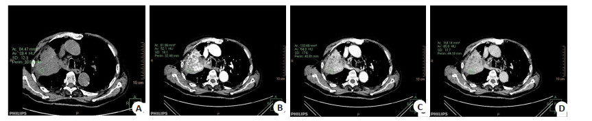
图 1 肺腺癌Ⅳ期患者光谱CT图像
A: CT平扫图像, 右肺下叶近肺门处见80 mm×49 mm不规则软组织肿块, 右肺下叶部分支气管局部狭窄闭塞, 部分肺组织不张, 相邻胸膜被牵连; B~D: 分别为动脉期图像、静脉期图像、延迟期图像, 病灶呈不均匀轻-中度进行性强化, 灶内见低密度坏死区, 邻近部分肺动、静脉显示不清.
Figure 1. Spectral CT imagings of patients with stage IV lung adenocarcinoma.

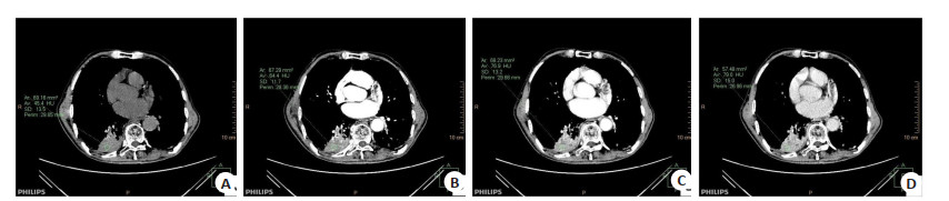
图 2 肺鳞癌Ⅳ期患者光谱CT图像
A: CT平扫图像,右肺上叶见一85 mm×71 mm不规则肿块, 邻近胸膜稍示增厚, 边缘见分叶及毛刺征, 内密度欠均匀,右肺上叶支气管狭窄截断; B~D: 分别为动脉期图像、静脉期图像、延迟期图像, 肿块呈不均匀中度延迟强化, 灶内见无强化低密度坏死区, 动脉期灶内见多发增粗迂曲血管影, 右肺上叶部分动、静脉局部受侵.
Figure 2. Spectral CT imagings of patients with stage IV squamous cell lung carcinoma.

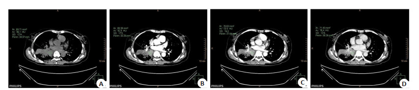
图 3 肺小细胞癌Ⅳ期患者光谱CT图像
A: CT平扫图像, 右肺下叶见一48 mm×32 mm软组织团块, 内见低密度坏死区, 边缘见浅分叶, 未见毛刺, 与相邻胸膜分界不清; B~D: 分别为动脉期图像、静脉期图像、延迟期图像, 病灶呈轻度不均匀延迟强化, 测三期CT值约64.4 Hu、76.9 Hu、79.0 Hu.
Figure 3. Spectral CT imagings of patients with stage IV small cell lung cancer.
表 1 两组常规CT参数比较
Table 1. Comparison of conventional CT parameters between the two groups [n(%)]
组别 分叶征 毛刺征 胸膜凹陷征 血管集束征 棘突征 CT值(Hu, Mean±SD) 平扫 动脉期 静脉期 肺癌组(n=87) 52(59.77) 49(56.32) 55(63.22) 48(55.17) 45(51.72) 43.56±8.06 61.99±12.27 87.43±16.95 对照组(n=36) 12(33.33) 10(27.78) 14(38.89) 11(30.56) 15(41.67) 44.69±7.35 66.07±13.44 94.05±18.22 χ2/t 7.130 8.312 6.120 6.182 1.031 0.725 1.631 1.928 P 0.008 0.004 0.013 0.013 0.310 0.470 0.105 0.056  下载: 导出CSV
表 2 两组光谱CT定量参数比较
Table 2. Comparison of quantitative parameters of spectral CT between the two groups (Mean±SD)
组别 IC(mg/mL) NIC Zeff 动脉期 静脉期 动脉期 静脉期 动脉期 静脉期 肺癌组(n=87) 1.31±0.24 2.09±0.22 0.13±0.03 0.41±0.05 8.09±0.16 8.23±0.15 对照组(n=36) 1.39±0.27 2.31±0.35 0.14±0.04 0.47±0.06 8.14±0.17 8.44±0.17 t 1.621 4.201 1.520 5.703 1.548 6.791 P 0.108 < 0.001 0.131 < 0.001 0.124 < 0.001 IC: 碘密度; NIC: 标准化碘密度; Zeff: 有效原子序列.
下载: 导出CSV
表 3 肺癌组不同病理特征者光谱CT定量参数比较
Table 3. Comparison of spectral CT quantitative parameters of patients with different pathological characteristics in lung cancer group (Mean±SD)
组别 IC(mg/mL) NIC Zeff 动脉期 静脉期 动脉期 静脉期 动脉期 静脉期 腺癌(n=48) 1.33±0.25 2.17±0.23 0.13±0.03 0.43±0.04 8.12±0.17 8.33±0.16 鳞癌(n=24) 1.30±0.22 2.04±0.22a 0.13±0.04 0.40±0.06a 8.08±0.16 8.14±0.15a 小细胞癌(n=15) 1.25±0.20 1.90±0.18ab 0.12±0.03 0.35±0.05ab 8.02±0.15 8.03±0.13ab F 0.685 9.411 0.569 16.370 2.206 27.311 P 0.507 < 0.001 0.568 < 0.001 0.116 < 0.001
下载: 导出CSV
-
[1] 郎中平, 罗柳林, 乐军, 等. 晚期非小细胞肺癌化疗患者医院感染病原学特点及影响因素分析[J]. 中华医院感染学杂志, 2019, 29(17): 2615-8. https://www.cnki.com.cn/Article/CJFDTOTAL-ZHYY201917015.htm [2] 谢馥淳, 侯阳. 双层探测器光谱CT在外周血管成像中的应用进展[J]. 中华放射学杂志, 2021, 55(12): 1341-4. doi: 10.3760/cma.j.cn112149-20210816-00760 [3] 罗昆, 董仟, 杨明, 等. 双层探测器光谱CT单能量成像联合个性化注射方案在颅脑CT血管成像中的应用研究[J]. 中华放射学杂志, 2022, 56(2): 196-200. [4] 郁义星, 王希明, 张妤, 等. 能谱CT影像组学特征鉴别肺癌结节与炎性结节的价值[J]. 中华放射学杂志, 2020, 54(12): 1167-72. doi: 10.3760/cma.j.cn112149-20200227-00281 [5] 陆璐, 盛茂, 李若梅, 等. 双层探测器光谱CT冠状动脉成像虚拟单能级图像与常规图像质量比较[J]. 中国医学影像学杂志, 2022, 30 (4): 335-40. doi: 10.3969/j.issn.1005-5185.2022.04.007 [6] 中华放射学杂志双层探测器光谱CT临床应用协作组. 双层探测器光谱CT临床应用中国专家共识(第一版)[J]. 中华放射学杂志, 2020, 54 (7): 635-43. doi: 10.3760/cma.j.cn112149-20200513-00679 [7] 袁海滨, 李新, 强金伟, 等. 双层探测器光谱CT诊断肝纤维化分期的最佳单能量研究[J]. 中国医学计算机成像杂志, 2022, 28(1): 44-9. https://www.cnki.com.cn/Article/CJFDTOTAL-YJTY202201008.htm [8] 李敏, 王娅菲, 姜文蓁, 等. 双层探测器光谱CT平扫定性联合定量参数预测肺纯磨玻璃结节侵袭性的价值[J]. 中华放射学杂志, 2022, 56 (3): 248-53. doi: 10.3760/cma.j.cn112149-20210419-00385 [9] 徐一铭, 袁梅, 邱琳, 等. 能谱CT及碘基图影像组学特征鉴别肺炎性及恶性病变[J]. 南京医科大学学报: 自然科学版, 2021, 41(7): 1063-8. https://www.cnki.com.cn/Article/CJFDTOTAL-NJYK202107021.htm [10] 翟晓静, 郭红红, 曹珊, 等. 双层探测器光谱CT肺动脉造影40 keV虚拟单能量图像优化窗口设置研究[J]. 临床放射学杂志, 2022, 41(1): 59-63. https://www.cnki.com.cn/Article/CJFDTOTAL-LCFS202201012.htm [11] 傅文悦, 朱广辉. Revolution CT能谱成像技术对不同性质肺结节鉴别诊断价值的应用研究[J]. 中国CT和MRI杂志, 2021, 19(6): 58-61. doi: 10.3969/j.issn.1672-5131.2021.06.018 [12] 罗久伟, 原卫民, 成官迅. 局灶性肺磨玻璃结节的低剂量胸部CT特征分析[J]. 中国CT和MRI杂志, 2021, 19(11): 51-2, 156. doi: 10.3969/j.issn.1672-5131.2021.11.016 [13] 徐茗, 张新日. 双层探测器光谱CT在肺部疾病应用中的研究进展[J]. 国际呼吸杂志, 2021, 41(23): 1836-40. doi: 10.3760/cma.j.cn131368-20201209-01116 [14] 付蓝琦, 潘馨梦, 刘思佳, 等. 双层探测器光谱CT虚拟平扫替代常规平扫评估甲状腺结节的可行性分析[J]. 放射学实践, 2022, 37(3): 302-6. https://www.cnki.com.cn/Article/CJFDTOTAL-FSXS202203006.htm [15] 戚元刚, 范明新, 殷月慧, 等. 双层探测器光谱CT区分中心型肺癌与肺不张区域的价值[J]. 中华放射学杂志, 2021, 55(11): 1167-71. [16] 于芹, 陈进, 夏茜, 等. 双能光谱CT定量分析对肺腺癌化疗后疗效的评估价值[J]. 海南医学, 2021, 32(14): 1846-9. https://www.cnki.com.cn/Article/CJFDTOTAL-HAIN202114021.htm [17] 傅奕铖, 余烨, 陈杏彪, 等. 双层探测器光谱CT鉴别诊断肺癌与炎性结节的价值[J]. 中华放射学杂志, 2021, 55(12): 1264-9. [18] 张莹, 傅奕铖, 余烨, 等. 基于2011及2020年版病理分级系统双层探测器光谱CT评估实性肺腺癌病理分级的价值[J]. 中华放射学杂志, 2022, 56(6): 623-30. -

 点击查看大图
计量
- 文章访问数: 166
- HTML全文浏览量: 58
- PDF下载量: 7
- 被引次数: 0