


Characteristics and clinical significance of thoracic aorta ct reconstruction in valvular heart disease
-
摘要:
目的 观察心脏瓣膜病胸主动脉CT重建特征及其临床意义。 方法 选取2018年10月~2021年12月北碚区中医医院放射科收治的300例患者作为研究对象,将95例心脏瓣膜病患者纳入病例组,205例非心脏瓣膜病变的普通患者纳入对照组,均行CT增强扫描和三维重建,比较两组的临床一般资料和CT测量值(包括胸主动脉长度、主动脉峡部直径和膈肌层面主动脉直径),采用Logistic分析患者胸主动脉长度的影响因素。 结果 病例组患者的胸主动脉长度、主动脉峡部直径和膈肌层面主动脉直径均高于对照组,差异有统计学意义(P < 0.05),心脏瓣膜病变患者主要CT特征为主动脉扩张,瓣膜增厚; 不同年龄、性别、高血压和主动脉壁斑块、心脏瓣膜病的患者胸主动脉长度比较,差异有统计学意义(P < 0.05);年龄(OR=2.121)、主动脉壁斑块(OR=2.234)、心脏瓣膜病(OR=1.964)是影响患者胸主动脉长度的因素,差异有统计学意义(P < 0.05)。 结论 心脏瓣膜病患者胸主动脉增长、主动脉峡部及膈肌层面增长增宽,同时胸主动脉长度受年龄、主动脉壁斑块和心脏瓣膜病的影响,了解其形态特征,对于主动脉手术治疗具有一定的指导意义。 Abstract:Objective To observe the characteristics and clinical significance of thoracic aorta CT reconstruction in valvular heart disease. Methods A total of 300 patients admitted to radiology department of Beibei District Hospital of Traditional Chinese Medicine were enrolled from October 2018 to December 2021, including 95 cases with valvular heart disease in case group and other 205 ordinary cases in control group. All patients underwent CT scan and multi-phase reconstruction. The general clinical data and CT measurement data (length of thoracic aorta, diameter of aortic isthmus, aortic diameter at diaphragm level) were compared between the two groups. The influencing factors of thoracic aorta length were analyzed by Logistics analysis. Results The length of thoracic aorta, diameter of aortic isthmus and aortic diameter at diaphragm level in cases were longer than those in control group (P < 0.05). The main CT characteristics in patients with heart valve disease included aortic dilatation and valve thickening. The differences in length of thoracic aorta among patients with different age, genders, hypertension, aortic wall plaques and valvular heart diseases were statistically significant (P < 0.05). Age (OR=2.121), aortic wall plaque (OR= 2.234) and valvular heart disease(OR=1.964) were influencing factors of thoracic aorta length. Conclusion The thoracic aorta is increased, aortic isthmus and aortic diameter at diaphragm level are thickened in patients with valvular heart disease. The length of thoracic aorta is affected by age, aortic wall plaques and heart valve disease. Understanding the morphological characteristics has certain guiding significance for aortic surgical treatment. -
Key words:
- valvular heart disease /
- thoracic aorta /
- multi-slice spiral CT /
- reconstruction
-

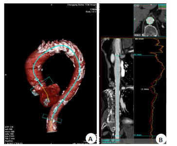
图 1 患者男,85岁,确诊为主动脉瓣及二尖瓣增厚、钙化伴狭窄
A: 容积再现图, 主动脉壁可见多发钙化, 粗细不均, 弯曲度增加; B: 曲面重建图, 胸主动脉较长、主动脉峡部扩大, 胸主动脉长376.5 mm, 主动脉峡部直径26.2 mm, 胸主动脉膈肌层面直径21.9 mm.
Figure 1. A 85-year-old male patient, aorta and mitral valve thickening, calcification combined with stenosis
表 1 两组临床资料比较
Table 1. Comparison of clinical data between the two groups (n)
临床资料 病例组(n=95) 对照组(n=205) t/χ2 P 年龄(岁, Mean±SD) 64.68±11.03 61.39±15.13 1.667 0.097 性别 0.223 0.637 男 62 128 女 33 77 高血压 6.098 0.014 有 31 98 无 64 107 糖尿病 0.514 0.474 有 11 30 无 84 175 主动脉壁斑块 6.813 0.009 有 67 112 无 28 93  下载: 导出CSV
表 2 两组患者胸主动脉CT相关测量值比较
Table 2. Comparison of thoracic aorta CT-related measurement data between two groups (mm, Mean±SD)
组别 胸主动脉长度 主动脉峡部直径 膈肌层面主动脉直径 病例组(n=95) 357.47±39.20 27.22±3.58 22.08±2.69 对照组(n=205) 335.69±37.90 25.01±3.31 21.00±2.94 t 4.580 5.241 3.039 P < 0.001 < 0.001 0.003
下载: 导出CSV
表 3 影响胸主动脉长度的单因素分析
Table 3. Univariate analysis on the influencing factors of thoracic aorta length (mm, Mean±SD)
因素 胸主动脉长度 t P 年龄(岁) 10.537 < 0.001 ≥62(n=176) 362.51±44.47 < 62(n=124) 314.31±29.57 性别 9.237 < 0.001 男(n=190) 358.28±42.28 女(n=110) 315.48±31.46 高血压 10.981 < 0.001 有(n=129) 370.61±35.57 无(n=171) 321.45±40.38 糖尿病 1.471 0.142 有(n=41) 350.78±33.02 无(n=259) 341.29±39.15 主动脉壁斑块 11.976 < 0.001 有(n=179) 364.38±39.32 无(n=121) 310.35±36.82 心脏瓣膜病 4.580 < 0.001 有(n=95) 357.47±39.20 无(n=205) 335.69±37.90
下载: 导出CSV
表 4 影响胸主动脉长度的多因素分析
Table 4. Multivariate analysis on the influencing factors of thoracic aorta length
因素 β SE wald χ2 OR 95% CI P 年龄 0.752 0.336 11.328 2.121 1.098~4.098 0.026 主动脉壁斑块 0.804 0.351 5.882 2.234 1.123~4.446 0.022 心脏瓣膜病 0.675 0.209 10.312 1.964 1.304~2.958 0.001 赋值情况:年龄(< 62岁=0,≥62岁=1),性别(女=0,男=1),高血压(无=0,有=1),主动脉壁斑块(无=0,有=1),心脏瓣膜病(无=0,有=1).
下载: 导出CSV
-
[1] 周钧, 孟春营, 刘海燕, 等. 生物瓣膜置换术治疗老年心脏瓣膜病的临床研究[J]. 罕少疾病杂志, 2019, 26(4): 10-1, 14. https://www.cnki.com.cn/Article/CJFDTOTAL-HSJB201904004.htm [2] 秦芸芸, 张连仲. 经食管实时三维超声心动图在心脏瓣膜病应用中的研究进展[J]. 中华实用诊断与治疗杂志, 2017, 31(11): 1139-41. https://www.cnki.com.cn/Article/CJFDTOTAL-HNZD201711029.htm [3] Belvroy VM, de Beaufort HWL, van Herwaarden JA, et al. Tortuosity of the descending thoracic aorta: normal values by age[J]. PLoS One, 2019, 14(4): e0215549. doi: 10.1371/journal.pone.0215549 [4] Vriz O, Driussi C, Bettio M, et al. Aortic root dimensions and stiffness in healthy subjects[J]. Am J Cardiol, 2013, 112(8): 1224-9. doi: 10.1016/j.amjcard.2013.05.068 [5] Lionakis N, Mendrinos D, Sanidas E, et al. Hypertension in the elderly[J]. World J Cardiol, 2012, 4(5): 135-47. doi: 10.4330/wjc.v4.i5.135 [6] Palatini P. Cardiovascular effects of exercise in young hypertensives [J]. Int J Sports Med, 2012, 33(9): 683-90. doi: 10.1055/s-0032-1304633 [7] 雷佳瑞, 郭瑞强. 超声心动图评价心脏瓣膜病患者心功能与心肌受损的研究进展[J]. 临床超声医学杂志, 2018, 20(2): 114-7. https://www.cnki.com.cn/Article/CJFDTOTAL-LCCY201802016.htm [8] 张蕾, 杨全新, 毛翠平, 等. 双源CT冠状动脉成像与超声心动图对左心室功能评价的对比研究[J]. 实用放射学杂志, 2019, 35(6): 884-6. [9] 吴小松, 龚波, 贺俊斌, 等. 双源CT冠状动脉联合左房-肺静脉"一站式"成像检查的应用[J]. 中国CT和MRI杂志, 2021, 19(10): 61-3. https://www.cnki.com.cn/Article/CJFDTOTAL-CTMR202110019.htm [10] Baumgartner H, Falk V, Bax JJ, et al. 2017 ESC/EACTS Guidelines for the management of valvular heart disease[J]. Eur Heart J, 2017, 38(36): 2739-91. [11] 赵萍, 潘闽, 龚亚驰, 等. 南通地区老年退行性心脏瓣膜病的危险因素[J]. 中国老年学杂志, 2019, 39(18): 4385-8. https://www.cnki.com.cn/Article/CJFDTOTAL-ZLXZ201918008.htm [12] Fukumoto R, Kawai M, Minai K, et al. Conflicting relationship between age-dependent disorders, valvular heart disease and coronary artery disease by covariance structure analysis: possible contribution of natriuretic peptide[J]. PLoS One, 2017, 12(7): e0181206. [13] 王戈楠, 李华. 心脏瓣膜置换术后影响患者死亡危险因素分析[J]. 医学临床研究, 2018, 35(12): 2426-8. https://cdmd.cnki.com.cn/Article/CDMD-11065-1021886036.htm [14] Polonskaya YV, Kashtanova EV, Murashov IS, et al. The influence of calcification factors and endothelial-dysfunction factors on the development of unstable atherosclerotic plaques[J]. Diagnostics (Basel), 2020, 10(12): 1074. [15] Lee SE, Budoff MJ, Conte E, et al. Association between the progression of aortic valve calcification and coronary atherosclerotic plaque volume[J]. Eur Heart J, 2020, 41 (Supplement_2): 79-86. [16] 曲浥晨, 李宜嘉, 李玉琳, 等. 二叶式主动脉瓣主动脉病变相关基因突变者的影像学特征与遗传学分析[J]. 临床超声医学杂志, 2020, 22 (4): 296-9. https://www.cnki.com.cn/Article/CJFDTOTAL-LCCY202004021.htm [17] Westerhof N, Stergiopulos N, Noble MIM, et al. Snapshots of Hemodynamics: An Aid for Clinical Research and Graduate Education[M]. Cham: Springer International Publishing, 2019. [18] Shahid L, Rice J, Berhane H, et al. Enhanced 4D flow MRI- based CFD with adaptive mesh refinement for flow dynamics assessment in coarctation of the aorta[J]. Ann Biomed Eng, 2022, 50(8): 1001- 16. [19] Ha H, Kim GB, Kweon J, et al. The influence of the aortic valve angle on the hemodynamic features of the thoracic aorta[J]. Sci Rep, 2016, 6: 32316. [20] Callahan S, Singam NS, Kendrick M, et al. Dual-Venc acquisition for 4D flow MRI in aortic stenosis with spiral readouts[J]. J Magn Reson Imaging, 2020, 52(1): 117-28. -

 点击查看大图
计量
- 文章访问数: 207
- HTML全文浏览量: 94
- PDF下载量: 10
- 被引次数: 0