


Effect of Bushen decoction for the treatment of ovariectomized osteoporosis model rats based on Micro-CT
-
摘要:
目的 基于Micro-CT探究补肾方对去卵巢骨质疏松大鼠股骨骨微结构、骨周围血管体积、骨力学和血清碱性磷酸酶(ALP)的影响。 方法 选择3月龄雌性SD大鼠30只,随机分为对照组、补肾方干预组和戊酸雌二醇干预组,10只/组,摘除双侧卵巢。造模3月后,分别用注射用0.9%NaCl溶液、补肾方、戊酸雌二醇灌胃干预4周,通过Micro-CT扫描观察股骨微结构和骨周围血管体积,小动物骨骼强度测定仪测量骨折应力和压碎力,并检测大鼠血清ALP活性。 结果 与对照组相比,Micro-CT扫描结果显示,补肾方干预组可明显改善骨密度(P=0.0114),提高骨体积分数(P=0.0006),增加骨小梁数目(P=0.0016),加厚骨小梁厚度(P=0.0050),血管体积(P < 0.0001);骨骼强度检测显示,补肾方干预组股骨骨折应力增强(P=0.0044);同时ALP活性增加(P= 0.0181)。 结论 补肾方能显著改善股骨骨微结构,提升骨周围血管体积,增强骨折应力,提高ALP水平,提示其可能成为今后临床治疗骨质疏松患者的配方。 Abstract:Objective To investigate the effects of Bushen decoction on femur's bone-microarchitecture, vascular volume around the bone, biomechanics and alkaline phosphatase (ALP) activity of ovariectomized rat with osteoporosis based on micro-CT. Methods Thirty healthy Sprague-Dawley female rats aged three months were randomly divided into control group, Bushen decoction intervention group and estradiol valerate intervention group, with 10 rats/group. Both ovaries of rats were removed. Rats were intragastrically administered for 4 weeks after the model creation 3 months. Femur's bone-microarchitecture and vascular volume around the bone were observed by Micro-CT scan. Bone fracture stress and structural strength of each femoral bone were measured. Serum ALP activity were detected. Results Micro-CT showed an increase in morphometry such as bone mineral density (P=0.0114), bone volume fraction (P=0.0006), trabecular number (P=0.0016), trabecular thickness (P= 0.0050), vascular volume (P < 0.0001) in Bushen decoction intervention group as compared with control group. Bushen decoction significantly enhanced bone fracture stress (P=0.0044) as compared with normal saline. Serum ALP activity in Bushen decoction group were improved (P=0.0181) as compared with control group. Conclusion Bushen decoction can improve bone-microarchitecture of femurs, increase vascular volume, strengthen bone fracture stress and higher the activity of ALP. Therefore, Bushen decoction should have a good effect on treating osteoporosis in future clinical studies. -
Key words:
- Bushen decoction /
- osteoporosis /
- ovary /
- femur /
- microcomputed tomography /
- vascular volume /
- bone mechanics
-

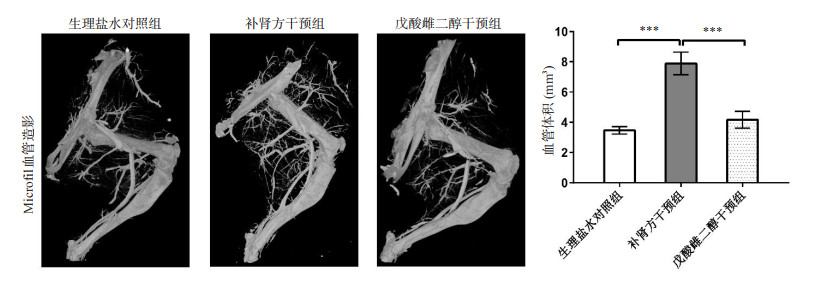
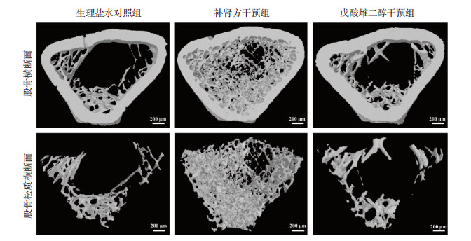
图 1 7月龄OVX大鼠股骨Micro-CT三维图
Figure 1. Three-dimensional microcomputed tomography images of the femurs of 7-month age OVX rat.

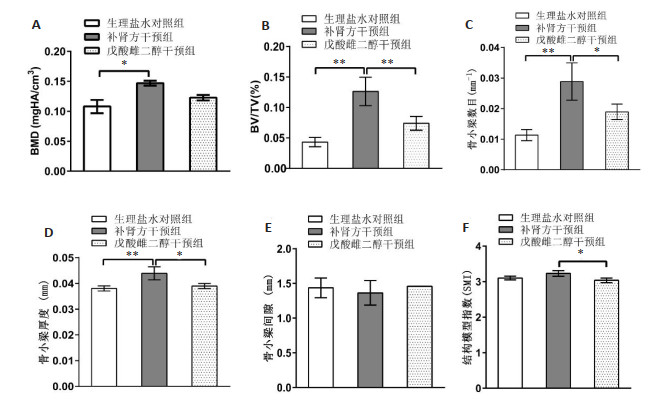
图 2 7月龄OVX大鼠各组股骨Micro-CT骨形态学分析
A: 骨密度分析; B: 骨体积分数分析; C: 骨小梁数目分析; D: 骨小梁厚度分析; E: 骨小梁间隙分析; F: 结构模型指数分析. n=3. *P < 0.05, **P < 0.01.
Figure 2. Micro-CT bone morphology analysis of the femurs of 7-month age OVX rat in each group.

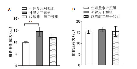
图 3 7月龄OVX大鼠各组股骨生物力学分析
A: 股骨骨折应力分析; B: 压碎力分析. n=3. **P < 0.01.
Figure 3. Bone biomechanics analysis of the femurs of 7-month age OVX rat in each group.

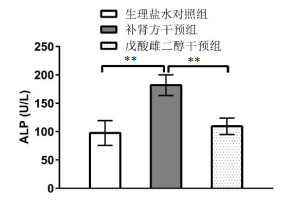
图 4 7月龄OVX大鼠各组血清碱性磷酸酶活性分析
Figure 4. Alkaline phosphatase activity in serum analysis of 7-month age OVX rat in each group. **P < 0.001.
-
[1] Lama A, Santoro A, Corrado B, et al. Extracorporeal shock waves alone or combined with raloxifene promote bone formation and suppress resorption in ovariectomized rats[J]. PLoS One, 2017, 12 (2): e0171276. doi: 10.1371/journal.pone.0171276 [2] Simon JA. What's new in hormone replacement therapy: focus on transdermal estradiol and micronized progesterone[J]. Climacteric, 2012, 15(sup1): 3-10. doi: 10.3109/13697137.2012.669332 [3] Makins R, Ballinger A. Gastrointestinal side effects of drugs[J]. Expert Opin Drug Saf, 2003, 2(4): 421-9. doi: 10.1517/14740338.2.4.421 [4] Stutz C, Batool F, Petit C, et al. Influence of parathyroid hormone on periodontal healing in animal models: a systematic review[J]. Arch Oral Biol, 2020, 120: 104932. doi: 10.1016/j.archoralbio.2020.104932 [5] 潘欣, 梁兴伦, 曾思良, 等. 补肾方含药血清干预大鼠椎间盘软骨细胞miRNA的差异表达[J]. 上海中医药杂志, 2016, 50(1): 65-9. https://www.cnki.com.cn/Article/CJFDTOTAL-SHZZ201601025.htm [6] 梁兴伦, 潘欣, 曾思良, 等. 补肾方含药血清处理的大鼠成骨细胞miRNA差异表达[J]. 安徽中医药大学学报, 2017, 36(4): 71-5. doi: 10.3969/j.issn.2095-7246.2017.04.021 [7] 潘欣, 曾思良, 梁兴伦, 等. microRNA-195-5p调节Bmpr1α表达对骨髓间充质干细胞成脂分化的影响[J]. 同济大学学报: 医学版, 2017, 38(3): 1-7, 13. doi: 10.3969/j.issn.1009-3060.2017.03.001 [8] Bouxsein ML, Boyd SK, Christiansen BA, et al. Guidelines for assessment of bone microstructure in rodents using micro-computed tomography[J]. J Bone Miner Res, 2010, 25(7): 1468-86. doi: 10.1002/jbmr.141 [9] Chen J, Yang XQ, Meng YZ, et al. In vitro and in vivo CT imaging using bismuth sulfide modified with a highly biocompatible Pluronic F127[J]. Nanotechnology, 2014, 25(29): 295103. doi: 10.1088/0957-4484/25/29/295103 [10] Bernard F, Mercier P, Chappard D. Microvascularization of the human central and peripheral nervous system: a new microcomputed tomography method[J]. Morphologie, 2020, 104 (347): 247-53. doi: 10.1016/j.morpho.2020.05.004 [11] Ghanavati S, Yu LX, Lerch JP, et al. A perfusion procedure for imaging of the mouse cerebral vasculature by X-ray micro-CT[J]. J Neurosci Methods, 2014, 221: 70-7. doi: 10.1016/j.jneumeth.2013.09.002 [12] Lyu HZ, Lee JH. Correlation between two-dimensional micro-CT and histomorphometry for assessment of the implant osseointegration in rabbit tibia model[J]. Biomater Res, 2021, 25(1): 11. doi: 10.1186/s40824-021-00213-x [13] Xie ZJ, Weng SJ, Li H, et al. Teriparatide promotes healing of critical size femur defect through accelerating angiogenesis and degradation of β-TCP in OVX osteoporotic rat model[J]. Biomed Pharmacother, 2017, 96: 960-7. doi: 10.1016/j.biopha.2017.11.141 [14] 嵇承栋, 钱淑雯, 秦海庆, 等. 咪达唑仑联合芬太尼静脉麻醉在实验大鼠的应用[J]. 实用疼痛学杂志, 2013, 9(1): 15-7. [15] Elmeliegy M, Udata C, Liao K, et al. Considerations on the calculation of the human equivalent dose from toxicology studies for biologic anticancer agents[J]. Clin Pharmacokinet, 2021, 60(5): 563-7. doi: 10.1007/s40262-021-00987-2 [16] 宋林奇, 杜先婕, 侯洁文, 等. 强筋健骨胶囊抗骨质疏松作用实验研究[J]. 中国药业, 2009, 18(6): 19-20. doi: 10.3969/j.issn.1006-4931.2009.06.014 [17] Balbaied T, Hogan A, Moore E. Electrochemical detection and capillary electrophoresis: comparative studies for alkaline phosphatase (ALP) release from living cells[J]. Biosensors, 2020, 10 (8): 95. doi: 10.3390/bios10080095 [18] 刘振海, 刘红, 王少君, 等. 防治骨质疏松症常用单味中药实验研究概况[J]. 环球中医药, 2013, 6(6): 473-9. doi: 10.3969/j.issn.1674-1749.2013.06.025 [19] 章喻, 张孙正远, 王利波, 等. 牛膝-杜仲成分组合干预糖皮质激素性骨质疏松模型小鼠的研究[J]. 中国骨质疏松杂志, 2022, 28(5): 643-7. doi: 10.3969/j.issn.1006-7108.2022.05.004 [20] 贾艳萍, 张国明. 补肾壮骨汤联合西药治疗围绝经期骨质疏松症的临床疗效及对骨代谢、炎症因子的影响[J]. 中医研究, 2021, 34(12): 37-40. https://www.cnki.com.cn/Article/CJFDTOTAL-ZYYJ202112011.htm [21] 李敏, 史晓林, 许超, 等. 右归丸抗骨质疏松症的中药化合物及靶点网络药理学作用机制[J]. 中国骨伤, 2020, 33(10): 933-7. doi: 10.12200/j.issn.1003-0034.2020.10.009 [22] 李慧文, 曾令烽, 陈海云. 基于网络药理学探讨菟丝子、女贞子治疗绝经后骨质疏松症的作用机制[J]. 医学综述, 2021, 27(23): 4746-53. doi: 10.3969/j.issn.1006-2084.2021.23.032 [23] 佟颖, 刁志惠, 孙乐, 等. 益肾壮骨法治疗系统性红斑狼疮患者继发的激素性骨质疏松症48例的临床研究[J]. 中国骨质疏松杂志, 2018, 24(9): 1214-8. https://www.cnki.com.cn/Article/CJFDTOTAL-ZGZS201809018.htm [24] Meng QX, Wang BL, Yu PS, et al. Extract of cornus officinalis SIEB ameliorates osteoporosis in spinal cord-injured rats[J]. Chin J Osteoporos, 2015, 21(5): 627-33. [25] 李秋江, 房晓敏, 王胤斌, 等. 枸杞子治疗骨质疏松症的分子机制[J]. 中国骨质疏松杂志, 2021, 27(12): 1757-62. https://www.cnki.com.cn/Article/CJFDTOTAL-ZGZS202112006.htm [26] 王飞达, 王娜妮, 刘亦杨. 糖尿病骨质疏松症验案举隅[J]. 浙江中医杂志, 2020, 55(7): 536. https://www.cnki.com.cn/Article/CJFDTOTAL-ZJZZ202007046.htm [27] Gregson CL, Armstrong DJ, Bowden J, et al. UK clinical guideline for the prevention and treatment of osteoporosis[J]. Arch Osteoporos, 2022, 17(1): 1-46. [28] London GM. Bone-vascular cross-talk[J]. J Nephrol, 2012, 25(5): 619-25. [29] Street J, Bao M, de Guzman L, et al. Vascular endothelial growth factor stimulates bone repair by promoting angiogenesis and bone turnover[J]. PNAS, 2002, 99(15): 9656-61. [30] Alagiakrishnan K, Juby A, Hanley D, et al. Role of vascular factors in osteoporosis[J]. J GerontolABiol Sci Med Sci, 2003, 58(4): M362-6. [31] Weng SJ, Yan DY, Tang JH, et al. Combined treatment with Cinnamaldehyde and β-TCP had an additive effect on bone formation and angiogenesis in critical size calvarial defect in ovariectomized rats[J]. Biomed Pharmacother, 2019, 109: 573-81. -

 下载:
下载:
 点击查看大图
计量
- 文章访问数: 485
- HTML全文浏览量: 167
- PDF下载量: 46
- 被引次数: 0