


Comparison of clinical value between enhanced CT and MRI in diagnosis of lumbar tuberculosis
-
摘要:
目的 比较增强CT和MRI诊断腰椎结核的应用价值。 方法 选取2019年6月~2021年6月在本院收治疑似60例腰椎结核患者,均行增强CT和MRI图像,以病理或治疗随访结果为金标准,观察其影像学表现,比较两种方法对腰椎结核的诊断效能。 结果 60例患者中,有42例确诊为腰椎结核。增强CT检出腰椎结核36例,与金标准比较,敏感度为85.71%,特异性为77.77%,准确度为83.33%,Kappa值为0.615;MRI检出腰椎结核40例,与金标准比较,敏感度为95.24%,特异性为88.89%,准确率为93.33%,Kappa值为0.841;MRI在椎间盘受累、椎旁脓肿、椎管受累中的检出率均高于增强CT(P<0.05),CT在死骨形成中的检出率高于MRI(P<0.05)。 结论 增强CT与MRI对诊断腰椎结核均具有一定的临床效能,MRI优于增强CT,MRI在椎间盘受累、椎旁脓肿、椎管受累的诊断有明显优势,增强CT在死骨形成检出率优于MRI。 Abstract:Objective To compare the value of enhanced CT and MRI in diagnosis of lumbar tuberculosis. Methods Sixty patients with suspected lumbar tuberculosis admitted to the hospital were enrolled between June 2019 and June 2021. All underwent examinations by contrast-enhanced CT and MRI images. Taking pathological or treatment follow-up results as the golden standard, their imaging manifestations were observed. The diagnostic efficiency of the two methods for lumbar tuberculosis was compared. Results There were 42 cases confirmed with lumbar tuberculosis out of 60 cases. The contrast-enhanced CT showed that there were 36 cases with lumbar tuberculosis. Compared with the golden standard, its sensitivity, specificity, accuracy and Kappa value were 85.71%, 77.77%, 83.33% and 0.615, respectively. MRI showed that there were 40 cases with lumbar tuberculosis. Compared with the golden standard, its sensitivity, specificity, accuracy and Kappa value were 95.24%, 88.89%, 93.33% and 0.841, respectively. The detection rates of intervertebral disc involvement, paravertebral abscess and spinal canal involvement by MRI were higher than those by contrast-enhanced CT (P<0.05). The detection rate of sequestrum formation by CT was higher than that by MRI (P<0.05). Conclusion Both contrast-enhanced CT and MRI are of certain clinical diagnostic efficiency for lumbar tuberculosis. MRI is superior to contrast-enhanced CT. MRI has obvious advantages in the diagnosis of intervertebral disc involvement, paravertebral abscess and spinal canal involvement, while enhanced CT is superior to MRI in the detection of sequestrum formation. -
Key words:
- contrast-enhanced CT /
- MRI /
- lumbar tuberculosis /
- lesion /
- diagnostic efficiency
-

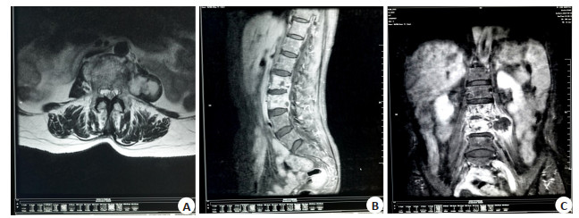
图 1 腰椎结核患者CT扫描图
A:CT轴位软组织窗;B:CT冠状位可见腰2~3锥体周围骨质破坏,破坏区可见局部硬化,周围软组织肿胀;C:CT增强静脉期可见腰2~4椎体虫噬样骨质破坏,脊柱旁见囊性低密影,囊壁较厚.
Figure 1. CT scans of a patient with lumbar tuberculosis.

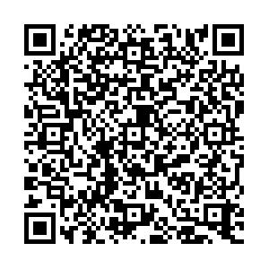
图 2 腰椎结核患者MRI扫描图
A:MRI轴位;B:MRI增强矢状位;C:MRI增强冠状位. MRI可见腰2、3椎体见斑片状长T1稍长T2异常信号影,增强扫描呈不均匀强化,局部压迫椎管,相应脊髓受压变细,相应椎间隙变窄,左侧腰大肌见类圆形长T1长T2信号影,增强扫描呈环形强化,边缘欠光整.
Figure 2. MRI scans of a patient with lumbar tuberculosis.
表 1 增强CT及MRI诊断腰椎结核临床效能
Table 1. Clinical efficacy of enhanced CT and MRI in diagnosis of lumbar tuberculosis
检测方法 金标准(n) 敏感度(%) 特异性(%) 准确度(%) 阳性预测值(%) 阴性预测值(%) Kappa值 阳性 阴性 增强CT 阳性 36 4 85.71 77.77 83.33 90.00 70.00 0.615 阴性 6 14 MRI 阳性 40 2 95.24 88.89 93.33 95.24 72.73 0.841 阴性 2 16  下载: 导出CSV
表 2 增强CT与MRI对腰椎结核病变检出情况比较
Table 2. Comparison between enhanced CT and MRI in the detection of lumbar tuberculosis changes [n(%)]
检测方法 椎间盘受累(n=52) 椎旁脓肿(n=56) 椎管受累(n=31) 骨质破坏(n=76) 死骨形成(n=21) 增强CT 46(88.46) 41(73.21) 17(54.84) 74(97.37) 21(100.00) MRI 52(100.00) 55(98.21) 28(90.32) 76(100.00) 10(47.62) χ2 6.367 14.292 9.807 2.027 14.903 P 0.012 <0.001 0.002 0.155 <0.001
下载: 导出CSV
-
[1] 黄炎, 张文志, 胡业丰, 等. 后路经单侧椎弓根入路病灶清除植骨内固定术治疗胸腰椎结核效果观察[J]. 山东医药, 2017, 57(30): 80-2. doi: 10.3969/j.issn.1002-266X.2017.30.025 [2] Dunn RN, Ben Husien M. Spinal tuberculosis: review of current management[J]. Bone Joint J, 2018, 100-B(4): 425-31. doi: 10.1302/0301-620X.100B4.BJJ-2017-1040.R1 [3] 高强, 李莹, 任翠萍, 等. 胸腰椎结核的MRI多序列诊断研究[J]. 中国CT和MRI杂志, 2018, 16(7): 144-6. doi: 10.3969/j.issn.1672-5131.2018.07.043 [4] 宋敏, 谢智恩, 杨宏志, 等. 成人脊柱结核并不全瘫的多层螺旋CT特征分析[J]. 实用放射学杂志, 2020, 36(2): 267-72. doi: 10.3969/j.issn.1002-1671.2020.02.024 [5] 李大伟, 乔娟, 唐国柯, 等. 经椎体强化术后胸腰椎结核患者行外科手术治疗的特点与效果分析[J]. 中国防痨杂志, 2019, 41(4): 383-8. doi: 10.3969/j.issn.1000-6621.2019.04.004 [6] Martrille L, de Angelis D, Blum A, et al. The potential of bone disease for personal identification: a case of tuberculosis[J]. Int J Legal Med, 2020, 134(5): 1957-62. doi: 10.1007/s00414-020-02348-3 [7] 郑欢露, 李白艳, 陈鹰, 等. HISTO及DWI序列在脊柱结核中的定量研究[J]. 磁共振成像, 2019, 10(5): 356-60. https://www.cnki.com.cn/Article/CJFDTOTAL-CGZC201905011.htm [8] 陈宋全, 姜剑榕, 王雪梅, 等. 探讨CT、MRI在胸腰椎结核诊断与鉴别诊断中的应用价值[J]. 新疆医学, 2018, 48(10): 1099-102. https://www.cnki.com.cn/Article/CJFDTOTAL-XJYI201810018.htm [9] 赵成孝. CT与MRI在胸腰椎结核诊断中的应用价值研究[J]. 影像研究与医学应用, 2019, 3(3): 254-5. doi: 10.3969/j.issn.2096-3807.2019.03.176 [10] 李愉, 蒋敏. MRI测量腰椎关节突角度在早期诊断椎间盘突出的应用价值[J]. 中国CT和MRI杂志, 2020, 18(2): 135-7, 141. doi: 10.3969/j.issn.1672-5131.2020.02.040 [11] Boruah DK, Gogoi BB, Prakash A, et al. Magnetic resonance imaging evaluation of posterior spinal tuberculosis: a crosssectional study[J]. Acta Radiol, 2021, 62(8): 1035-44. doi: 10.1177/0284185120948496 [12] 张鹤亭, 吴永光, 张勇刚, 等. X线、多层CT、MRI影像检查对脊柱结核的诊断价值比较[J]. 中国CT和MRI杂志, 2017, 15(2): 137-9. doi: 10.3969/j.issn.1672-5131.2017.02.043 [13] 王筱璇, 马晓文, 张玉婷, 等. MRI、MSCT及两者联合应用对单椎体骨肿瘤的诊断效能比较研究[J]. 实用放射学杂志, 2018, 34(7): 1073-6. doi: 10.3969/j.issn.1002-1671.2018.07.023 [14] 曾继先, 梁志凌. 腰3-5椎棘结核并冷脓肿的MRI影像特征研究[J]. 中国CT和MRI杂志, 2017, 15(10): 132-4, 138. doi: 10.3969/j.issn.1672-5131.2017.10.041 [15] 曾金光. 脊柱结核CT和MRI影像学特征比较研究[J]. 中国CT和MRI杂志, 2017, 15(1): 116-8. doi: 10.3969/j.issn.1672-5131.2017.01.037 [16] 袭龙祥, 袁秀娟, 田孝彬, 等. X线和多层CT及MRI对胸腰椎体结核感染的诊断效果研究[J]. 中华医院感染学杂志, 2018, 28(6): 883-6. https://www.cnki.com.cn/Article/CJFDTOTAL-ZHYY201806022.htm [17] 贾兆刚. X线和多层CT及MRI对胸腰椎体结核感染的诊断效果分析[J]. 中国急救医学, 2018, 38(S1): 127. https://www.cnki.com.cn/Article/CJFDTOTAL-ZHYY201806022.htm [18] 孙伟, 马东, 姜加学, 等. 脊椎结核的CT、MRI诊断与鉴别[J]. 分子影像学杂志, 2020, 43(3): 481-4. doi: 10.12122/j.issn.1674-4500.2020.03.23 -

 点击查看大图

计量
- 文章访问数: 320
- HTML全文浏览量: 193
- PDF下载量: 9
- 被引次数: 0