


Value of transrectal contrast-enhanced ultrasound combined with shear wave elastography in the differential diagnosis of benign and malignant prostatic lesions
-
摘要:
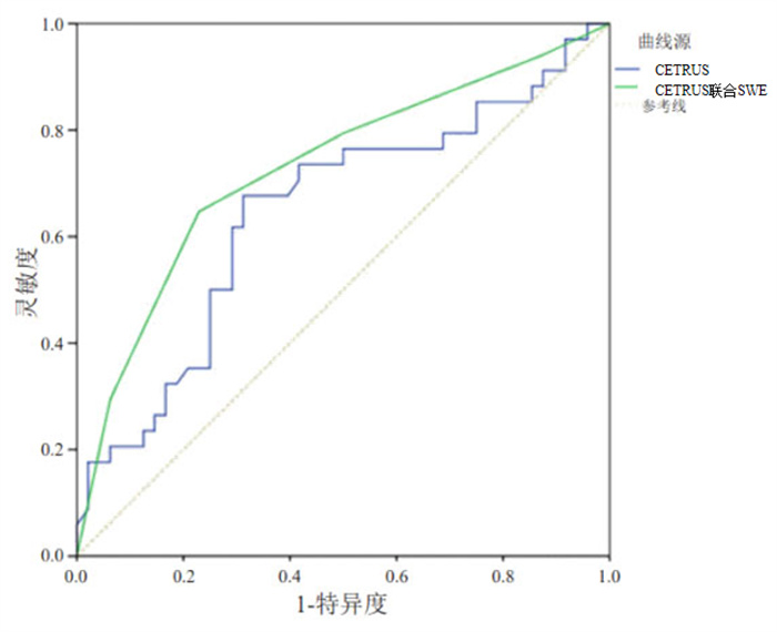
目的 分析经直肠超声造影(CETRUS)联合剪切波弹性成像鉴别诊断前列腺良恶性病变的价值。 方法 选取2020年5月~2021年12月我院收治的102例前列腺疾病患者,以经直肠超声(TRUS)引导前列腺穿刺活检为金标准,将其分为前列腺癌组(n=37)和前列腺增生组(n=65),对所有患者进行经直肠超声造影联合剪切波弹性成像检查,比较两组患者的Eration值和Emax值,比较不同方法的诊断准确度。 结果 前列腺癌组患者Eration、Emax值明显高于前列腺增生组患者,差异有统计学意义(P < 0.05);CETRUS联合剪切波弹性成像诊断前列腺良恶性病变的敏感度、特异性、准确率明显高于单一CETRUS检查,单一技术诊断(81.08%、80.00%、80.39% vs 56.76%、64.62%、61.76%,P < 0.05);CETRUS联合剪切波弹性成像的曲线下面积为0.748(95%CI:0.534~0.760),大于单一CETRUS曲线下面积0.685(95%CI:0.626~0.851)。 结论 经直肠超声造影联合剪切波弹性成像可通过杨氏模量值来反应组织的硬度,进而对前列腺良恶性病变进行诊断,在临床上具有一定的应用价值。 Abstract:Objective To analyze the value of transrectal contrast-enhanced ultrasound (CETRUS) combined with shear wave elastography in the differential diagnosis of benign and malignant prostate lesions. Methods A total of 102 patients with prostate diseases admitted to our hospital from May 2020 to December 2021 were selected, and divided into prostate cancer group (n=37) divided into prostate cancer group (n=37). All patients underwent transrectal contrast-enhanced ultrasound combined with shear wave elastography. Eration value and Emax were compared between the two groups, and the diagnostic accuracy of different methods was compared. Results The Eration value and Emax in prostate cancer group were significantly higher than those in benign prostatic hyperplasia group(P < 0.05). The sensitivity, specificity and accuracy of CETRUS combined with shear wave elastography in diagnosing benign and malignant prostate lesions were significantly higher than those of CETRUS alone(81.08%、80.00%、80.39% vs 56.76%、64.62%、61.76%, P < 0.05). The area under the curve of CETRUS combined with shear wave elastography was 0.748 (95%CI: 0.534-0.760), which was larger than that of CETRUS alone 0.685(95%CI: 0.266-0.851). Conclusion Transrectal contrast-enhanced ultrasound combined with shear-wave elastography can reflect tissue hardness through Young's modulus value, and then diagnose benign and malignant prostate lesions, which has certain clinical value. -
Key words:
- transrectal ultrasound /
- shear-wave elastic imaging /
- prostate gland /
- malignant lesions /
- value
-

图 2 不同方法诊断前列腺良恶性病变的ROC曲线
Figure 2. ROC curves of benign and malignant prostate lesions diagnosed by different methods.
表 1 两组患者Eration值、Emax比较
Table 1. Comparison of Eration value and Emax between the two groups (Mean±SD)
组別 Eration Emax 前列腺癌组(n=37) 6.54±1.42 87.26±15.31 前列腺增生组(n=65) 2.60±0.53 40.35±8.24 t 20.103 20.146 P < 0.001 < 0.001  下载: 导出CSV
表 2 不同方法诊断前列腺良恶性病变结果比较
Table 2. Comparison of results of benign and malignant prostate lesions diagnosed by different methods
检杳方法 前列腺穿刺活检(n) 合计(n) 敏感度(%) 特异性(%) 准确率(%) 前列腺癌 前列腺增生 CETRUS 56.76 64.62 61.76 前列腺癌 21 23 44 前列腺增生 16 42 58 CETRUS联合剪切波弹性成像 81.08 80.00 80.39 前列腺癌 30 13 43 前列腺增生 7 52 59 合计 37 65 102
下载: 导出CSV
-
[1] 薛念余, 程跃, 张盛敏, 等. 经直肠剪切波弹性成像技术在前列腺外周带低回声结节诊断中的价值[J]. 中华泌尿外科杂志, 2019, 40(1): 62- 3. doi: 10.3760/cma.j.issn.1000-6702.2019.01.012 [2] 梁蕾, 孙铮, 孙亚, 等. 经直肠超声剪切波弹性成像在鉴别前列腺外周带良恶性病变中的应用[J]. 中国超声医学杂志, 2021, 37(9): 1049- 52. doi: 10.3969/j.issn.1002-0101.2021.09.027 [3] Sigrist RMS, Liau J, Kaffas AE, et al. Ultrasound elastography: review of techniques and clinical applications[J]. Theranostics, 2017, 7(5): 1303-29. doi: 10.7150/thno.18650 [4] 阿吉古丽·玉山, 张利, 布阿依夏木·艾比. SWE成像技术联合血流灌注参数Peak、Tp、MTT对甲状腺良恶性结节的诊断价值[J]. 中国临床医学影像杂志, 2021, 32(10): 708-13. https://www.cnki.com.cn/Article/CJFDTOTAL-LYYX202110006.htm [5] Hou J, Wang G, Wang F, et al. Guideline of prevention and treatment for chronic hepatitis B (2015 Update)[J]. J Clin Transl Hepatol, 2017, 5(4): 297-318 doi: 10.14218/JCTH.2016.00019 [6] 高莉, 史红梅, 白彩花. 超声造影与弹性成像在肝脏良恶性肿瘤的诊断价值分析[J]. 中国药物与临床, 2018, 18(7): 1124-5. https://www.cnki.com.cn/Article/CJFDTOTAL-YWLC201807020.htm [7] Siegel RL, Miller KD, Jemal A. Cancer statistics, 2018[J]. CA Cancer J Clin, 2018, 68(1): 7-30. doi: 10.3322/caac.21442 [8] Wang YR, Yao BW, Li HF, et al. Assessment of tumor stiffness with shear wave elastography in a human prostate cancer xenograft implantation model[J]. J Ultrasound Med, 2017, 36(5): 955-63. doi: 10.7863/ultra.16.03066 [9] Porsch M, Wendler J, Liehr U, et al. New aspects in shear- wave elastography of prostate cancer[J]. J Ultrason, 2015, 15: 5-14. doi: 10.15557/JoU.2015.0001 [10] Gandhi J, Zaidi S, Shah J, et al. The evolving role of shear wave elastography in the diagnosis and treatment of prostate cancer[J]. Ultrasound Q, 2018, 34(4): 245-9. doi: 10.1097/RUQ.0000000000000385 [11] Good DW, Stewart GD, Hammer S, et al. Elasticity as a biomarker for prostate cancer: a systematic review[J]. BJU Int, 2014, 113(4): 523-34. doi: 10.1111/bju.12236 [12] 毕建斌, 白遵光, 陈兴发, 等. 超声引导下经直肠前列腺穿刺安全共识[J]. 现代泌尿外科杂志, 2018, 23(11): 814-9. https://www.cnki.com.cn/Article/CJFDTOTAL-MNWK201811003.htm [13] Boehm K, Budäus L, Tennstedt P, et al. Prediction of significant prostate cancer at prostate biopsy and per core detection rate of targeted and systematic biopsies using real- time shear wave elastography[J]. Urol Int, 2015, 95(2): 189-96. doi: 10.1159/000431233 [14] 石波, 徐可, 李景, 等. 剪切波弹性成像联合超声造影对甲状腺小结节的诊断价值[J]. 成都医学院学报, 2020, 15(5): 578-82. doi: 10.3969/j.issn.1674-2257.2020.05.008 [15] Berg WA, Cosgrove DO, Doré CJ, et al. Shear-wave elastography improves the specificity of breast US: the BE1 multinational study of 939 masses[J]. Radiology, 2012, 262(2): 435-49. doi: 10.1148/radiol.11110640 [16] 武忆东. 高分辨率超声联合声脉冲辐射力弹性成像和超声造影在甲状腺良恶性结节中的诊断价值分析[J]. 中国医学工程, 2017, 25(11): 55-7. https://www.cnki.com.cn/Article/CJFDTOTAL-YCGC201711018.htm [17] 刘月, 郭丽苹, 刘瑜, 等. 超声造影联合实时弹性成像在浅表肿大淋巴结良恶性鉴别诊断中的应用价值[J]. 肿瘤预防与治疗, 2020, 33 (3): 224-8. doi: 10.3969/j.issn.1674-0904.2020.03.004 [18] 刘晓芳, 陈武, 李淳, 等. 超声造影联合实时弹性成像在浅表肿大淋巴结良恶性鉴别诊断中的应用价值[J]. 中国超声医学杂志, 2019, 35 (5): 426-9. doi: 10.3969/j.issn.1002-0101.2019.05.014 [19] 周璇, 李青, 郝晓勇. 超声造影联合超声弹性成像鉴别诊断甲状腺良恶性结节的临床意义[J]. 贵州医药, 2019, 43(5): 807-9. doi: 10.3969/j.issn.1000-744X.2019.05.055 [20] Yan P, Wang XY, Huang W, et al. Local anesthesia for pain control during transrectal ultrasound- guided prostate biopsy: a systematic review and meta-analysis[J]. J Pain Res, 2016, 9: 787-96. doi: 10.2147/JPR.S117451 [21] Siegel RL, Miller KD, Jemal A. Cancer statistics, 2018[J]. CA Cancer J Clin, 2018, 68(1): 7-30. doi: 10.3322/caac.21442 [22] Wei C, Li CH, Szewczyk- Bieda M, et al. Performance characteristics of transrectal shear wave elastography imaging in the evaluation of clinically localized prostate cancer: a prospective study[J]. J Urol, 2018, 200(3): 549-58. doi: 10.1016/j.juro.2018.03.116 [23] 魏杰, 杨晓, 魏灿, 等. 超声剪切波弹性成像引导前列腺穿刺的应用及其在前列腺良恶性病变诊断中的价值[J]. 中华男科学杂志, 2019, 25(9): 792-6. https://www.cnki.com.cn/Article/CJFDTOTAL-NKXB201909006.htm [24] 陆健斐, 冯蕾, 卜锐, 等. 磁共振—经直肠超声认知融合引导下前列腺靶向穿刺联合系统穿刺对血清前列腺特异性抗原水平4~20 ng/mL患者的前列腺癌诊断有效性[J]. 分子影像学杂志, 2021, 44(6): 932- 6. doi: 10.12122/j.issn.1674-4500.2021.06.09 [25] 赵蕾, 王纯, 李霞. 经直肠超声联合弹性成像与超声造影在早期前列腺癌诊断的应用价值[J]. 国际泌尿系统杂志, 2018, 38(6): 896-9. doi: 10.3760/cma.j.issn.1673-4416.2018.06.005 -

 点击查看大图

图(2) / 表(2)
计量
- 文章访问数: 198
- HTML全文浏览量: 145
- PDF下载量: 8
- 被引次数: 0