


Clinical value of MRI combined with multi-slice spiral CT in evaluating the therapeutic effect of interventional therapy for primary liver cancer
-
摘要:
目的 评价MRI联合多层螺旋CT(MSCT)用于原发性肝癌介入治疗患者疗效评定中的临床价值。 方法 选取2019年5月~2021年5月130例原发性肝癌介入治疗患者作为研究对象,其中50例患者接受MSCT单一检查,40例患者接受MRI单一检查,40例患者接受MRI、MSCT联合检查,所有患者均以数字减影血管造影验证。以数字减影血管造影结果为金标准,比较MRI单一检查、MSCT单一检查以及联合检查患者介入治疗后残留病灶、复发病灶的敏感度、特异性及准确性,比较3种检查方式的病灶检出率。 结果 MRI、MSCT联合检查残留病灶的敏感度为89.3%、准确性为96.2%,高于MSCT单一检查(77.9%、91.4%)和MRI单一检查(83.5%、95.9%)(P < 0.05);MRI、MSCT联合检查复发病灶的敏感度为88.6%、准确性为95.1%,高于MSCT单一检查(70.7%、91.4%)和MRI单一检查(74.2%、93.9%)(P < 0.05)。 结论 MRI联合MSCT检查用于原发性肝癌介入治疗患者疗效评定可更好的检出残留以及复发病灶,检出阳性病灶能力比单一影像学检查更高。 Abstract:Objective To evaluate the clinical value of MRI combined with multi-slice spiral CT (MSCT) in evaluating the efficacy of interventional therapy for primary liver cancer. Methods A total of 130 patients with primary liver cancer undergoing interventional therapy from May 2019 to May 2021 were selected as the study subjects, of which 50 patients received single MSCT examination, 40 patients received single MRI examination, and 40 patients received combined MRI and MSCT examination. All patients were verified by digital subtraction angiography. The sensitivity, specificity and accuracy of MRI single examination, MSCT single examination and combined examination of residual lesions and recurrent lesions after interventional therapy were compared using the results of digital subtraction angiography as the gold standard, and the lesion detection rates of three examination methods were compared. Results The sensitivity and accuracy of MRI and MSCT combined examination of residual lesions were 89.3% and 96.2%, which were higher than those of MSCT single examination (77.9%, 91.4%) and MRI alone (83.5%, 95.9%) (P < 0.05). The sensitivity and accuracy of MRI combined with MSCT were 88.6% and 95.1%, which were higher than those of MSCT single examination (70.7%, 91.4%) and MRI single examination (74.2%, 93.9%) (P < 0.05). Conclusion MRI combined with MSCT can better detect residual and recurrent lesions in patients with primary liver cancer undergoing interventional therapy, and the ability to detect positive lesions is more in line with the actual clinical needs than single imaging examination. -

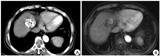
图 1 原发性肝癌患者介入治疗后行MSCT及MRI检查图像
A: MSCT图像; B: MRI图像.
Figure 1. MSCT and MRI imaging of patients with primary liver cancer after interventional therapy.
表 1 MRI、MSCT单一检查及联合检查病灶检出情况
Table 1. Detection of lesions by single MRI, single MSCT and joint inspection (n)
检查方式 残留病灶 复发病灶 阴性残留病灶 阴性复发病灶 单一MSCT(n=50 81 58 27 26 单一MRI(n=40) 74 49 18 19 MRI、MSCT联合(n=40) 52 41 10 12 MSCT: 多层螺旋CT.  下载: 导出CSV
表 2 MSCT、MRI单一检查及联合检查诊断残留病灶的敏感度、特异性及准确性
Table 2. Sensitivity, specificity and accuracy of MRI alone, MSCT alone and joint inspection in the diagnosis of residual lesions (%)
检查方式 残留病灶 敏感度 特异性 准确性 MSCT 77.9(74/95)a 46.2(6/13) 91.4(74/81)a MRI 83.5(71/85)b 57.1(4/7) 95.9(71/74)b MRI、MSCT联合 89.3(50/56)ab 66.7(4/6) 96.2(50/52)ab aP < 0.05 vs MRI; bP < 0.05 vs MSCT.
下载: 导出CSV
表 3 MSCT、MRI单一检查及联合检查诊断复发病灶的敏感度、特异性及准确性
Table 3. Sensitivity, specificity and accuracy of MRI alone, MSCT alone and joint inspection in the diagnosis of residual lesions (%)
检查方式 复发病灶 敏感度 特异性 准确性 MSCT 70.7(53/75)a 44.4(4/9) 91.4(53/58)a MRI 74.2(46/62)b 50.0(3/6) 93.9(46/49)b MRI、MSCT联合 88.6(39/44)ab 77.8(7/9) 95.1(39/41ab aP < 0.05 vs MRI; bP < 0.05 vs MSCT.
下载: 导出CSV
-
[1] 郑新闻, 刘丹, 李振平, 等. 3.0T MRI与64排CT评价原发性肝癌介入治疗后疗效的价值[J]. 中国CT和MRI杂志, 2019, 17(2): 32-4, 56. doi: 10.3969/j.issn.1672-5131.2019.02.010 [2] 樊建朝, 赵香田. 核磁共振、增强CT及超声造影对肝癌介入治疗疗效评估的对比研究[J]. 中西医结合肝病杂志, 2019, 29(5): 463-5. https://www.cnki.com.cn/Article/CJFDTOTAL-ZXGB201905025.htm [3] 孙瑜. CT和MRI对肝癌介入手术患者术后肿瘤活性的判定价值[J]. 中国CT和MRI杂志, 2020, 18(7): 85-7. doi: 10.3969/j.issn.1672-5131.2020.07.027 [4] 张宝国. CT与MRI在诊断原发性肝癌介入术后残余肿瘤活性的评估价值比较[J]. 中国医疗器械信息, 2022, 28(1): 67-9. https://www.cnki.com.cn/Article/CJFDTOTAL-ZGQX202201016.htm [5] 朱甲峰, 刘敏, 安宁. 探讨CT扫描联合磁共振诊断原发性肝癌及评估其介入治疗术后效果的临床效果[J]. 影像研究与医学应用, 2020, 4(18): 198-200. doi: 10.3969/j.issn.2096-3807.2020.18.107 [6] 杨青松. CT扫描联合磁共振诊断原发性肝癌及评估其介入治疗术后的临床效果评价[J]. 当代医学, 2019, 25(30): 110-2. doi: 10.3969/j.issn.1009-4393.2019.30.046 [7] 赵宝贵. 原发性肝癌运用多层螺旋CT多期增强扫描诊断的价值评价[J]. 中国实用医药, 2020, 15(5): 44-6. https://www.cnki.com.cn/Article/CJFDTOTAL-ZSSA202005018.htm [8] Li S, Shi SL, Li AP, et al. Diffusion-weighted magnetic resonance imaging in assessment of primary liver cancer after HIFU treatment [J]. J Coll Physicians Surg Pak, 2019, 29(4): 305-8. doi: 10.29271/jcpsp.2019.04.305 [9] 彭川, 罗鹰, 夏玉梅. 多层螺旋CT与核磁共振在原发性肝癌介入术后病灶残留及复发中的应用价值[J]. 中国CT和MRI杂志, 2018, 16 (5): 87-9, 106. doi: 10.3969/j.issn.1672-5131.2018.05.027 [10] 张志坚, 雷志丹. 多层螺旋CT在诊断鉴别肝转移瘤、原发性肝癌患者中的应用[J]. 中国CT和MRI杂志, 2018, 16(5): 90-3. doi: 10.3969/j.issn.1672-5131.2018.05.028 [11] 胡元清, 赵春, 刘颖. CT扫描联合磁共振诊断原发性肝癌及评估其介入治疗术后效果的临床研究[J]. 全科口腔医学电子杂志, 2019, 6 (1): 153, 157. doi: 10.3969/j.issn.2095-7882.2019.01.115 [12] 朱芳成, 陈鸿光, 郎清. CT增强扫描在评估原发性肝癌介入治疗近期疗效中的应用价值分析[J]. 中国CT和MRI杂志, 2019, 17(2): 29-31, 97. doi: 10.3969/j.issn.1672-5131.2019.02.009 [13] 邬振国, 陈永永, 李楠. 射频消融联合TACE治疗原发性肝癌患者疗效及对外周血T细胞亚群的影响[J]. 实用肝脏病杂志, 2017, 20(3): 324-7. doi: 10.3969/j.issn.1672-5069.2017.03.017 [14] Chen SL, Feng ST, Wei JW, et al. Pretreatment prediction of immunoscore in hepatocellular cancer: a radiomics-based clinical model based on Gd-EOB-DTPA-enhanced MRI imaging[J]. Eur Radiol, 2019, 29(8): 4177-87. doi: 10.1007/s00330-018-5986-x [15] 孙文杰, 高知玲, 高雨佳, 等. 基于多层螺旋CT全肝灌注成像小肝癌射频消融术后早期血流状态变化的定量评价[J]. 中华肝脏病杂志, 2020, 28(6): 488-93. doi: 10.3760/cma.j.cn501113-20200317-00120 [16] 刘小玲, 阮君, 朱敬松. MRI与CT在原发性肝癌介入治疗术后残余复发的应用分析[J]. 医学影像学杂志, 2020, 30(11): 2135-8. https://www.cnki.com.cn/Article/CJFDTOTAL-XYXZ202011053.htm [17] 刘帅, 许亚飞. CT扫描联合MRI在原发性肝癌的诊断及介入治疗术后的评价应用[J]. 浙江创伤外科, 2019, 24(2): 411-2. doi: 10.3969/j.issn.1009-7147.2019.02.102 [18] 潘利, 郑大伟. 多层螺旋CT与磁共振成像对原发性肝癌经肝动脉化疗栓塞术后疗效评价[J]. 中国医学装备, 2018, 15(8): 45-8. doi: 10.3969/J.ISSN.1672-8270.2018.08.014 [19] Liu YZ, Lei Y, Wang YN, et al. MRI-based treatment planning for proton radiotherapy: dosimetric validation of a deep learning-based liver synthetic CT generation method[J]. Phys Med Biol, 2019, 64 (14): 145015. doi: 10.1088/1361-6560/ab25bc [20] 王敬敏, 李潜, 王雁, 等. 超声造影与增强CT对原发性肝癌诊断准确率对比[J]. 实用癌症杂志, 2020, 35(5): 853-5, 859. doi: 10.3969/j.issn.1001-5930.2020.05.041 [21] 唐亚琴, 卿仁强, 杨松, 等. MRI联合血清ApoB、γ-GT用于原发性肝癌临床价值探讨[J]. 中国CT和MRI杂志, 2022, 20(1): 110-2. doi: 10.3969/j.issn.1672-5131.2022.01.035 [22] 吴建刚, 雷雪梅, 马清明. Gd-EOB-DTPA增强MRI与增强CT诊断原发性肝癌价值比较[J]. 中国CT和MRI杂志, 2021, 19(9): 100-1, 134 doi: 10.3969/j.issn.1672-5131.2021.09.031 -

 点击查看大图
计量
- 文章访问数: 198
- HTML全文浏览量: 138
- PDF下载量: 5
- 被引次数: 0