


Application of amide proton transfer imaging in amnestic mild cognitive impairment
-
摘要:
目的 探讨采用磁共振酰胺质子转移成像技术(APT)研究遗忘型轻度认知障碍(aMCI)患者各感兴趣脑区的病理生理学变化及其与部分临床认知评分量表相关性的可行性。 方法 选取2021年1月~2022年1月神经内科确诊的21例遗忘型轻度认知障碍患者作为病例组,另外选择同期本院体检中心21例健康人员作为对照组,对两组分别进行临床资料分析、临床心理学量表评估,对各感兴趣脑区进行常规磁共振成像及酰胺质子转移成像。 结果 与对照组相比,病例组右侧海马、双侧杏仁核区域APT值升高(P < 0.05),左侧海马区APT值升高(P < 0.01);病例组蒙特利尔认知评估量表、简易精神状态评价量表评分低于对照组(P < 0.05);简易精神状态评价量表评分与右侧海马、左侧杏仁核的APT值呈负相关(P < 0.05);在蒙特利尔认知评估量表评分的各个子项目评分与各脑区域APT值比较中,aMCI组右侧海马的APT值与视空间及执行功能得分呈负相关(P < 0.05),左侧杏仁核及双侧海马区域APT值与记忆力分值呈负相关(P < 0.05)。 结论 aMCI患者部分脑组织存在一系列病理生理的改变,APT技术作为新兴的功能磁共振成像技术,能够在一定程度上帮助临床医生对aMCI患者进行早期诊断。 Abstract:Objective To investigate the feasibility of using magnetic resonance amide proton transfer imaging (APT) to study the pathophysiological changes in various brain regions of interest in patients with amnestic mild cognitive impairment (aMCI) and its correlation with some clinical cognitive score scales. Methods Twenty-one patients with aMCI diagnosed in the Department of Neurology from January 2021 to January 2022 were selected as the case group, and another twenty-one healthy subjects in the physical examination center of our hospital during the same period were selected as the control group. The clinical data of the two groups were analyzed, the clinical psychological scale was assessed, and routine magnetic resonance imaging and amide proton transfer imaging were performed in each brain region of interest. Results Compared with the control group, APT values in the right hippocampus and bilateral amygdala were significantly increased (P < 0.05), and left hippocampus had statistical significance (P < 0.01). The clinical scores of MoCA and MMSE in case group were significantly lower than those in control group (P < 0.05). MMSE score scale was negatively correlated with APT values of right hippocampus and left amygdala (P < 0.05). In the comparison of the APT values in each subitem score of the Montreal Cognitive Assessment score and each brain region, the APT values in the right hippocampus were negatively correlated with visual spatial and executive function scores in the aMCI group (P < 0.05), and the APT value of left amygdala and bilateral hippocampus were negatively correlated with memory scores (P < 0.05). Conclusion Forgetting in patients with aMCI part of the brain is a series of pathophysiological changes. APT technique, as an emerging functional magnetic resonance imaging technique, can help clinicians to make an early diagnosis of patients with aMCI to a certain extent. -
表 1 病例组与对照组临床资料比较
Table 1. Comparison of clinical data between case group and control group(n=21, Mean±SD)
项目 病例组 对照组 T/Z P 性别(n, 男/女) 13/8 10/11 - 1.000 年龄(岁) 66.52±4.84 65±3.74 1.141 0.261 吸烟史(n, 有/无) 12/9 8/13 - 1.000 受教育程度(年) 5.0±1.64 5.9±1.48 -1.946 0.052 MMSE得分(分) 23.86±2.03 27.43±0.75 -2.319 0.020 MoCA得分(分) 22.1±2.14 22.86±0.79 -3.355 0.001 蒙特利尔认知评估量表;MMSE:简易精神状态评价量表.  下载: 导出CSV
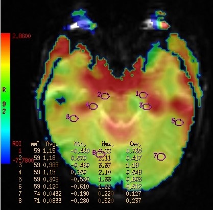
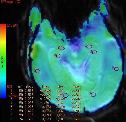
表 2 病例组与对照组各感兴趣脑区APT值比较
Table 2. Comparison of APT values in each brain region of interest between the case group and the control group(n=21, Mean±SD)
部位 病例组 对照组 t P 右侧海马 1.01±0.46 0.67±0.39 2.552 0.015 左侧海马 1.11±0.48 0.74±0.35 2.868 0.007 右侧杏仁核 0.97±0.19 0.76±0.20 -2.844 0.004 左侧杏仁核 0.93±0.14 0.77±0.14 -3.141 0.002 右侧颞叶白质 0.44±0.12 0.37±0.16 -1.661 0.097 左侧颞叶白质 0.43±0.12 0.41±0.16 0.479 0.634 右侧枕叶白质 0.42±0.14 0.38±0.15 0.938 0.354 左侧枕叶白质 0.35±0.20 0.34±0.10 0.379 0.706
下载: 导出CSV
表 3 病例组不同感兴趣区域APT值与临床评分量表的相关性
Table 3. Correlation of APT values in different regions of interest and clinical rating scale in case group
部位 MOCA MMSE r P r P 右侧海马 -0.395 0.077 -0.474 0.030 左侧海马 -0.256 0.263 -0.390 0.081 右侧杏仁核 -0.374 0.095 -0.252 0.270 左侧杏仁核 -0.227 0.323 -0.462 0.035 右侧颞叶白质 0.082 0.723 0.119 0.608 左侧颞叶白质 -0.073 0.754 0.055 0.813 右侧枕叶白质 -0.110 0.636 0.028 0.905 左侧枕叶白质 -0.128 0.579 0.241 0.293
下载: 导出CSV
表 4 病例组不同感兴趣区APT值与MoCA子项目分数的相关性
Table 4. Correlations between APT values and MoCA sub-item scores in different regions of interest in case group
部位 视空间和执行功能 抽象思维能力 记忆力 定向力 命名力 注意力 语言能力 r P r P r P r P r P r P r P 右侧海马 -0.492 0.023 0.040 0.081 -0.531 0.013 -0.078 0.624 -0.102 0.061 -0.405 0.069 0.202 0.379 左侧海马 0.309 0.173 0.095 0.549 0.483 0.026 0.171 0.458 0.166 0.471 0.236 0.304 0.049 0.832 右侧杏仁核 0.426 0.054 0.029 0.857 -0.342 0.129 0.185 0.423 0.074 0.750 -0.242 0.291 0.026 0.910 左侧杏仁核 -0.222 0.333 -0.018 0.420 0.453 0.039 0.184 0.424 -0.213 0.353 -0.102 0.661 0.143 0.537 右侧颞叶白质 0.064 0.784 0.053 0.740 -0.173 0.453 0.000 0.000 0.028 0.905 0.001 0.998 -0.204 0.374 左侧颞叶白质 0.011 0.961 0.029 0.145 -0.106 0.647 -0.028 0.905 -0.037 0.874 -0.192 0.405 -0.028 0.904 右侧枕叶白质 -0.162 0.484 0.177 0.263 -0.221 0.337 -0.209 0.364 0.139 0.549 0.260 0.255 0.064 0.782 左侧枕叶白质 -0.175 0.447 0.080 0.616 -0.183 0.427 -0, 240 0.295 0.231 0.314 0.177 0.444 0.005 0.983
下载: 导出CSV
-
[1] Lin H, Li MW, Zhan Y, et al. Disrupted white matter functional connectivity in aMCI APOEε4 carriers: a resting- state study[J]. Brain Imaging Behav, 2021, 15(4): 1739-47. doi: 10.1007/s11682-020-00367-7 [2] Zhang JH, Liu YF, Lan K, et al. Gray matter atrophy in amnestic mild cognitive impairment: a voxel-based meta-analysis[J]. Front Aging Neurosci, 2021, 13: 627919. doi: 10.3389/fnagi.2021.627919 [3] 孔令伟, 梅利. 遗忘型轻度认知障碍在磁共振波谱应用中的研究进展[J]. 临床医药文献电子杂志, 2020, 7(16): 193. https://www.cnki.com.cn/Article/CJFDTOTAL-LCWX202016168.htm [4] Jiménez-Bonilla JF, Quirce R, de Arcocha-Torres M, et al. A 5-year longitudinal evaluation in patients with mild cognitive impairment by 11C- PIB PET/CT: a visual analysis[J]. Nucl Med Commun, 2019, 40(5): 525-31. doi: 10.1097/MNM.0000000000001004 [5] Petersen RC, Parisi JE, Dickson DW, et al. Neuropathologic features of amnestic mild cognitive impairment[J]. Arch Neurol, 2006, 63 (5): 665-72. doi: 10.1001/archneur.63.5.665 [6] 2021 Alzheimer's disease facts and figures[J]. Alzheimers Dement, 2021, 17(3): 327-406[J]. Alzheimers Dement 2021, 17 (3): 327-406. https://www.cnki.com.cn/Article/CJFDTOTAL-ZGGL202102002.htm [7] Li X, Zhang ZJ. Neuropsychological and neuroimaging characteristics of amnestic mild cognitive impairment subtypes: a selective overview[J]. CNS Neurosci Ther, 2015, 21(10): 776-83. doi: 10.1111/cns.12391 [8] Zhou JY, Heo HY, Knutsson L, et al. APT- weighted MRI: Techniques, current neuro applications, and challenging issues[J]. J Magn Reson Imaging, 2019, 50(2): 347-64. doi: 10.1002/jmri.26645 [9] 魏平. 酰胺质子转移成像在脑疾病中的研究进展[J]. 医学影像学杂志, 2020, 30(3): 496-9. https://www.cnki.com.cn/Article/CJFDTOTAL-XYXZ202003043.htm [10] Petrella JR. Neuroimaging and the search for a cure for Alzheimer disease[J]. Radiology, 2013, 269(3): 671-91. doi: 10.1148/radiol.13122503 [11] Chen J, Zhang ZJ, Li SJ. Can multi-modal neuroimaging evidence from Hippocampus provide biomarkers for the progression of amnestic mild cognitive impairment?[J]. Neurosci Bull, 2015, 31 (1): 128-40. doi: 10.1007/s12264-014-1490-8 [12] Jaroudi W, Garami J, Garrido S, et al. Factors underlying cognitive decline in old age and Alzheimer's disease: the role of the Hippocampus[J]. Rev Neurosci, 2017, 28(7): 705-14. doi: 10.1515/revneuro-2016-0086 [13] 武文博, 刘任远, 张鑫, 等. 在AD病程中海马代谢水平与体积改变的研究[J]. 东南大学学报: 医学版, 2015, 34(4): 489-96. https://www.cnki.com.cn/Article/CJFDTOTAL-NJTD201504001.htm [14] He QL, Brown TI. Heterogeneous correlations between Hippocampus volume and cognitive map accuracy among healthy young adults[J]. Cortex, 2020, 124: 167-75. doi: 10.1016/j.cortex.2019.11.011 [15] Panza F, Lozupone M, Logroscino G, et al. A critical appraisal of amyloid- β- targeting therapies for Alzheimer disease[J]. Nat Rev Neurol, 2019, 15(2): 73-88. doi: 10.1038/s41582-018-0116-6 [16] Zhang ZW, Zhang CQ, Yao J, et al. Protein- based amide proton transfer- weighted MR imaging of amnestic mild cognitive impairment[J]. Neuroimage Clin, 2020, 25: 102153. doi: 10.1016/j.nicl.2019.102153 [17] Guo ZX, Jiang YC, Qin XY, et al. Amide proton transfer-weighted MRI might help distinguish amnestic mild cognitive impairment from a normal elderly population[J]. Front Neurol, 2021, 12: 707030. doi: 10.3389/fneur.2021.707030 [18] Wirt RA, Hyman JM. Integrating spatial working memory and remote memory: interactions between the medial prefrontal cortex and Hippocampus[J]. Brain Sci, 2017, 7(4): 43. [19] 张鸿, 王经泰, 何英杰, 等. 海马萎缩异质性的轻度认知障碍患者皮质下核团体积变化的研究[J]. 中华老年医学杂志, 2021, 40 (8): 1055-6. doi: 10.3760/cma.j.issn.0254-9026.2021.08.025 [20] 陈钱, 张雯, 青钊, 等. 轻度认知障碍患者海马和皮质下核团形状特征预测AD病理改变的研究[J]. 阿尔茨海默病及相关病, 2019, 2(4): 523-30. https://www.cnki.com.cn/Article/CJFDTOTAL-AAEC201904013.htm [21] 肖宁, 热米拉·玉山, 陆鹏, 等. 磁共振NODDI技术在帕金森认知障碍患者海马微观结构的应用[J]. 分子影像学杂志, 2022, 45 (1): 35-9. doi: 10.12122/j.issn.1674-4500.2022.01.07 [22] Ciesielska N, Sokołowski R, Mazur E, et al. Is the Montreal Cognitive Assessment (MoCA) test better suited than the MiniMental State Examination (MMSE) in mild cognitive impairment (MCI) detection among people aged over 60? Meta- analysis[J]. Psychiatr Pol, 2016, 50(5): 1039-52 doi: 10.12740/PP/45368 -

 点击查看大图
计量
- 文章访问数: 168
- HTML全文浏览量: 148
- PDF下载量: 11
- 被引次数: 0